The immune system plays an essential role in the organism's response to cancer. Several hematological markers can influence prognosis and survival of patients. The objective of this study is to determine their prognostic value in testicular germ cell tumors.
Material and methodsRetrospective cohort study on 164 patients with germ cell tumors. Clinical, analytical, histological and evolutionary data were collected. The absolute neutrophil and absolute platelet counts, neutrophil–lymphocyte (NLR), platelet–lymphocyte and lymphocyte–monocyte ratios were estimated at diagnosis. The association that these markers can have with the classic prognostic factors, as well as their effect on prognosis and survival, have been analyzed.
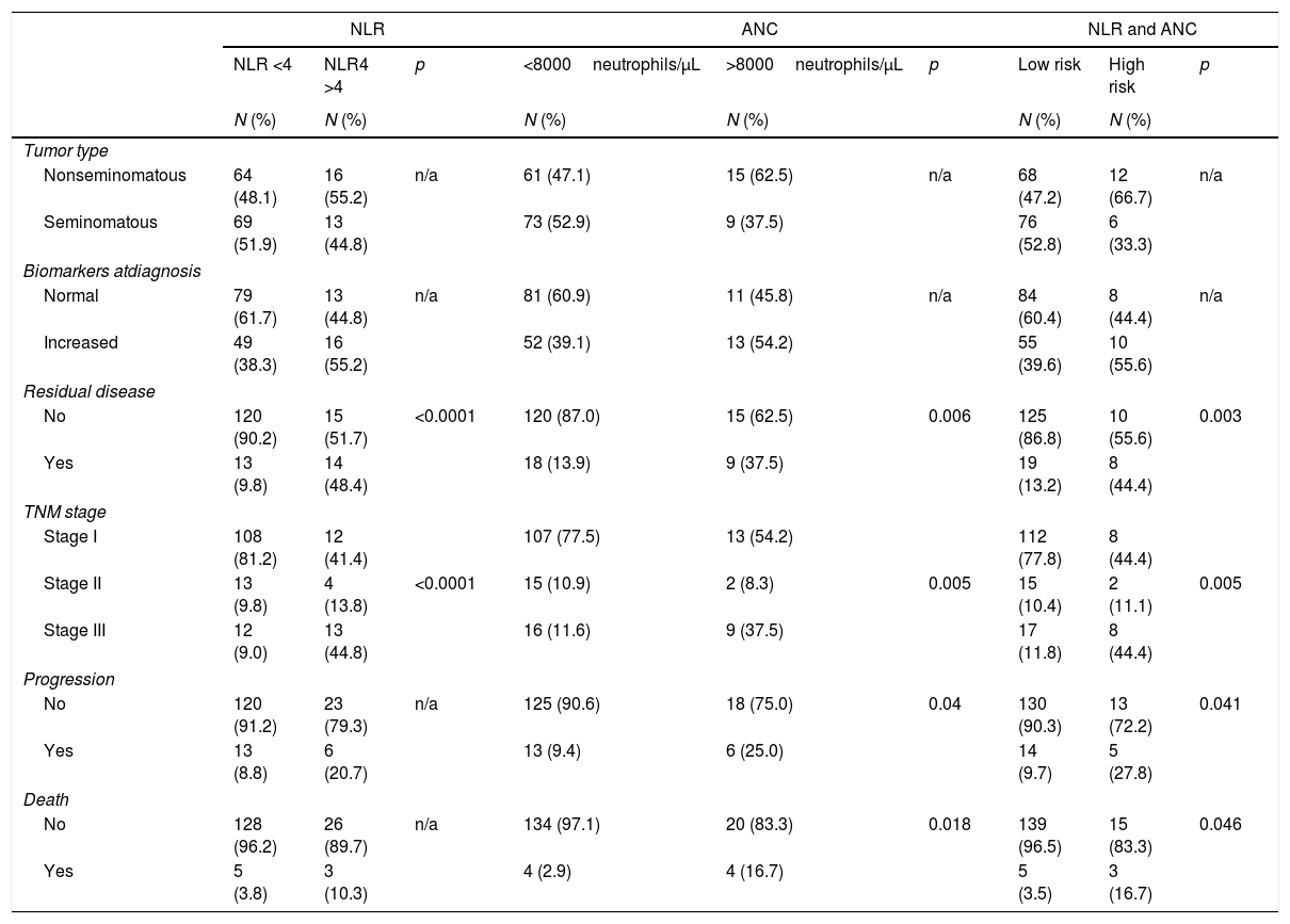
Results17.7% had NLR >4 and 14.6% ANC >8000/μL. These patients presented higher percentages of residual disease and stage II–III tumors. Patients with elevated absolute neutrophil showed also higher percentages of progression and exitus.
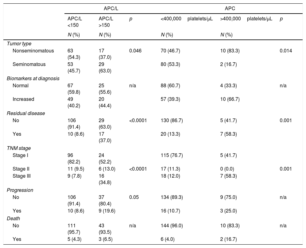
7.3% presented absolute platelet >400,000/μL. These patients obtained higher rates of residual disease, nonseminomatous and stage III tumors. 28.4% showed platelet–lymphocyte values >150. This data was associated to higher percentages of residual disease, progression, stage II and III tumors and seminomatous tumors.
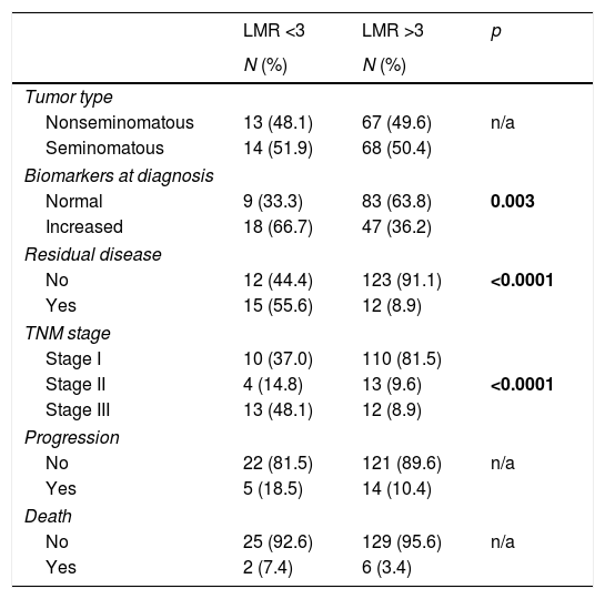
83.3% had a lymphocyte–monocyte >3. These patients presented: higher tumor markers in normal range, decreased residual disease rates and higher percentages of stage I and II tumors.
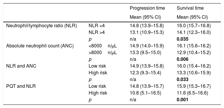
The mean survival time was shorter in patients with NLR >4 and absolute neutrophil >8000/μL.
The ROC curves showed significance in the prediction of progression and values of lymphocyte–monocyte >3, and prediction of survival and values NLR >4.
ConclusionOur results indicate that the analyzed hematological markers are associated with poor prognoses at diagnosis. Therefore, their use in daily clinical practice can be a valuable tool in the diagnosis and prognosis of patients with testicular germ cell tumors.
El sistema inmune ejerce un papel clave en la respuesta del organismo frente al cáncer. Existen diversos marcadores hematológicos que pueden influir en el pronóstico y supervivencia de los pacientes. El objetivo de este estudio es determinar su valor pronóstico en tumores testiculares de células germinales.
Material y métodosEstudio de cohortes retrospectivo sobre 164 pacientes con tumores testiculares de células germinales. Se recogieron datos clínicos, analíticos, histológicos y evolutivos. Se estimaron, al diagnóstico, el recuento total de neutrófilos y plaquetas, la ratio neutrófilo-linfocito (RN/L), plaqueta-linfocito(RP/L) y linfocito-monocito (RL/M). Se analizó la relación que estos marcadores pueden tener sobre los factores pronósticos clásicos, así como sobre el pronóstico y supervivencia.
ResultadosUn 17,7% tuvieron una RN/L>4 y un 14,6% un RNT>8000/μL. Estos enfermos, presentaron mayor porcentaje de enfermedad residual y tumores en estadios II y III. Los enfermos con recuento total de neutrófilos elevado también tuvieron mayor porcentaje de progresión y éxitus.
Un 7,3%, tenían un recuento total de plaquetas >400000/μL. Estos enfermos tuvieron un mayor porcentaje de tumores no seminomatosos, de enfermedad residual y tumores en estadio III. El 28,4% mostraron valores RP/L>150, asociándose este dato a mayor porcentaje de tumores seminomatosos, enfermedad residual, estadios II y III y progresión.
El 83,3% tuvieron una RL/M>3. Estos enfermos presentaron: mayor porcentaje de marcadores tumorales en rango normal, menor porcentaje de enfermedad residual y mayor porcentaje de pacientes en estadio I y II.
El tiempo medio de supervivencia fue menor en pacientes con RN/L>4 y con recuento total de neutrófilos >8.000/μL.
Las curvas ROC mostraron significación en la predicción de progresión y valores de RL/M>3, y predicción de supervivencia y valores RN/L>4.
ConclusiónNuestros resultados indican que los marcadores hematológicos analizados se asocian situaciones de mal pronóstico en el momento del diagnóstico. Por tanto, su utilización en la práctica clínica diaria puede ser considerada como una herramienta más en el diagnóstico y pronóstico de pacientes con tumores testiculares de células germinales de testículo.


