The aim of the study was to describe the clinical drivers that lead physicians to perform imaging tests in search of metastasis in non-metastasic castration prostate resistant cancer (nmCRPC) patients.
MethodsObservational, cross-sectional study conducted at the Departments of Urology of 38 Spanish hospitals. The study included 188 patients diagnosed with nmCRPC who underwent an imaging test for the assessment of metástasis. In one study visit, physicians were requested to specify the clinical factors that led them to perform these tests. The results of the imaging tests and the clinical characteristics of the patients since the time of prostate cancer (PC) diagnosis, were reported. Regression analyses were used to determine predictors of imaging test results.
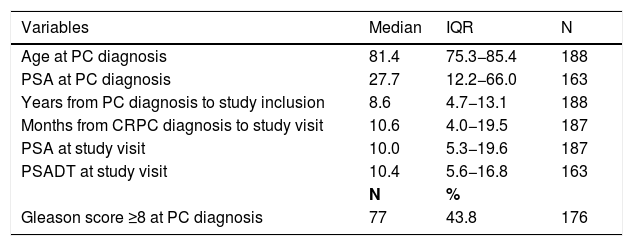
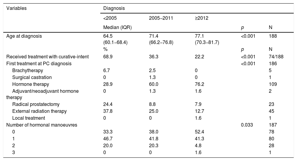
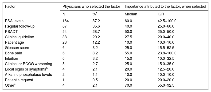
ResultsProstate-specific antigen (PSA) level was the most important driver to order imaging tests (57.1%), followed by regular follow-up (16.5%) and PSA doubling time (PSADT) (12.0%). Although these drivers were not associated to detection of metastasis, patients with PSA levels ≥20 ng/mL had a greater risk of metastasis than patients with PSA levels <4 ng/mL (p = 0.004) and CRPC patients diagnosed with metastasis (mCRPC) had higher median PSA levels (20.9; interquartile range [IQR]: 6.7–38.6) than nmCRPC (9.1; IQR: 5.0–18.0) (p = 0.005). Sixty-six percent of the patients did not undergo any imaging test after CRPC diagnosis until the study visit (10.6, IQR: 4.0−19.5 months). Curative-intent treatment at PC diagnosis and Gleason score predicted longer time from PC to CRPC diagnosis.
ConclusionsPhysicians based their decisions to order imaging tests for metastasis detection in nmCRPC patients mainly on PSA and PSA kinetics, including the regular follow-up stated by guideline recommendations.
El objetivo del estudio consistió en describir los factores clínicos que llevan a los médicos a realizar pruebas de imagen para identificar metástasis en pacientes con cáncer de próstata (CP) resistente a la castración no metastásico (CPRCnm).
MétodosEstudio observacional transversal realizado en los servicios de Urología de 38 hospitales españoles; 188 pacientes diagnosticados con CPRCnm sometidos una prueba de imagen para evaluar la presencia de metástasis fueron incluidos. Se solicitó a los médicos, en una única visita del estudio, que especificaran los factores clínicos que los llevaron a realizar estas pruebas. Se presentaron los resultados de las pruebas de imagen y las características clínicas de los pacientes desde el diagnóstico de CP. Se utilizaron análisis de regresión para determinar factores predictivos de los resultados de las pruebas de imagen.
ResultadosEl valor del «prostate-specific antigen» (por sus siglas en inglés, PSA), fue el factor más importante que determinó la solicitud de pruebas de imagen (57,1%), seguido de un seguimiento habitual (16,5%) y del tiempo de duplicación del PSA (TDPSA) (12,0%). Aunque estos factores no guardaron relación con la detección de metástasis, los pacientes con una concentración de PSA ≥20 ng/mL tuvieron un mayor riesgo de metástasis que aquellos con una concentración <4 ng/mL (p = 0,004), mientras que los pacientes con CPRC diagnosticados de metástasis (CPRCm) tuvieron una mayor mediana de concentración de PSA (20,9; intervalo intercuartílico [IIC]: 6,7–38,6) que aquellos con CPRCnm (9,1; IIC: 5,0–18,0) (p = 0,005). Un 66% no se sometió a ninguna prueba de imagen entre el diagnóstico de CPRC y la visita del estudio (10,6, IIC: 4,0–19,5 meses). El tratamiento con intención curativa en el momento del diagnóstico de CP y la puntuación de Gleason predijeron un mayor tiempo transcurrido entre los diagnósticos de CP y CPRC.
ConclusionesLos médicos basaron sus decisiones de solicitar pruebas de imagen para detectar metástasis en pacientes con CPRCnm principalmente en el PSA y su cinética, incluido el seguimiento habitual establecido por las guías clínicas.


