


La Aniridia: Actualización en Diagnóstico, Complicaciones y Tratamiento
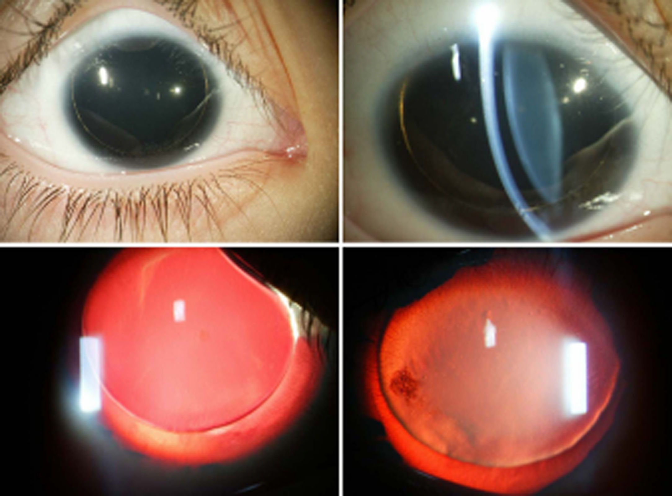
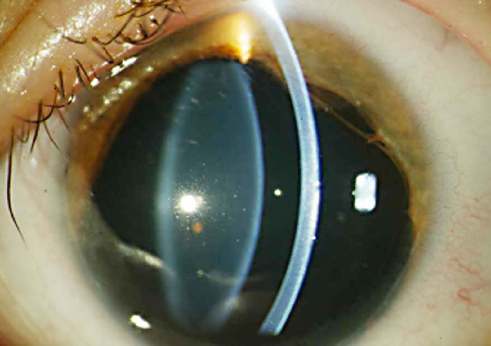
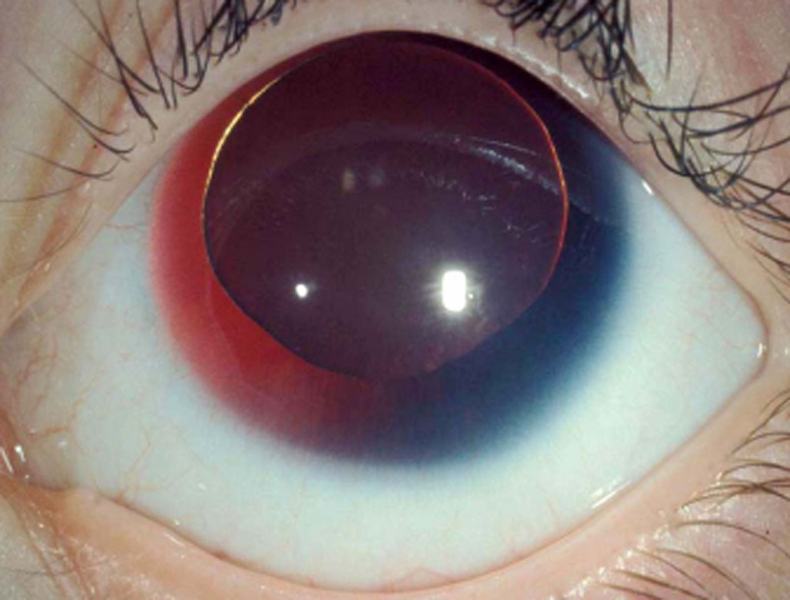
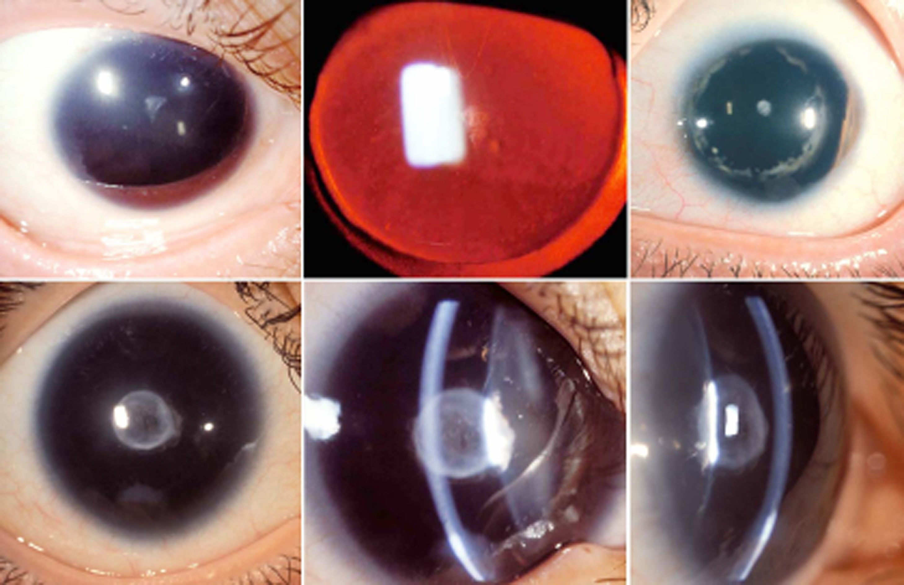
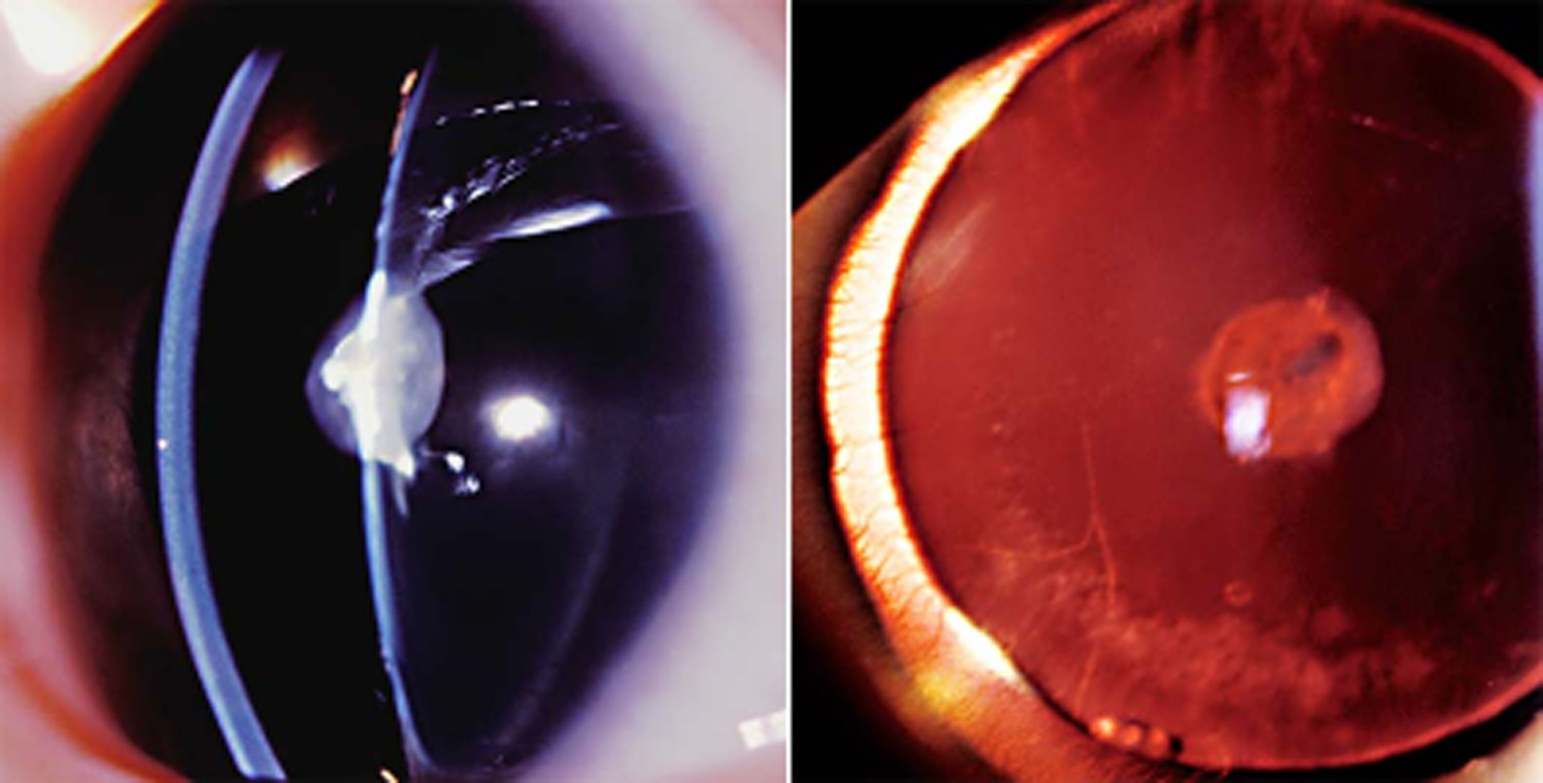
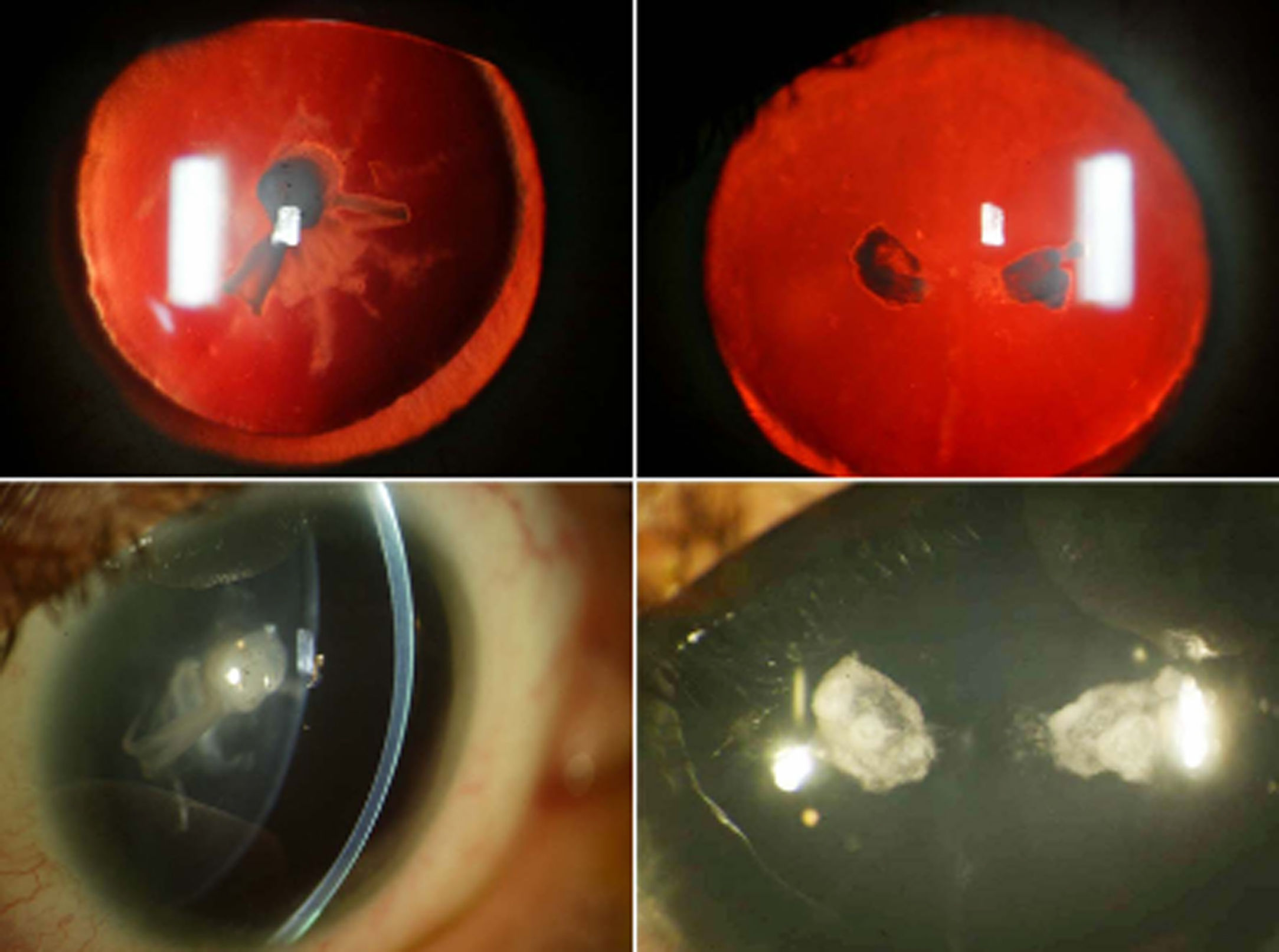
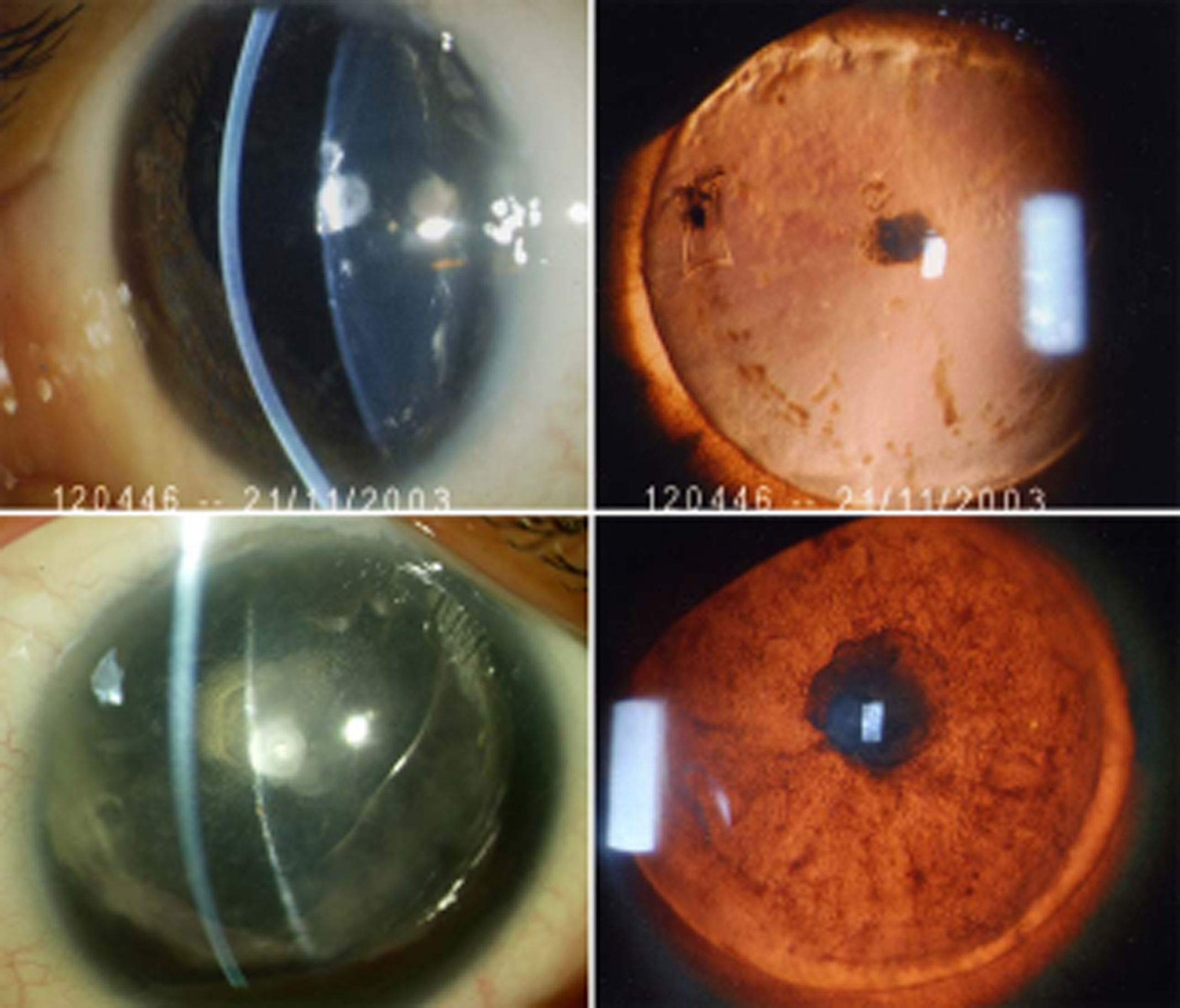
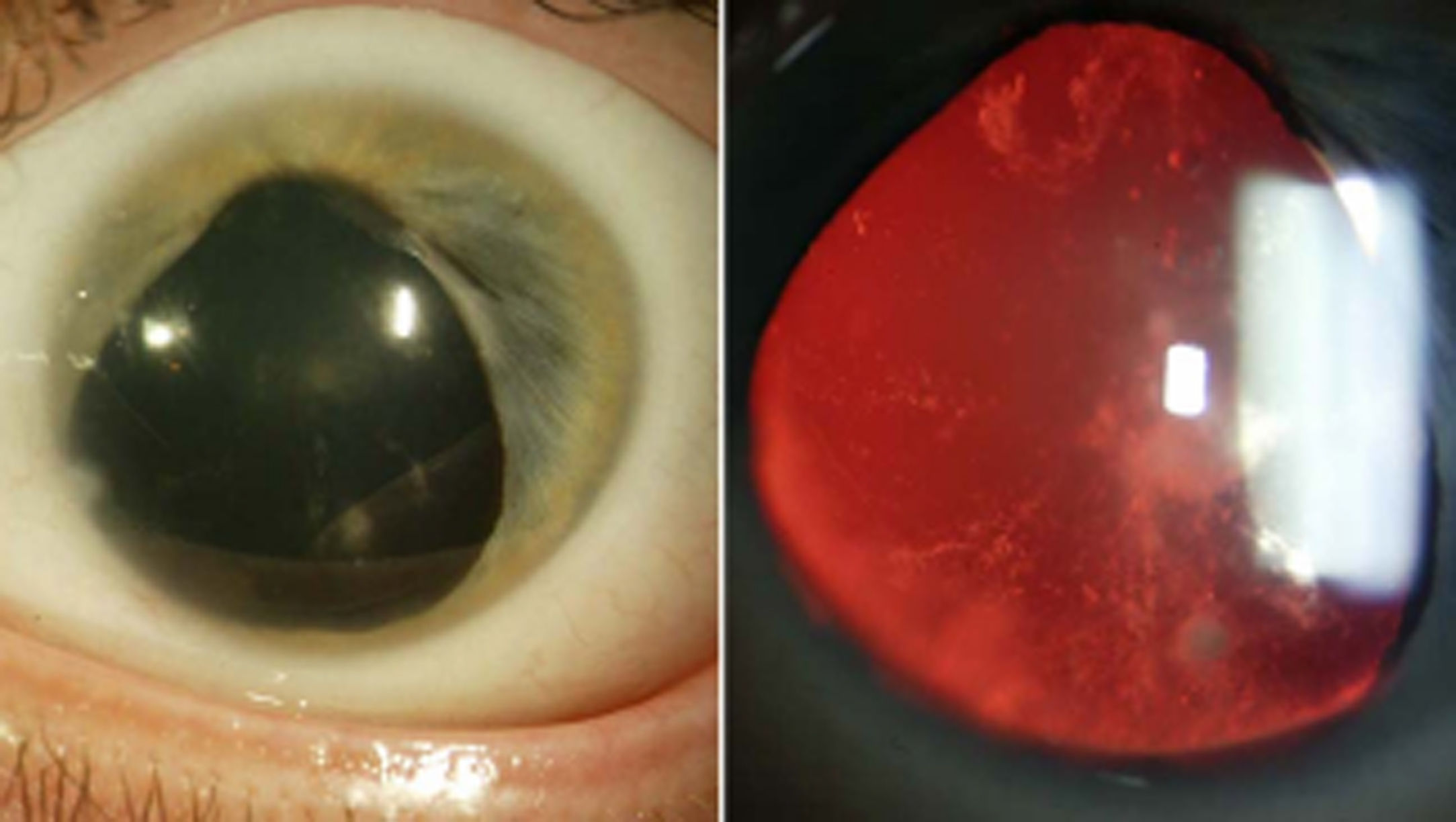
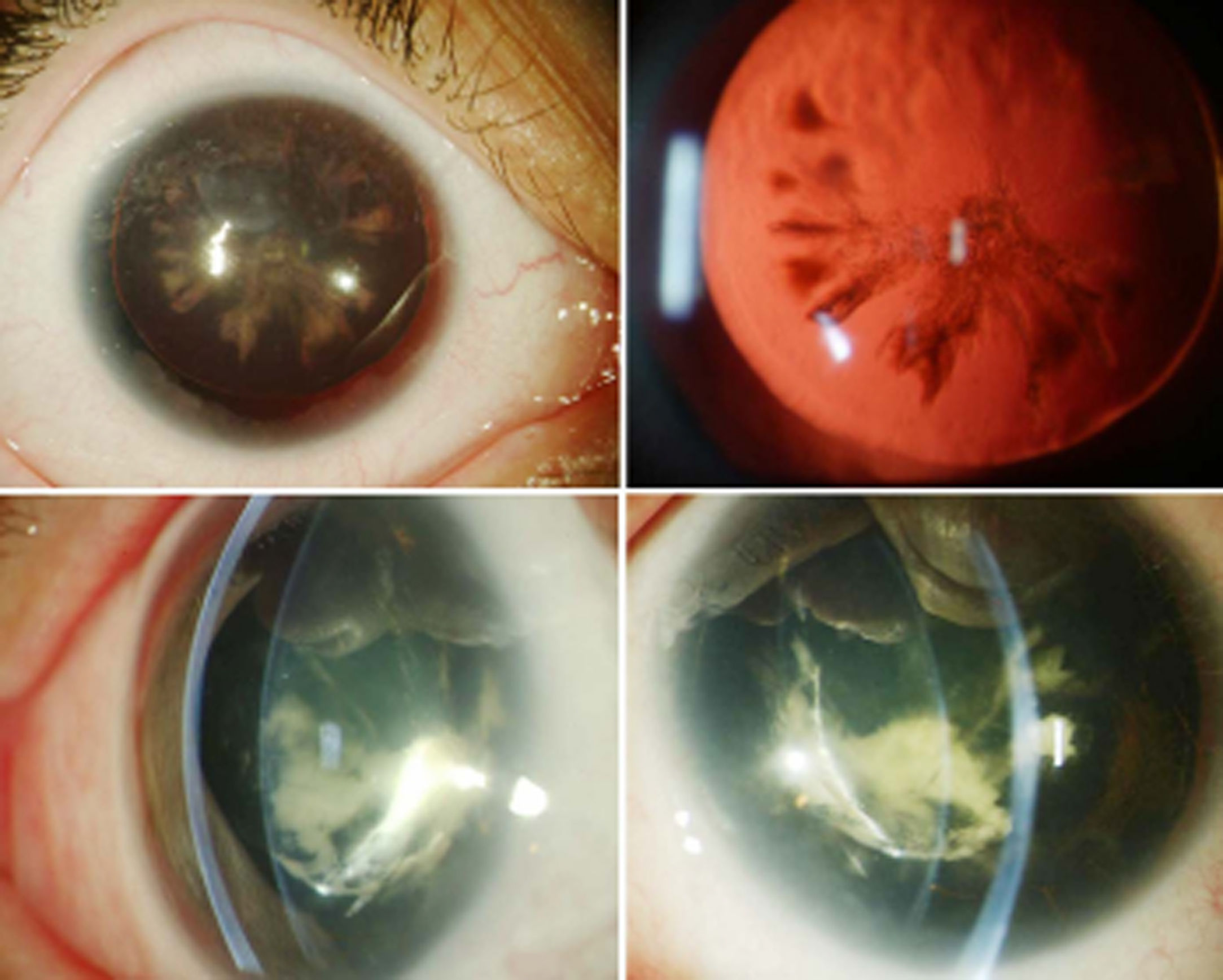
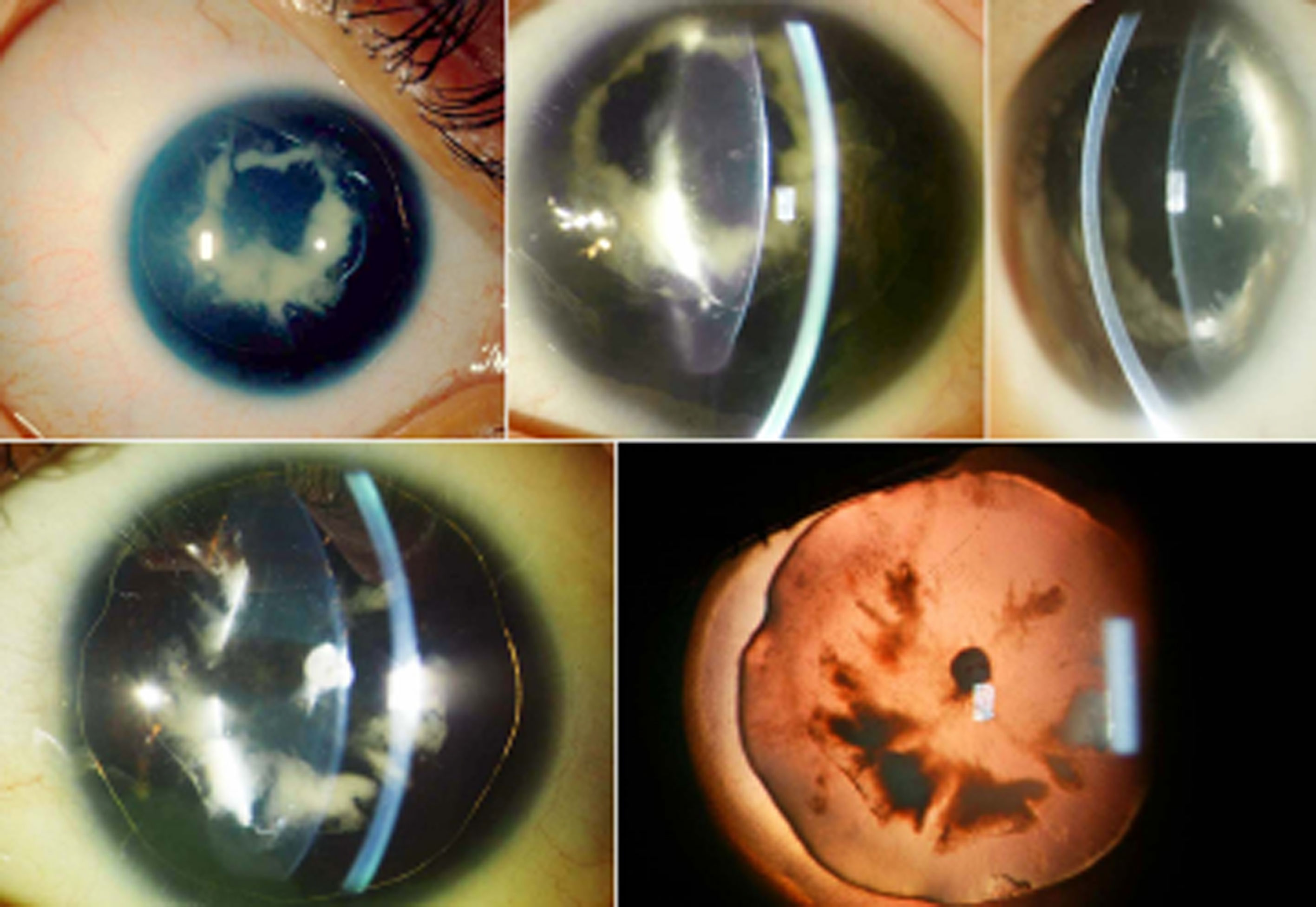
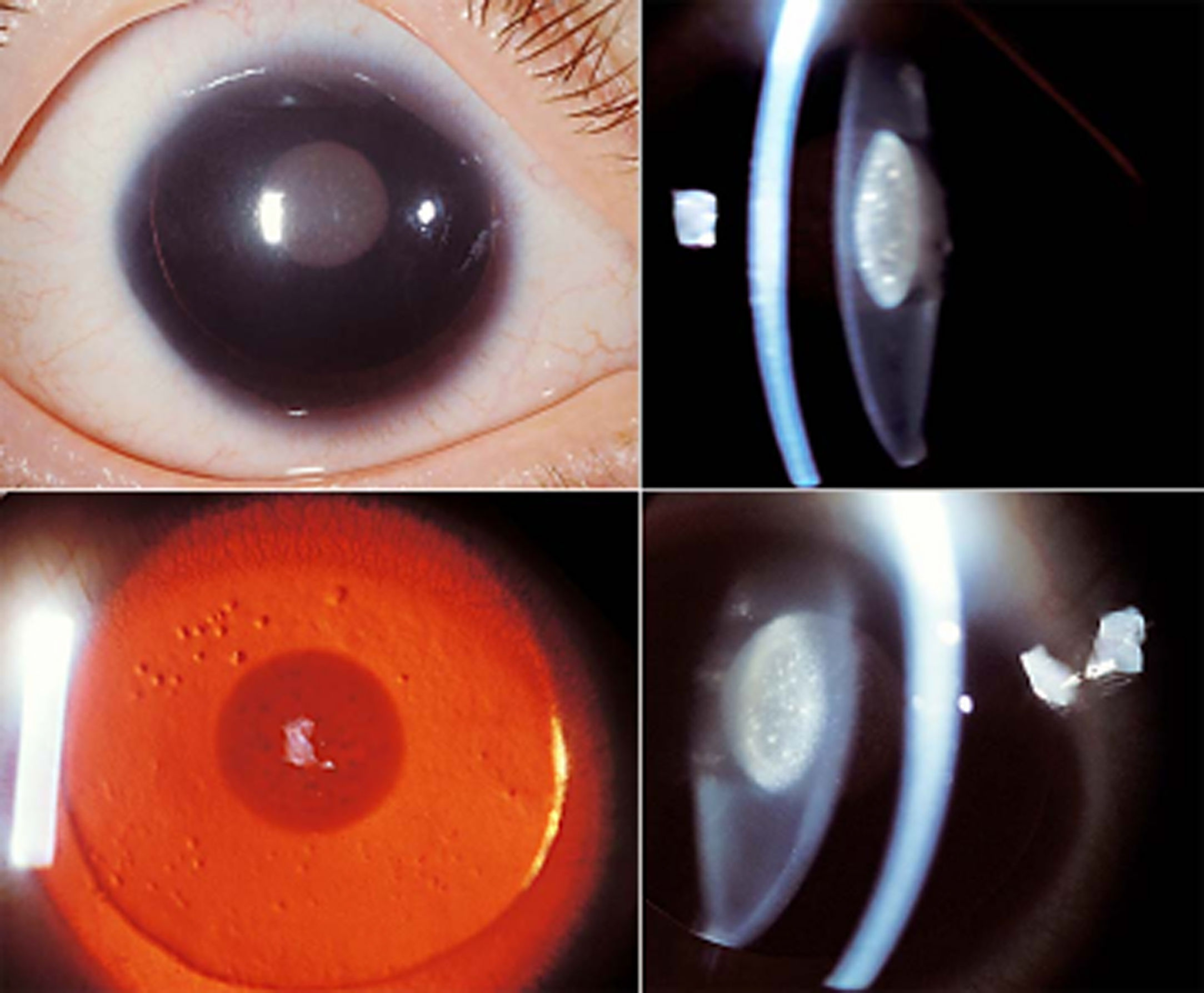
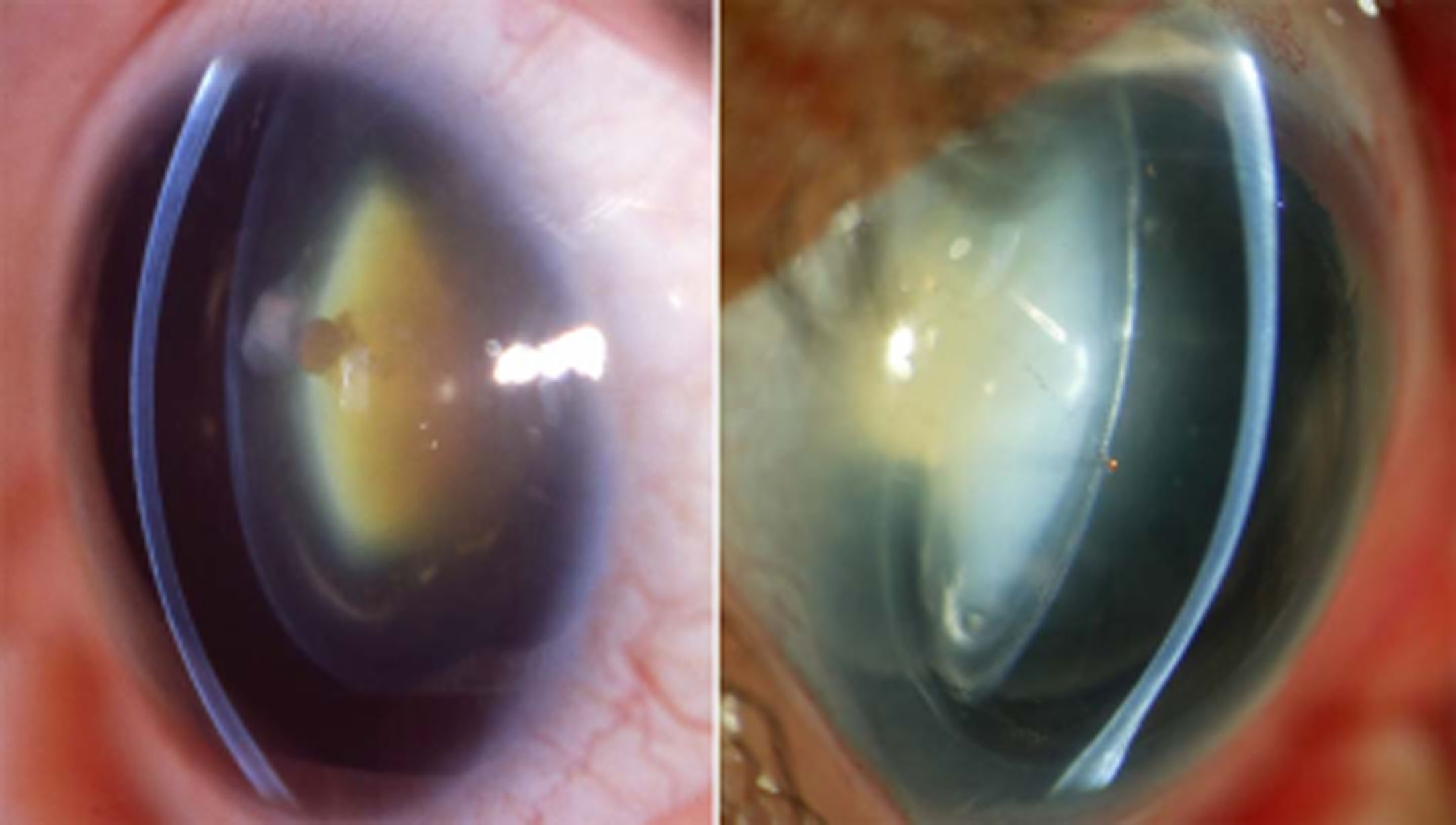
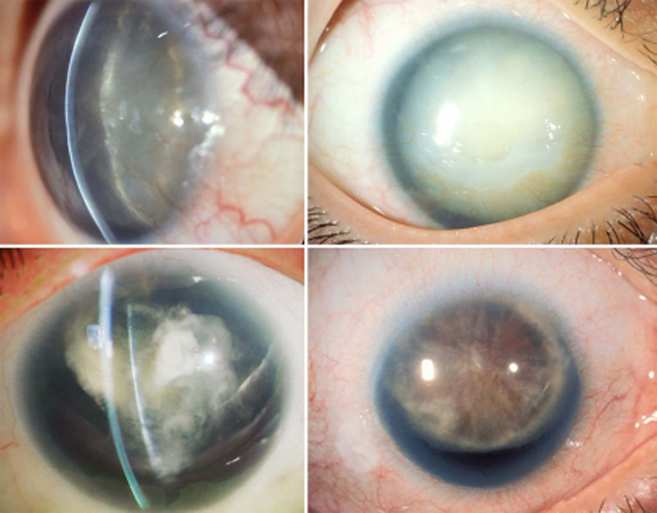
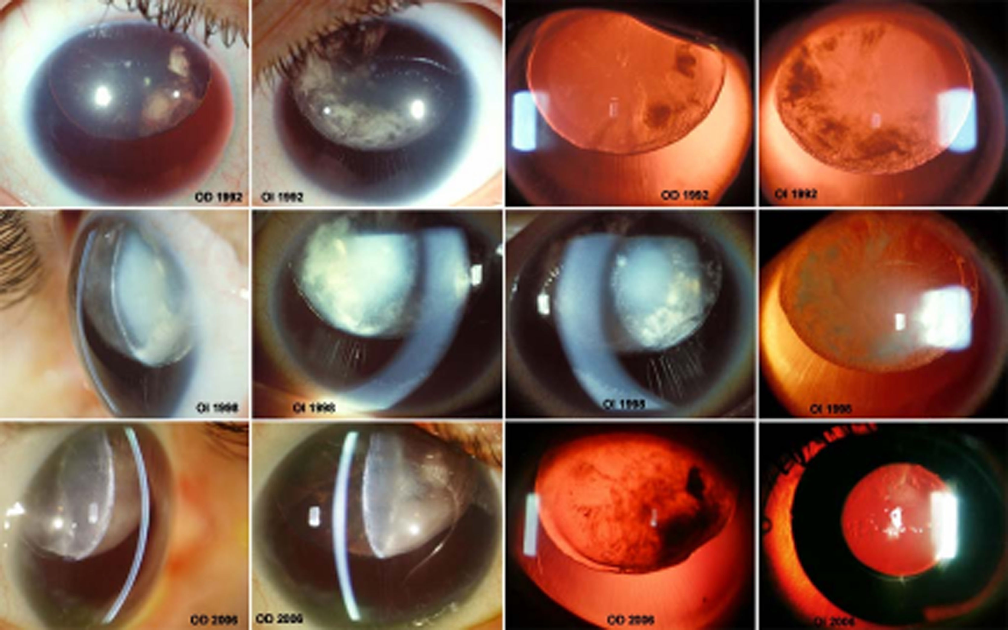
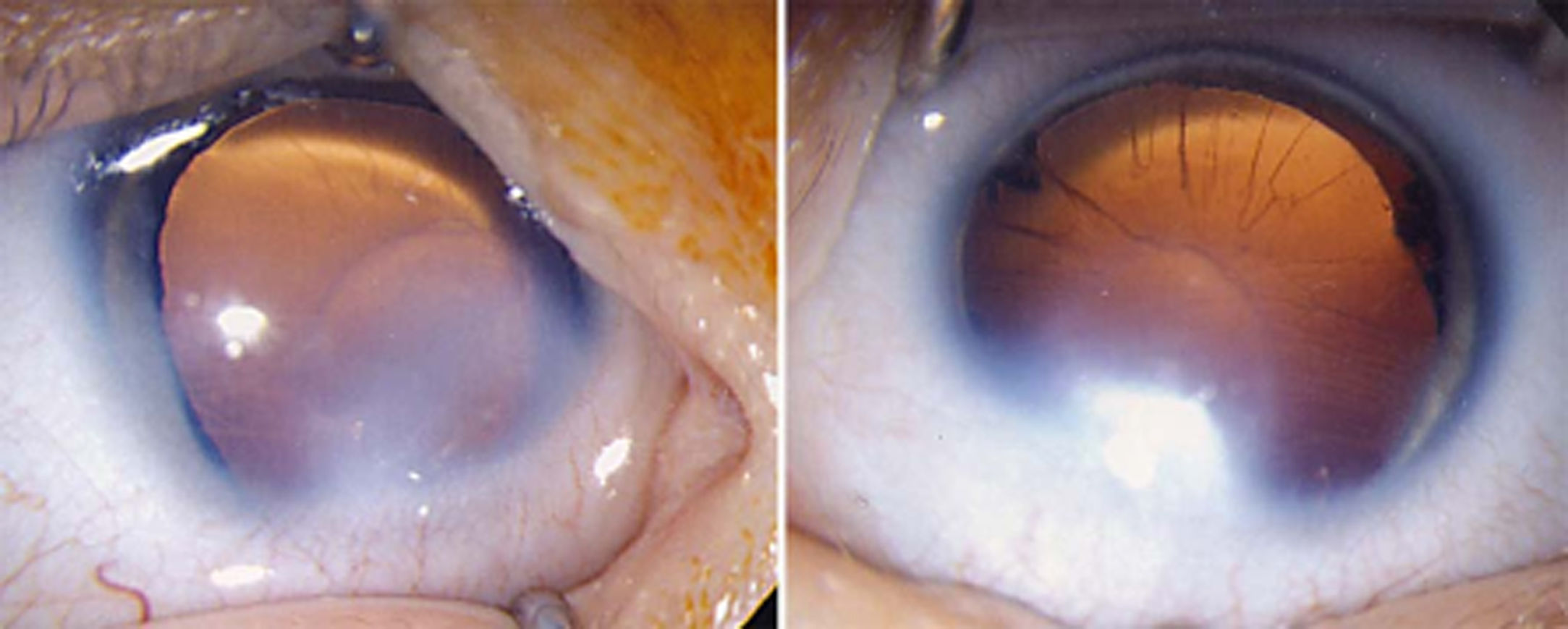
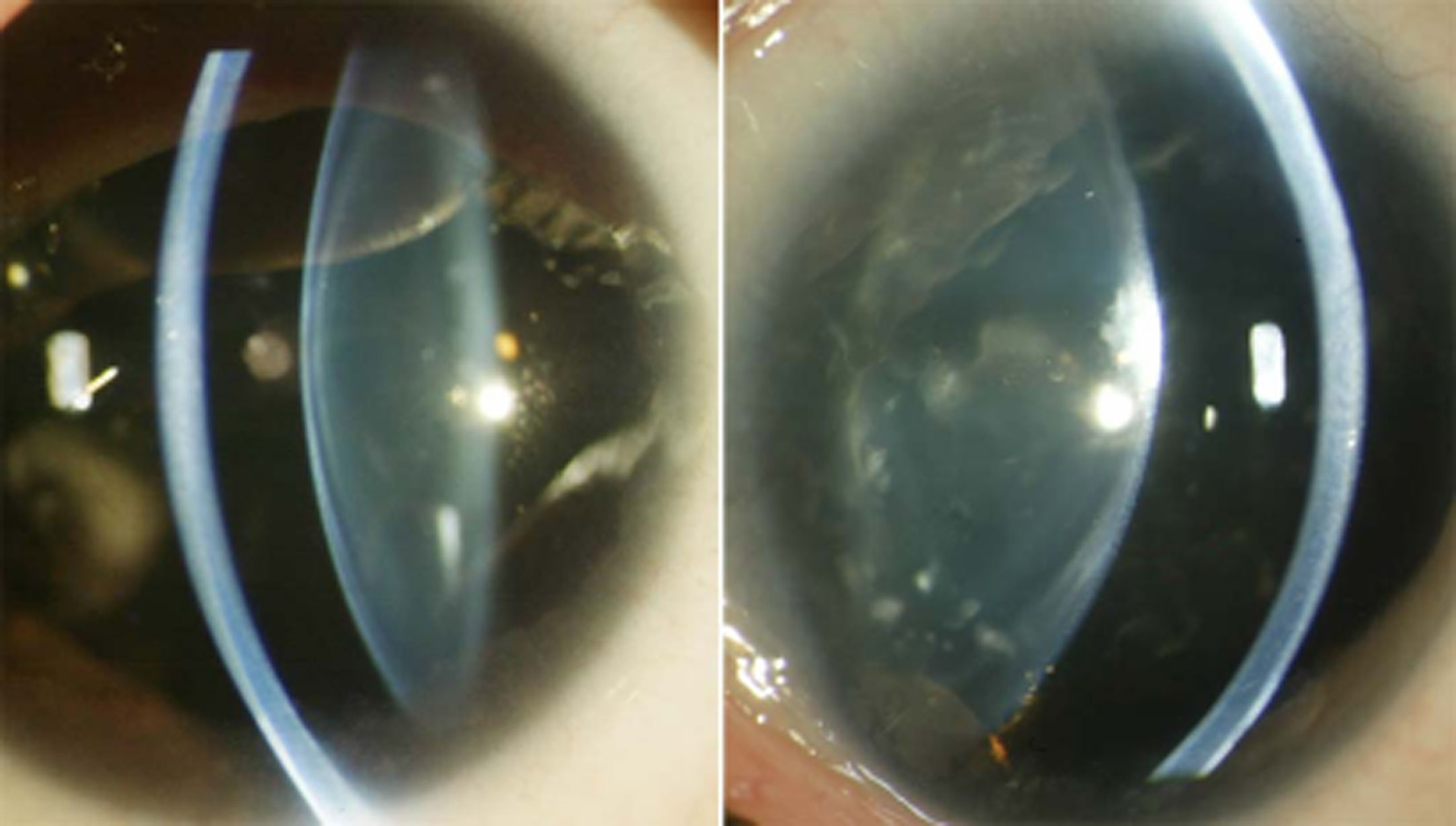
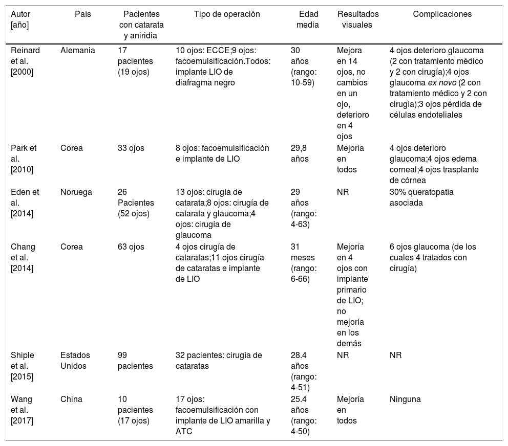
More infoLa aniridia congénita es una enfermedad genética rara asociada a mutaciones del gen PAX6. Las alteraciones del cristalino en aniridia pueden ser de tamaño y forma, de posición —que en general revelan debilidad zonular y posible subluxación del cristalino— y principalmente alteraciones de transparencia. Pueden presentar cataratas con morfología variable desde polares, corticales, subcapsulares y lamelares hasta más infrecuentemente nucleares. La agudeza visual y la calidad de la visión en pacientes con aniridia congénita asociada a cataratas se pueden mejorar mediante una cirugía cuidadosamente planificada cuando la falta de transparencia de los medios justifique la indicación quirúrgica. La cirugía de cataratas con aniridia se complica por alteraciones patológicas asociadas a la aniridia. Los desafíos incluyen opacificación corneal, cápsula friable y sobre todo reconstrucción de iris y pupila; además, pueden aparecer complicaciones tardías como glaucoma secundario o deterioro de glaucoma preexistente y descompensación endotelial corneal. Tras la cirugía cristaliniana en estos pacientes, bien por catarata o luxación, para la rehabilitación visual existen diversas técnicas como la queratopigmentación, los dispositivos protésicos iridianos o las lentes de Morcher con diafragma negro. Debe seleccionarse un método quirúrgico individualizado, según el paciente y la experiencia quirúrgica, que sea apropiado para minimizar las complicaciones y obtener mayores tasas de éxito postoperatorio.
Congenital aniridia is a rare genetic disease associated with mutations in the PAX6 gene. Changes in the lens in aniridia can be alterations of size and shape, of position — which generally reveal zonular weakness and determines subluxation of the lens — and mainly changes in transparency, cataracts, with variable morphology of polar, cortical, subcapsular, lamellar, and more rarely, nuclear cataract. Visual acuity and quality of vision in patients with congenital aniridia complicated by cataracts can be improved by carefully planned surgery, when lack of media transparency justifies surgical indication. Most patients have some improvement in visual acuity and quality of retinal image. Cataract surgery with aniridia is complicated by pathological changes due to the underlying cause of the aniridia. Challenges include corneal opacification, friable capsule and, above all, iris and pupil reconstruction. It can also determine late complications, such as secondary glaucoma or deterioration of pre-existent glaucoma, and corneal endothelial decompensation. After crystalline lens surgery in these patients, either by cataract or dislocation, for visual rehabilitation there are various techniques such as keratopigmentation, prosthetic iris devices or Morcher intraocular lenses with a black diaphragm. An appropriate individualised surgical plan should be selected depending on patient and surgical experience, in order to minimise complications and give the best chance of postoperative success.

