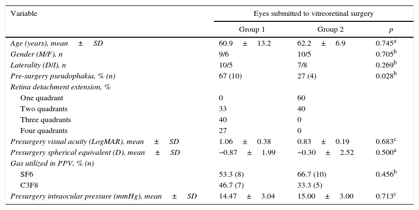
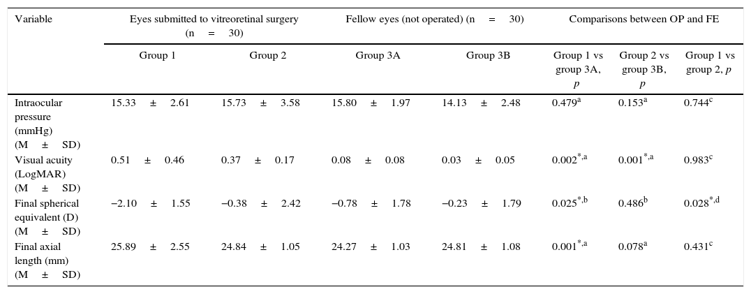
To evaluate the macular choroidal thickness (CT) of eyes subjected to pars plana vitrectomy (PPV) whether or not combined with encircling scleral buckling (ESB) surgery for primary rhegmatogenous retinal detachment repair at 6 months or more after surgery.
MethodsThis observational study included: 15 eyes (15 patients) submitted to combined ESB+PPV; 15 eyes submitted to PPV and their respective 30 normal fellow eyes (FE). Two 6mm lineal perpendicular optical coherence tomography B-scans centered on the fovea with enhanced depth imaging were performed on each eye. CT was measured at several macular locations: subfoveal (SF-CT) and at a radius of 1, 2, and 3mm from the fovea. CTs of the eyes in the CE+PPV group were compared to CT in the PPV group and the CTs of all operated eyes were compared to the CTs of their FE.
ResultsSF-CT of the eyes in the ESB+PPV group was significantly increased compared to their FE (p=0.001). CT at a radius of 1, 2, and 3mm from the fovea of the ESB+PPV group were significantly increased (p=0.001, p=0.005, and p=0.001, respectively). The SF-CT of the PPV group was similar to their FE (p=0.691). The SF-CT of the ESB+PPV group was significantly increased compared to SF-CT of the PPV group (p=0.019).
ConclusionsThe CT of the eyes subjected to combined ESB and PPV was significantly increased at 6 months or more after surgery compared to the CT of their FE and to the CT of the eyes subjected to PPV alone, which could be explained by a venous engorgement caused by the ESB.
Evaluar el espesor coroideo (EC) macular a largo plazo tras vitrectomía pars plana (VPP) con o sin cerclaje escleral (CE) para la reparación del desprendimiento de retina regmatógeno primario, después de al menos 6 meses de la cirugía.
MétodosEstudio observacional que incluyó: 15 ojos (15 pacientes) que se sometieron al CE añadido a VPP; 15 ojos (15 pacientes) que se sometieron a VPP y los 30 ojos contralaterales (OC) normales. Se obtuvo, en cada ojo, con la tomografía de coherencia óptica enhanced depth imaging, 2 escáneres de una línea de 6mm perpendiculares, centrados en la fóvea. Se midió el EC en varios puntos maculares: subfoveal (EC-SF) y en un radio de 1, 2 y 3mm respecto a la fóvea. El EC de los ojos sometidos a CE+VPP se comparó con el EC de los OC respectivos y con el EC de aquellos sometidos a VPP.
ResultadosEl EC-SF de los ojos del grupo CE+VPP fue significativamente mayor en comparación con los OC (p=0,001). Los EC en un radio de 1, 2 y 3mm respecto a la fóvea de los ojos operados estaban significativamente aumentados en los ojos del grupo CE+VPP (p=0,001, p=0,005 y p=0,001, respectivamente). El EC-SF de los ojos del grupo VPP y el EC-SF de los OC fue similar (p=0,691). El EC-SF de los ojos del grupo CE+VPP fue significativamente mayor que el EC-SF de los ojos del grupo VPP (p=0,019).
ConclusionesEl EC de los ojos sometidos a CE+VPP estaba aumentado después de al menos 6 meses de la cirugía, en comparación con el EC de los respectivos ojos adelfos y el EC de los ojos sometidos a VPP, lo que podría deberse a una reducción del drenaje venoso causada por el CE.
Artículo
Comprando el artículo el PDF del mismo podrá ser descargado
Precio 19,34 €
Comprar ahora


