


El síndrome de Silver-Russell es un trastorno congénito que cursa con déficit de crecimiento intrauterino y posnatal, macrocefalia relativa, frente prominente, cara triangular, clinodactilia, asimetría esquelética, problemas de alimentación y bajo índice de masa corporal. Entre las causas genéticas más comunes se encuentran la hipometilación del alelo paterno en la región de control de impronta 1 (ICR1) localizado en 11p15.5 (50% de los casos) y la disomía uniparental materna en el cromosoma 7 (7-10%).
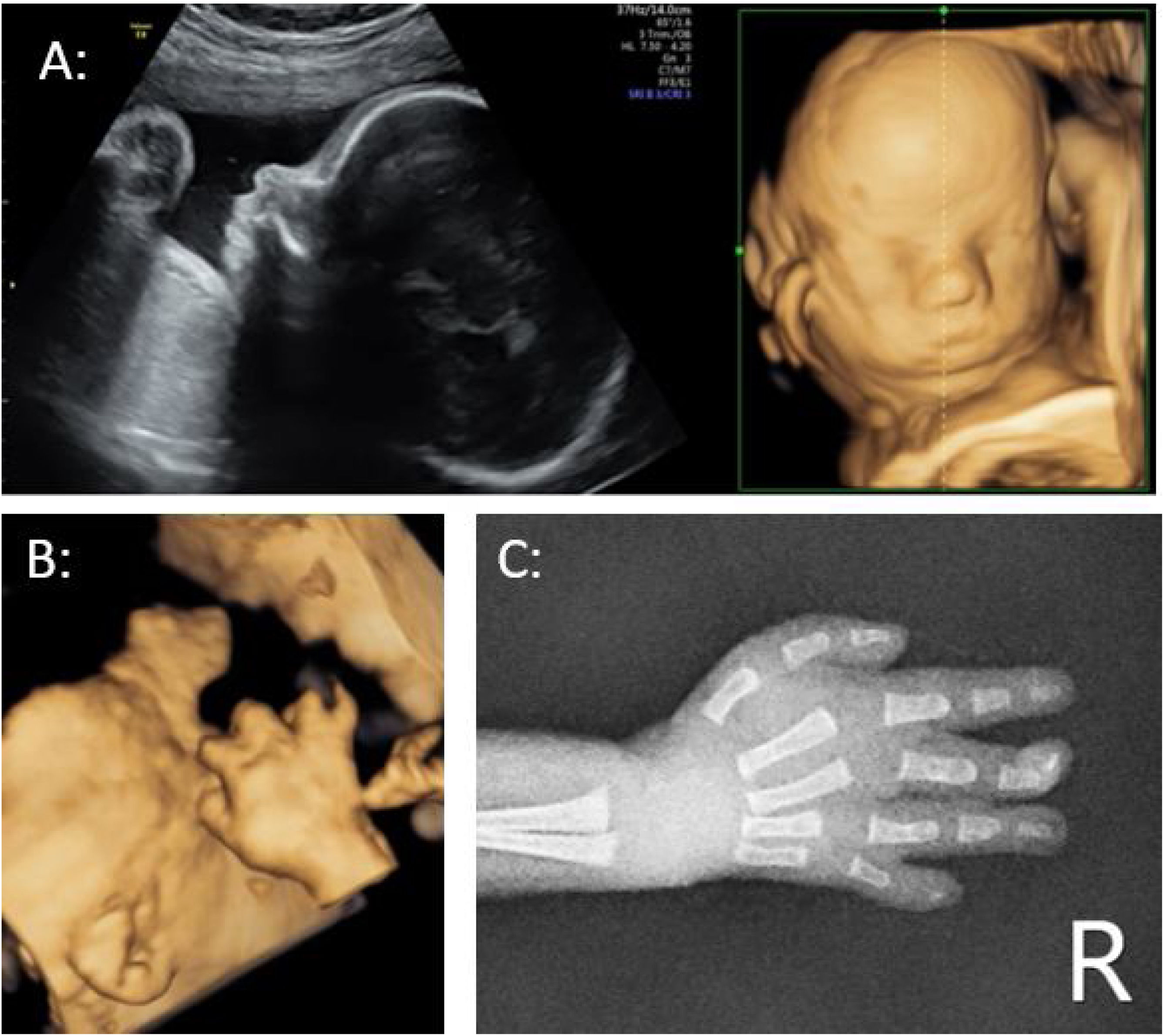
Hallazgos clínicosPresentamos el caso de una gestante de 29 años con un cribado de cromosomopatías de primer trimestre de bajo riesgo. En la ecografía selectiva, realizada con 20+4 semanas, se evidencia un crecimiento intrauterino restringido (CIR) precoz. Se realiza amniocentesis con QF-PCR, cariotipo y array-CGH normales. A las 31+3 semanas persiste CIR tipo I con un peso fetal estimado, circunferencia abdominal y longitud de fémur inferiores al percentil 1, siendo el diámetro biparietal y la circunferencial cefálica normales. Se evidencia prominencia frontal, facies pequeña y clinodactilia del quinto dedo de la mano derecha. A las 37 semanas nace mediante cesárea un varón de 1.410g.
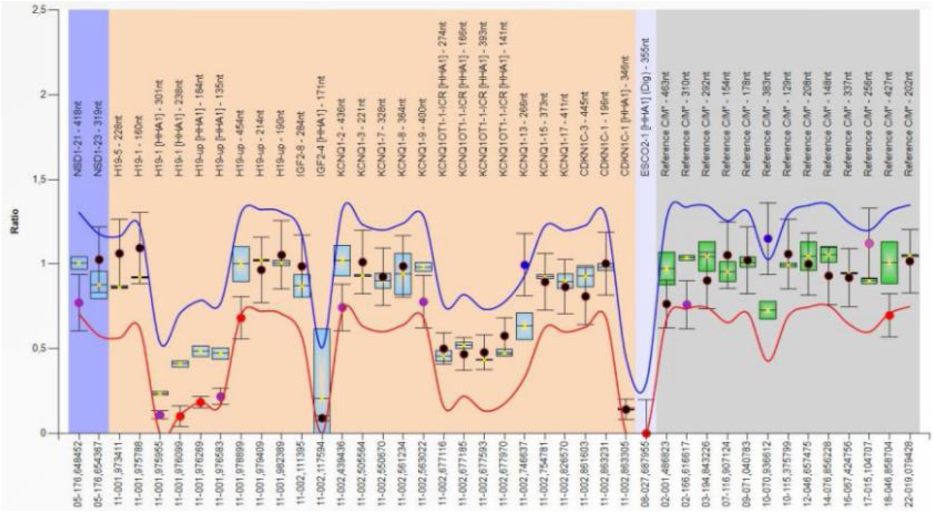
Diagnóstico, intervención terapéutica y resultadosA la exploración física destaca fenotipo peculiar sugestivo de síndrome de Silver-Russell. El estudio genético confirma hipometilación del ICR1 en la región 11p15.5. Se incluye iconografía del estudio ecográfico prenatal.
ConclusiónEs importante llegar al diagnóstico de esta entidad y conocer la correlación genotipo-fenotipo para poder ofrecer las mejores opciones terapéuticas, un adecuado seguimiento y realizar asesoramiento genético familiar.
Silver-Russell syndrome is a congenital disorder that causes prenatal and postnatal growth restriction, relative macrocephaly, prominent forehead, triangular facies, clinodactyly, body asymmetry, severe feeding difficulties, and low body mass index. The most common underlying mechanisms are hypomethylation of the paternal allele at the imprinting control region 1 (ICR 1) located at 11p15.5 (seen in 50% of patients) and maternal uniparental disomy for chromosome 7 (seen in 7%–10% of patients).
Clinical findingsWe present the case of a 29-year-old pregnant woman with low risk for chromosomal abnormalities at the first trimester screening. The 20-week ultrasound shows early intrauterine growth restriction (IUGR). We performed an amniocentesis with normal QF-PCR, foetal karyotype and array-CGH. Intrauterine growth restriction Type I persists at 31+4 weeks with estimated foetal weight, abdominal circumference, and femur length below the 1st centile. The biparietal diameter and head circumference centiles were normal. Prominent forehead, small face, and fifth finger clinodactyly of right hand were detected. At 37 weeks, a boy weighing 1,410g was born by caesarean section.
Diagnosis, therapeutic intervention, and resultsPhysical examination revealed a peculiar phenotype suggestive of Silver-Russell syndrome. The genetic study confirmed hypomethylation of ICR1 in the 11p15.5 region. Prenatal ultrasound images are shown.
ConclusionsIt is important to diagnose this entity and determine genotype-phenotype correlations in order to provide the best therapeutic options, ensure adequate follow-up, and offer timely family genetic counselling.

