


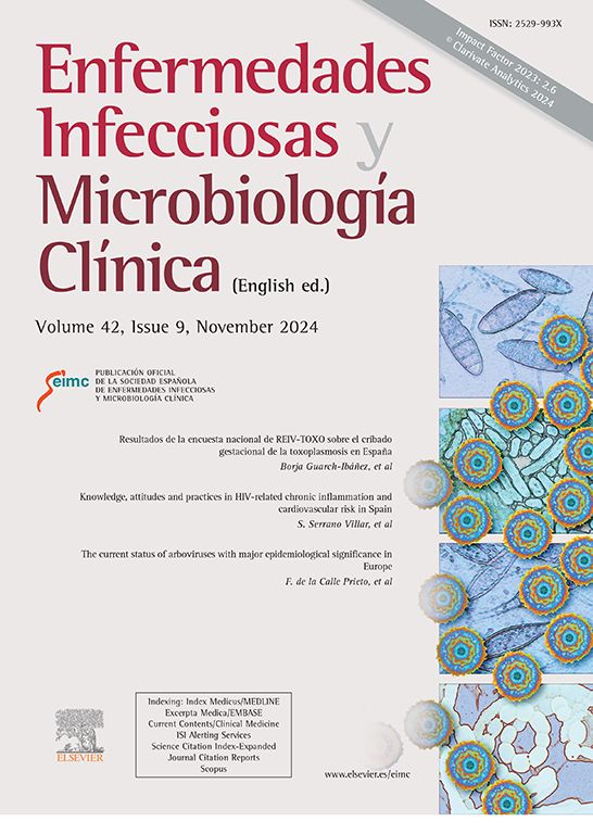

This was a 48-year-old man from Spain, with no relevant previous medical history, who went on a leisure trip to Gambia in the first week of March. While there, he noticed an injury to the external malleolus of his left ankle after visiting a fish market typical of the region with poor sanitary conditions. Initially he thought it was chafing from his footwear, but he later developed intense itching, which led to scratching and inflammation of the affected area. He decided to clean the wound with cologne as it was the only hygiene product available to him during the trip. On his return to Spain, the itching and swelling continued, increasing at night because of rubbing from his bedsheets. He tried to ease the symptoms by applying topical ointment and disinfecting the wound with 96° alcohol, after applying pressure and causing pus to come out (Fig. 1).
Clinical course and diagnosis
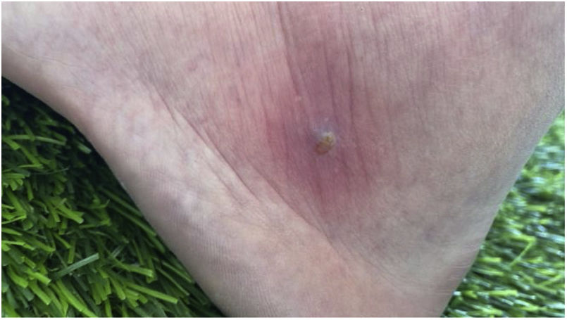
None of the measures he applied relieved the above symptoms, so he went to a private centre, where a larva was manually removed and he was prescribed amoxicillin/clavulanic acid for 10 days. The larva was sent to the Microbiology Department at our hospital. Tests were completed with serology for hepatitis A, B and C, syphilis and HIV, and analysis for Plasmodium falciparum in blood. All were negative. The patient improved immediately and is currently asymptomatic, awaiting a review by Dermatology in a few months.
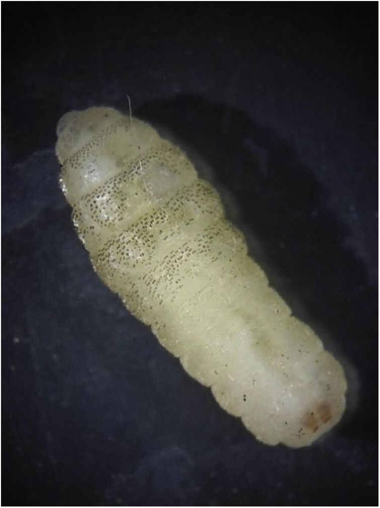
At the Microbiology Department, the larva was placed in 70° alcohol. It was then examined under a magnifying glass (Fig. 2) and microscope and the species was identified from its morphological characteristics. Of note are the numerous small spicules on the ventral and dorsal side of the anterior segments, the pair of toothed, spade-like mouth hooks and identification of the posterior spiracles (Fig. 3) with the three striae characteristic of the species Cordylobia anthropophaga. To better visualise the internal structures, the larvae were subsequently placed in 10% KOH for three days, followed by an alcohol dehydration process, adding xylol and suspending the larva in a few drops of Entellan on a slide for viewing by microscope.


C. anthropophaga (tumbu fly) belongs to the family Calliphoridae. Its natural habitat is the continent of Africa, in the regions south of the Sahara.1 It is particularly abundant in West Africa, with numerous cases of infestation having been reported in travellers from countries such as Senegal, Gambia and Sierra Leone.2,3 It causes skin-specific myiasis, as its larvae are obligate endoparasites of dermal and subdermal tissue.1
There is no specific treatment for C. anthropophaga infestation. It is recommended that the orifice of the boil be plugged with a substance such as petroleum jelly or paraffin oil, to create a hypoxic environment for the larva; this causes the larva to partially exit and facilitates its manual removal with tweezers.1
Although cases of cutaneous myiasis are not as serious as other imported diseases such as malaria, it is important to be aware this type of disease and its causal agents, in order to make an early and differential diagnosis between cases of imported myiasis and those caused by larvae of flies native to our environment here in Spain, such as Oestrus ovis and Hypoderma spp., and to establish appropriate treatment and indicate measures to prevent it from spreading.
To Dr Caroline Anne Reichelt, General Practitioner at the José Ramón Muñoz Fernández Health Centre, who put us in touch with the patient.
