metricas
covid
Buscar en
Gastroenterología y Hepatología (English Edition)
Toda la web
Inicio Gastroenterología y Hepatología (English Edition) Gastric linitis: A diagnostic challenge
Journal Information
Vol. 45. Issue 5.
Pages 375-376 (May 2022)
Vol. 45. Issue 5.
Pages 375-376 (May 2022)
Image of the month
Full text access
Gastric linitis: A diagnostic challenge
Linitis gástrica: un reto diagnóstico
Visits
76
Fernando Berdugo-Hurtadoa,
Corresponding author
fernando_bh94@hotmail.com

Corresponding author.
, Alicia Martín-Lagos-Maldonadoa, Ana Patricia Martínez-Domínguezb, Begoña Vidal-Vílcheza, Alberto Benavente-Fernándezc
a Servicio Aparato Digestivo, Hospital Universitario San Cecilio, Granada, Spain
b Servicio Cirugía General y Digestiva, Hospital Universitario San Cecilio, Granada, Spain
c Servicio Medicina Interna, Hospital Universitario San Cecilio, Granada, Spain
This item has received
Article information
Full Text
Download PDF
Statistics
Figures (4)
Show moreShow less
Full Text

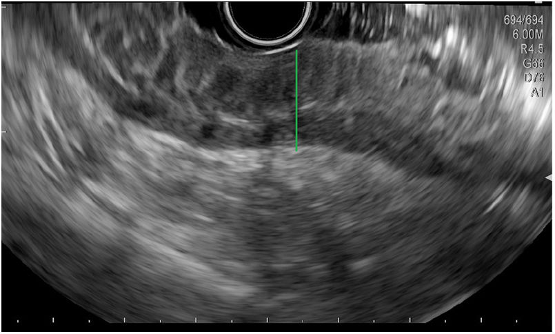
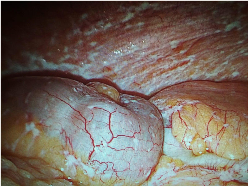
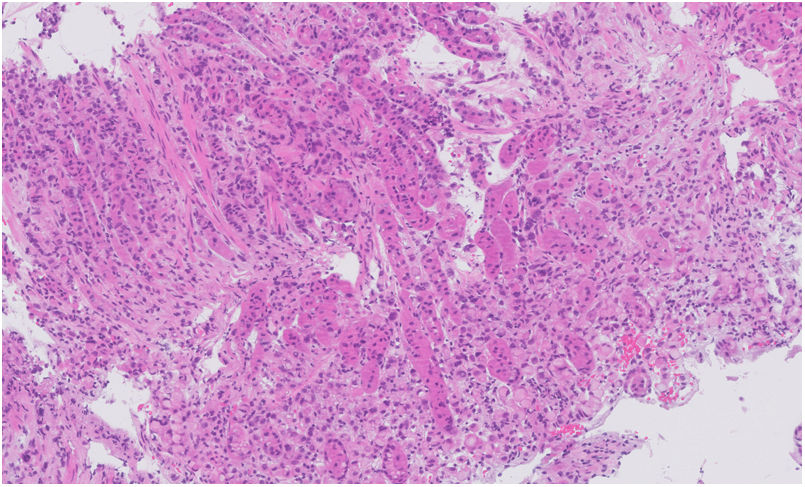
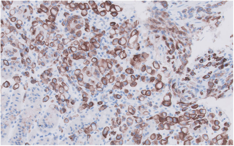
We present the case of a 65-year-old woman with no noteworthy history who came in for symptoms of dyspepsia lasting months consisting of postprandial distress, with vomiting and absolute oral intolerance in the days prior to admission. There were no other symptoms or noteworthy laboratory test abnormalities. Her physical examination was unremarkable. Gastroscopy was ordered in which thickened gastric folds were observed from the distal body through the subcardial region, with scarce distensibility in the gastric chamber. The biopsies were compatible with follicular lymphoid hyperplasia. The computed tomography (CT) scan did not show any further lesions. The study was completed with oral endoscopic ultrasound and a guided biopsy which confirmed the thickening of all the gastric wall layers (Fig. 1) and, histologically, showed the presence of mild chronic gastritis with oedema and hyperplasia, with no signs of malignancy. Given the persistent oral intolerance, no noteworthy laboratory test abnormalities, and the progressive clinical deterioration, it was decided to perform exploratory laparoscopy which found diffuse peritoneal nodules suggestive of peritoneal carcinomatosis (Fig. 2). The intraoperative biopsy of the gastric wall confirmed the final diagnosis of gastric adenocarcinoma of the linitis plastica phenotype (Figs. 3 and 4). The patient's malnutrition and frailty prevented chemotherapy, and she died three weeks later.

Figure 1.

Endoscopic ultrasound. Thickening of all layers of the gastric wall, with no clear signs of infiltration.

(0.06MB).
Figure 2.

Exploratory laparoscopy. Miliary seeds from peritoneal nodules in the small intestine and mesentery.

(0.1MB).
Figure 3.

Optical microscopy, H-E stain. Fragment of gastric mucosa with isolated and incohesive cells with signet-ring cell morphology.

(0.14MB).
Figure 4.

Optical microscopy, CK7 stain. Cancer cells positive for keratins.

(0.11MB).
Conflicts of interest

The authors declare that they have no conflicts of interest.

Please cite this article as: Berdugo-Hurtado F, Martín-Lagos-Maldonado A, Martínez-Domínguez AP, Vidal-Vílchez B, Benavente-Fernández A. Linitis gástrica: un reto diagnóstico. Gastroenterol Hepatol. 2022;45:375–376.

Copyright © 2020. Elsevier España, S.L.U.. All rights reserved
Article options
es en pt

¿Es usted profesional sanitario apto para prescribir o dispensar medicamentos?

Are you a health professional able to prescribe or dispense drugs?

Você é um profissional de saúde habilitado a prescrever ou dispensar medicamentos

Quizás le interese:
10.1016/j.gastre.2020.05.005
No mostrar más