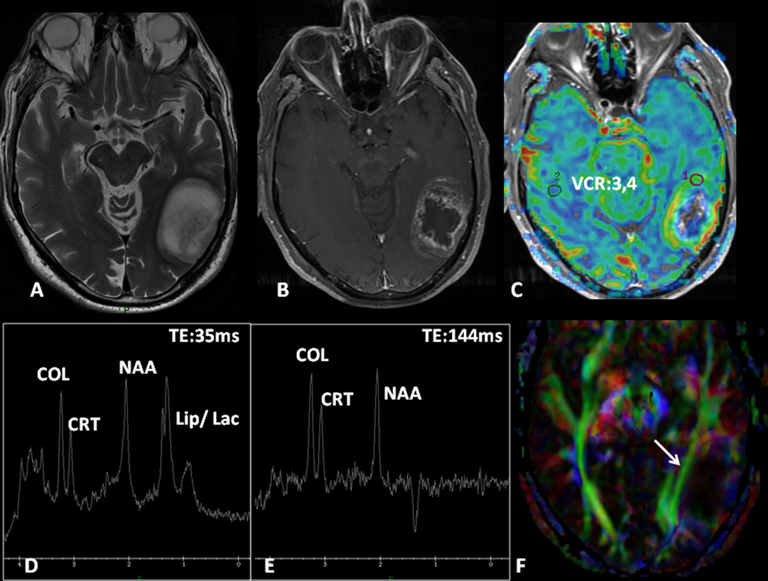
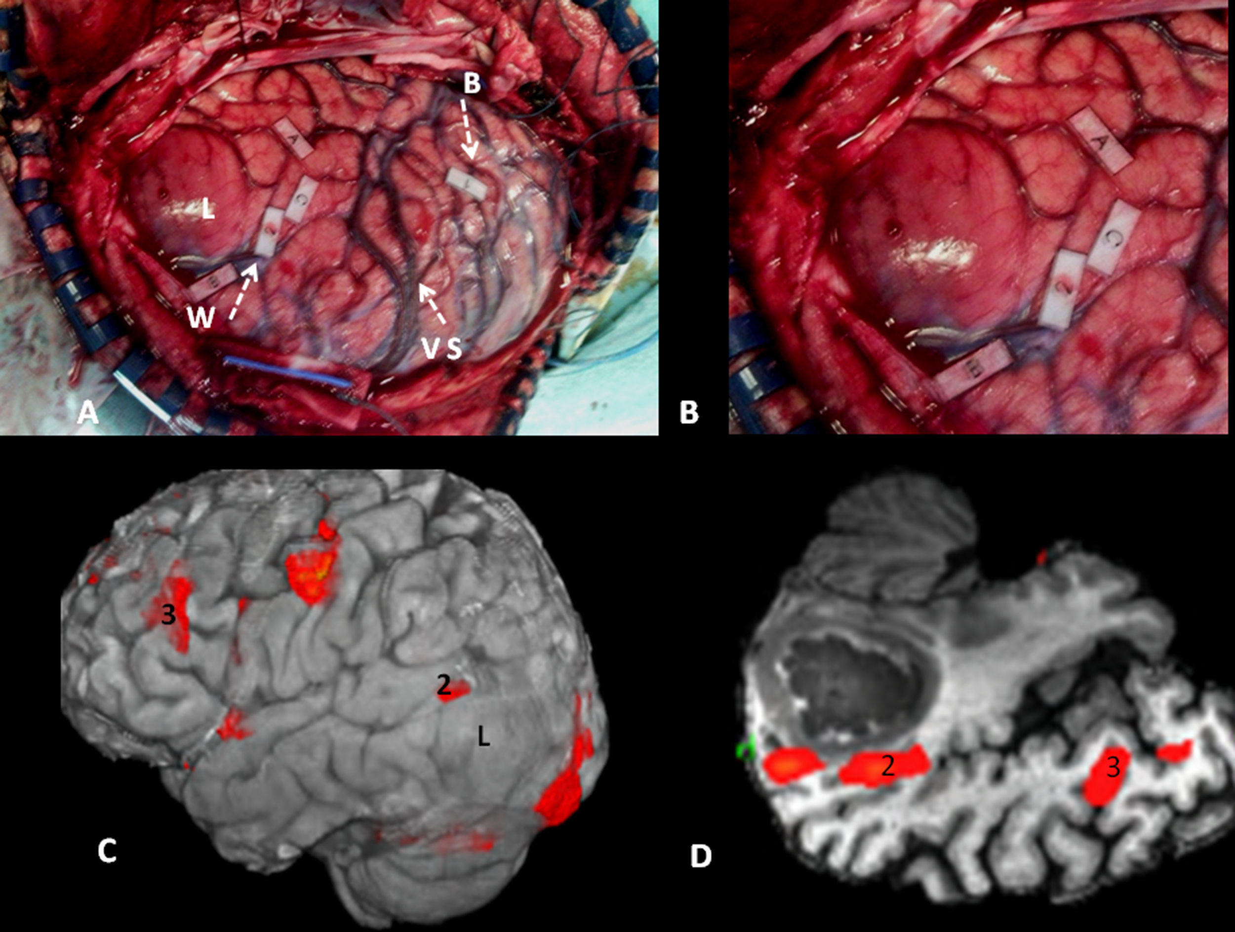
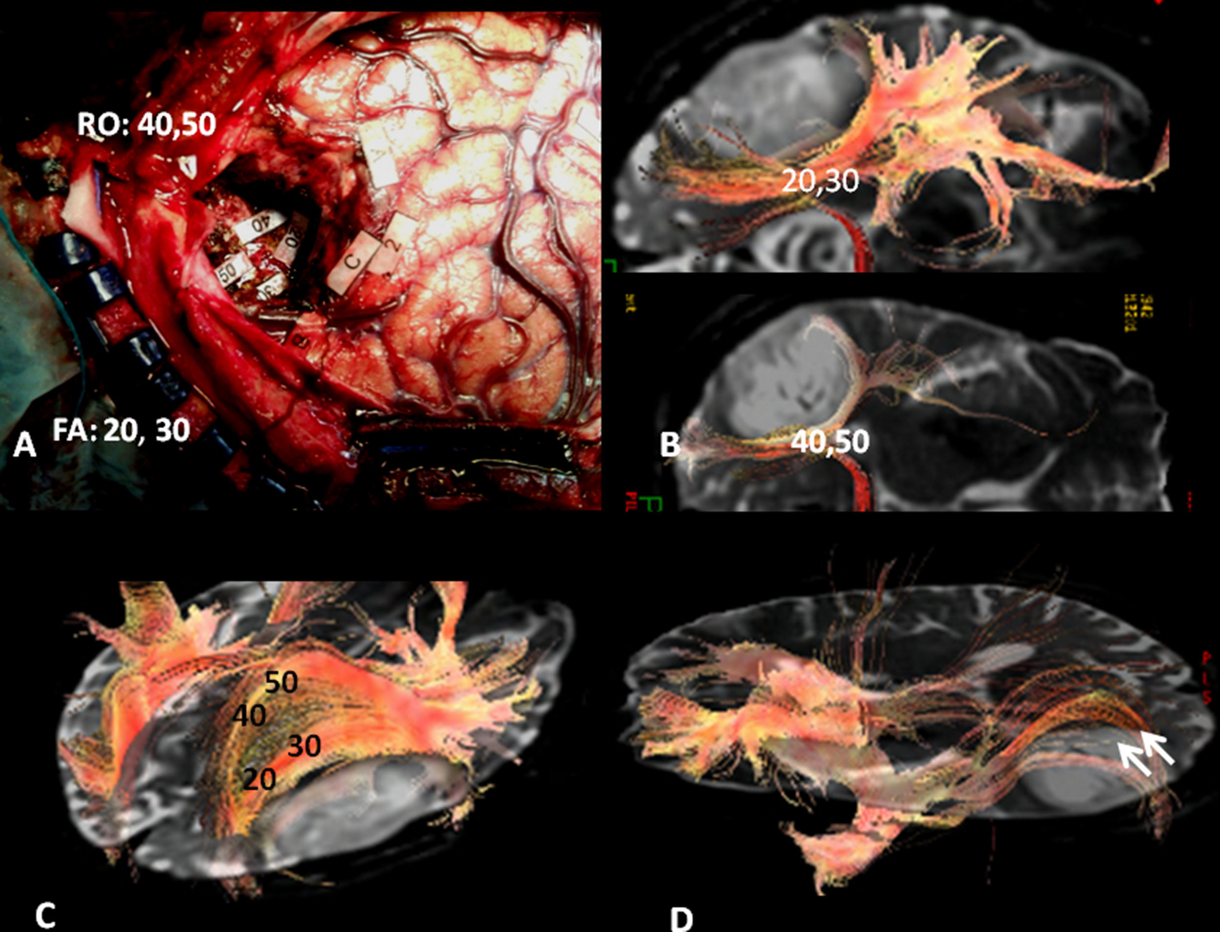
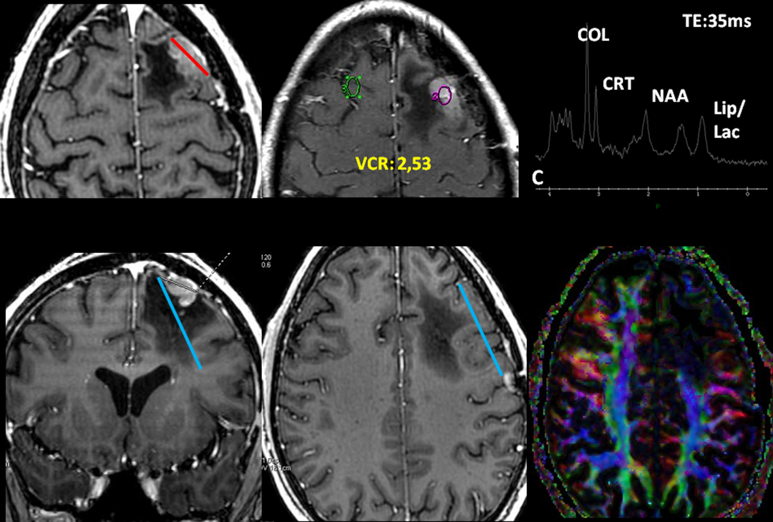
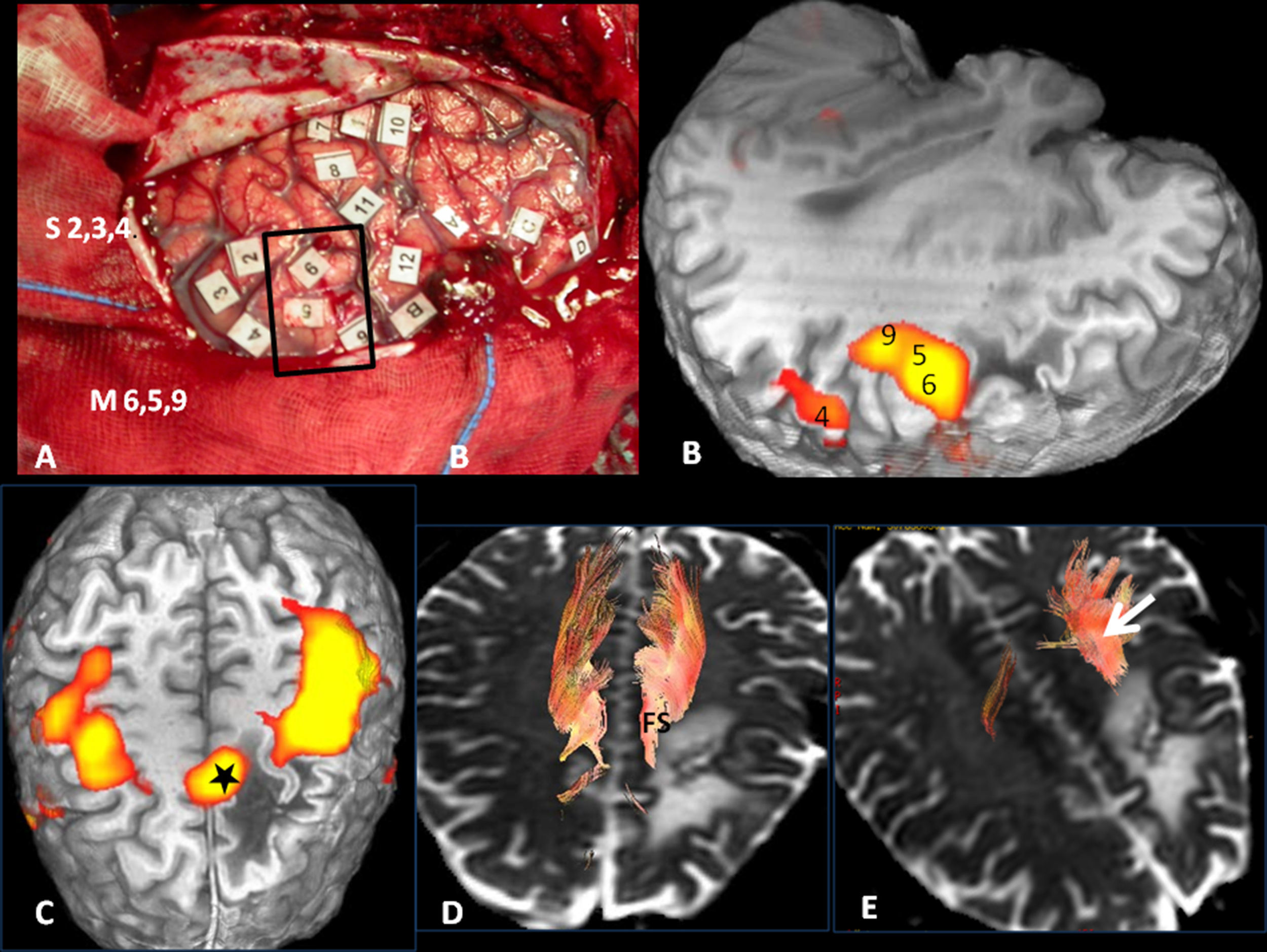
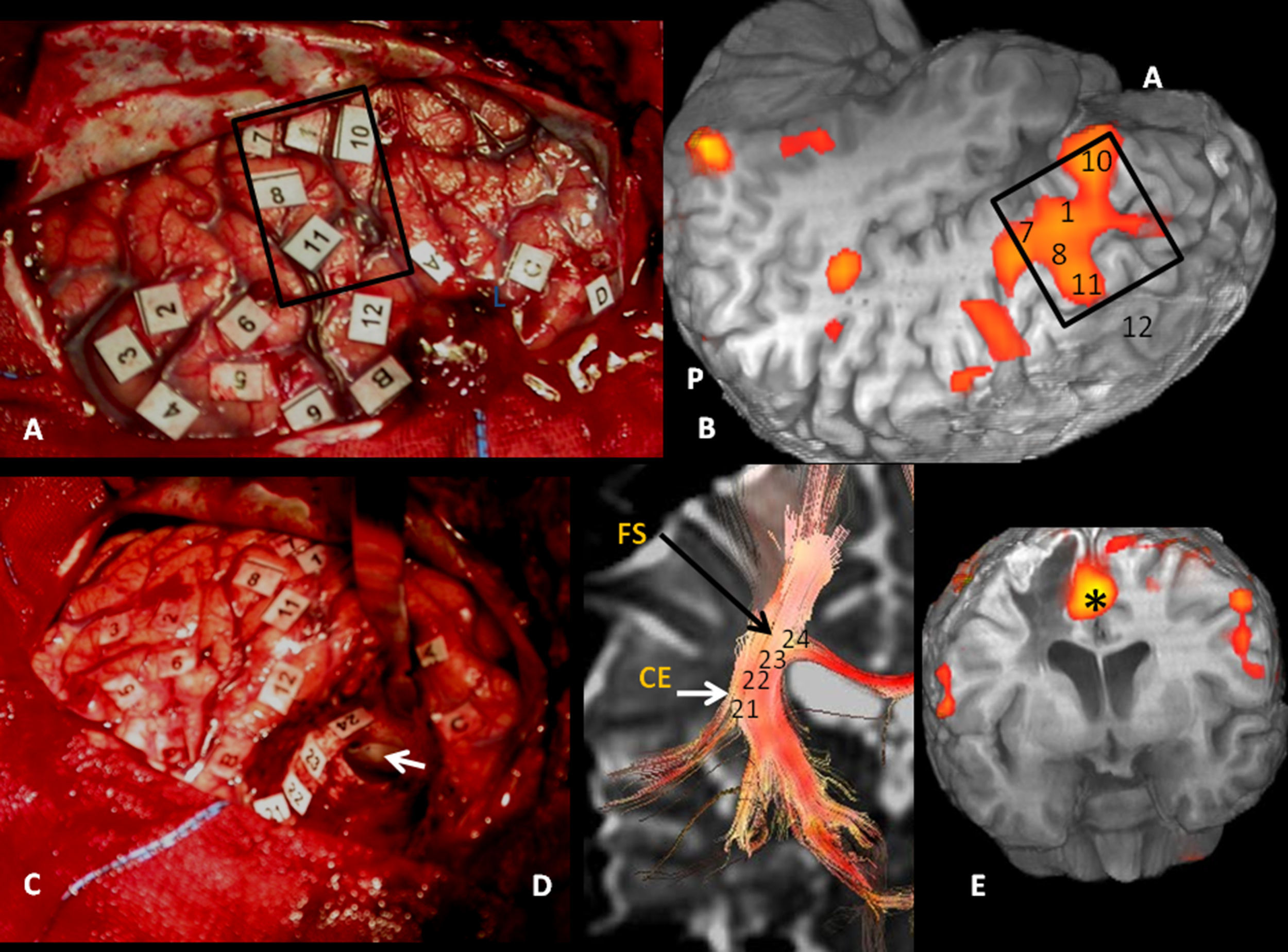
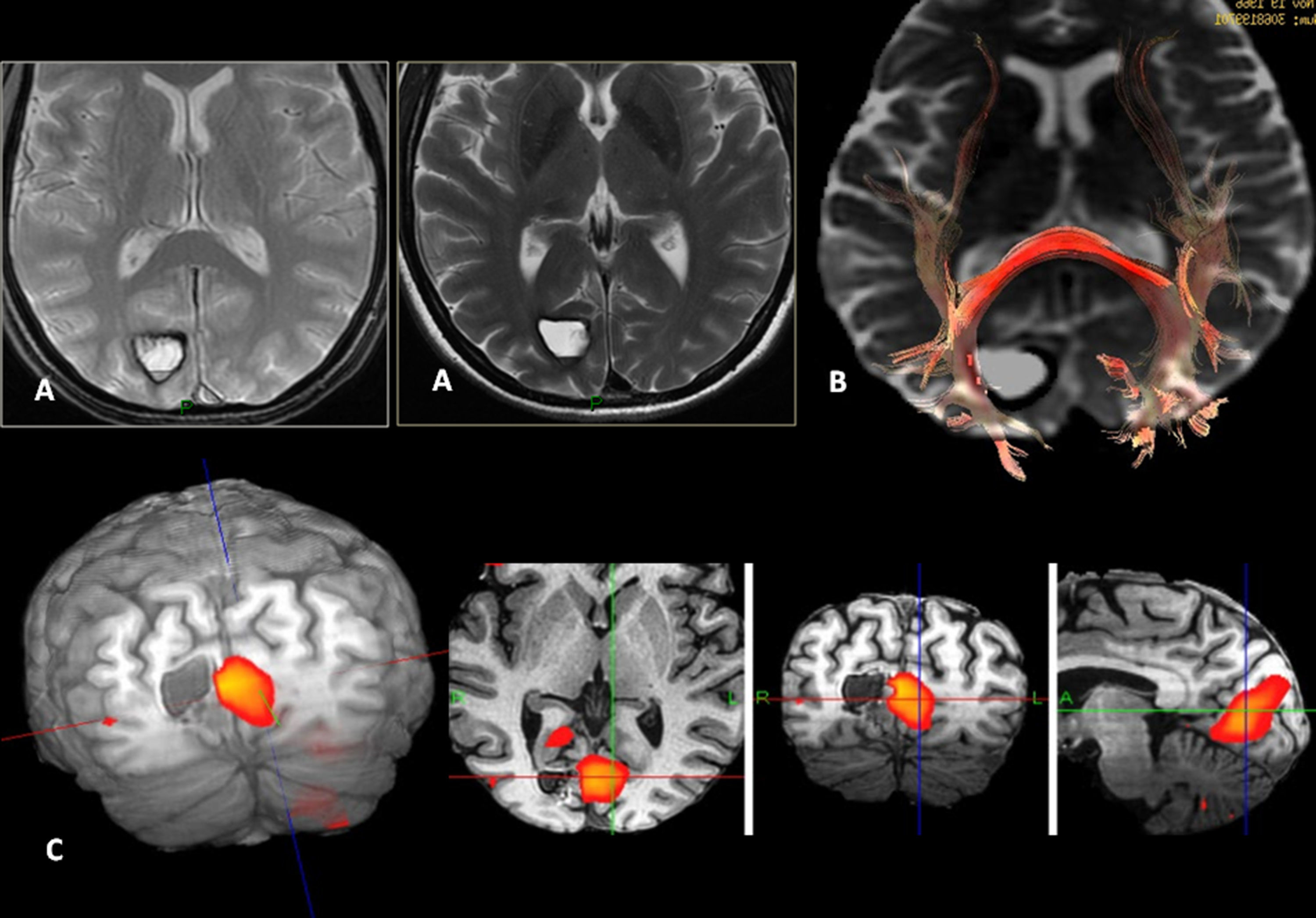
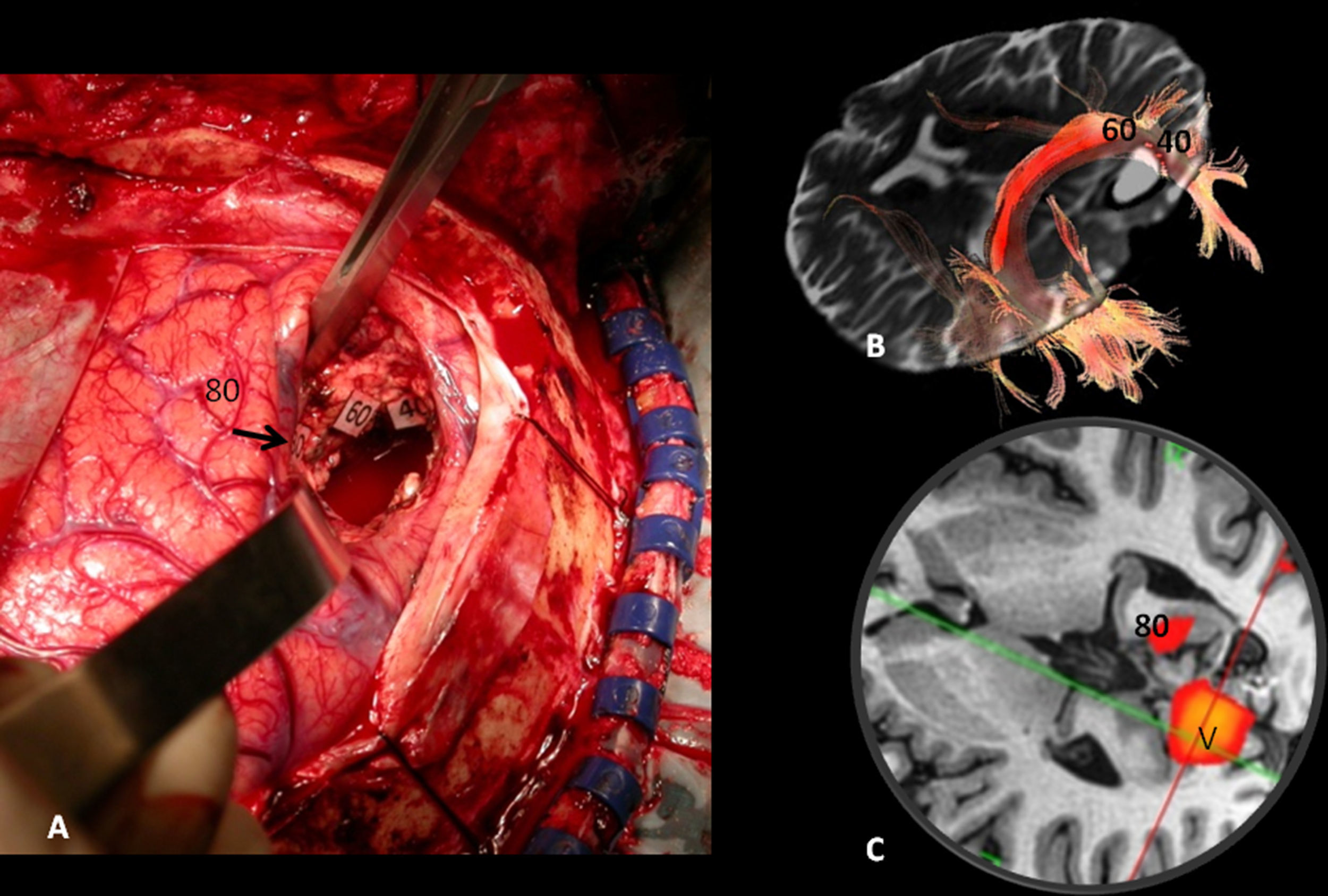
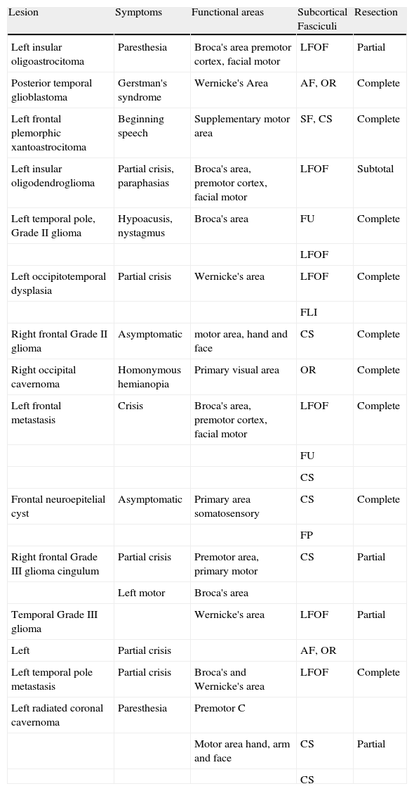
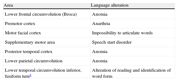
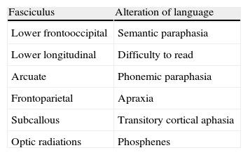
To describe the detection of cortical areas and subcortical pathways involved in language observed in MRI activation studies and tractography in a 3T MRI scanner and to correlate the findings of these functional studies with direct intraoperative cortical and subcortical stimulation.
Materials and methodsWe present a series of 14 patients with focal brain tumors adjacent to eloquent brain areas. All patients underwent neuropsychological evaluation before and after surgery. All patients underwent MRI examination including structural sequences, perfusion imaging, spectroscopy, functional imaging to determine activation of motor and language areas, and 3D tractography. All patients underwent cortical mapping through cortical and subcortical stimulation during the operation to resect the tumor. Postoperative follow-up studies were done 24h after surgery.
ResultsThe correlation of motor function and of the corticospinal tract determined by functional MRI and tractography with intraoperative mapping of cortical and subcortical motor areas was complete. The eloquent brain areas of language expression and reception were strongly correlated with intraoperative cortical mapping in all but two cases (a high grade infiltrating glioma and a low grade glioma located in the frontal lobe). 3D tractography identified the arcuate fasciculus, the lateral part of the superior longitudinal fasciculus, the subcallosal fasciculus, the inferior fronto-occipital fasciculus, and the optic radiations, which made it possible to mark the limits of the resection. The correlation with the subcortical mapping of the anatomic arrangement of the fasciculi with respect to the lesions was complete.
ConclusionThe best treatment for brain tumors is maximum resection without associated deficits, so high quality functional studies are necessary for preoperative planning.
Describir, con estudios funcionales de activación y tractografía en una RM de 3 Teslas (3T), las áreas corticales y vías subcorticales implicadas en el lenguaje, y mostrar la buena correlación de estos estudios funcionales con la estimulación directa cortical y subcortical intraoperatoria.
Material y métodosPresentamos una serie de 14 pacientes con lesiones focales cerebrales junto a áreas elocuentes. Todos los pacientes se evaluaron neuropsicológicamente antes y después de la cirugía, se estudiaron con RM con secuencias estructurales, de perfusión, espectroscopia, resonancia magnética funcional y del lenguaje y tractografía 3D, y se sometieron a un mapeo cortical de estimulación cortical y subcortical y resección de la lesión. Se hizo un control posquirúrgico a las 24h.
ResultadosLa correlación funcional motora y del haz corticoespinal con el mapeo intraoperatorio cortical y subcortical motor fue completa. Las áreas elocuentes del lenguaje expresivo y del lenguaje receptivo presentaron una alta correlación con el mapeo cortical intraoperatorio en todos los casos menos 2, un glioma infiltrativo de alto grado y un glioma de bajo grado frontal. La tractografía 3D identificó los fascículos arcuato, frontoparietal, subcalloso, frontooccipital inferior y las radiaciones ópticas, lo que permitió marcar los límites de la resección. La correlación con el mapeo subcortical en la disposición anatómica de los fascículos con respecto a las lesiones, fue completa.
ConclusiónLa máxima resección tumoral sin déficits asociados es el mejor tratamiento posible ante un tumor cerebral, lo que resalta la necesidad de estudios funcionales de alta calidad en la planificación prequirúrgica.
Artículo
Comprando el artículo el PDF del mismo podrá ser descargado
Precio 19,34 €
Comprar ahora


