


Suplemento “Avances en Radiología Tóracica”
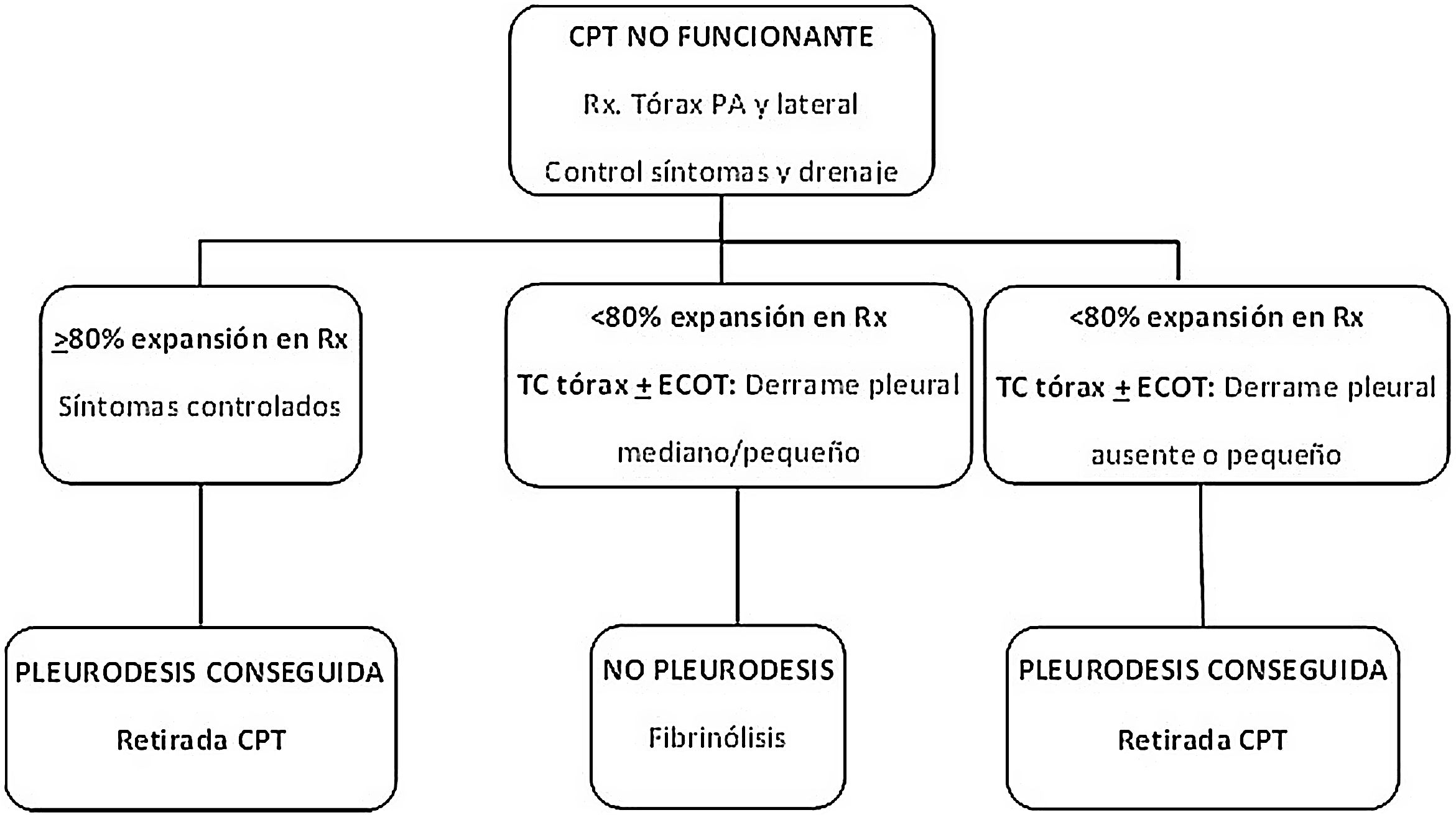
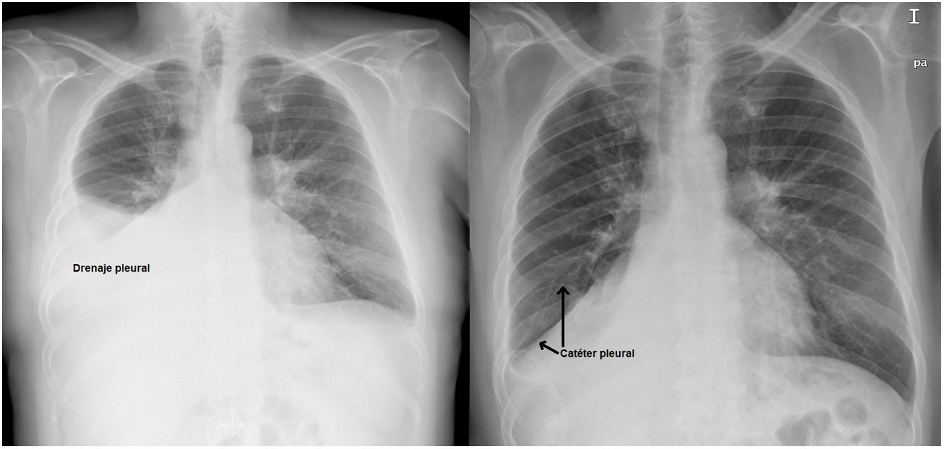
More infoNo existen criterios definidos para decidir la retirada de un catéter pleural tunelizado (CPT) no funcionante cuando la reexpansión pulmonar en la radiografía de tórax es incompleta. En muchas ocasiones se utiliza la tomografía computarizada (TC) de tórax. El objetivo de este trabajo es validar la utilidad de la ecografía torácica realizada por un neumólogo y por un radiólogo comparada con la TC de tórax.
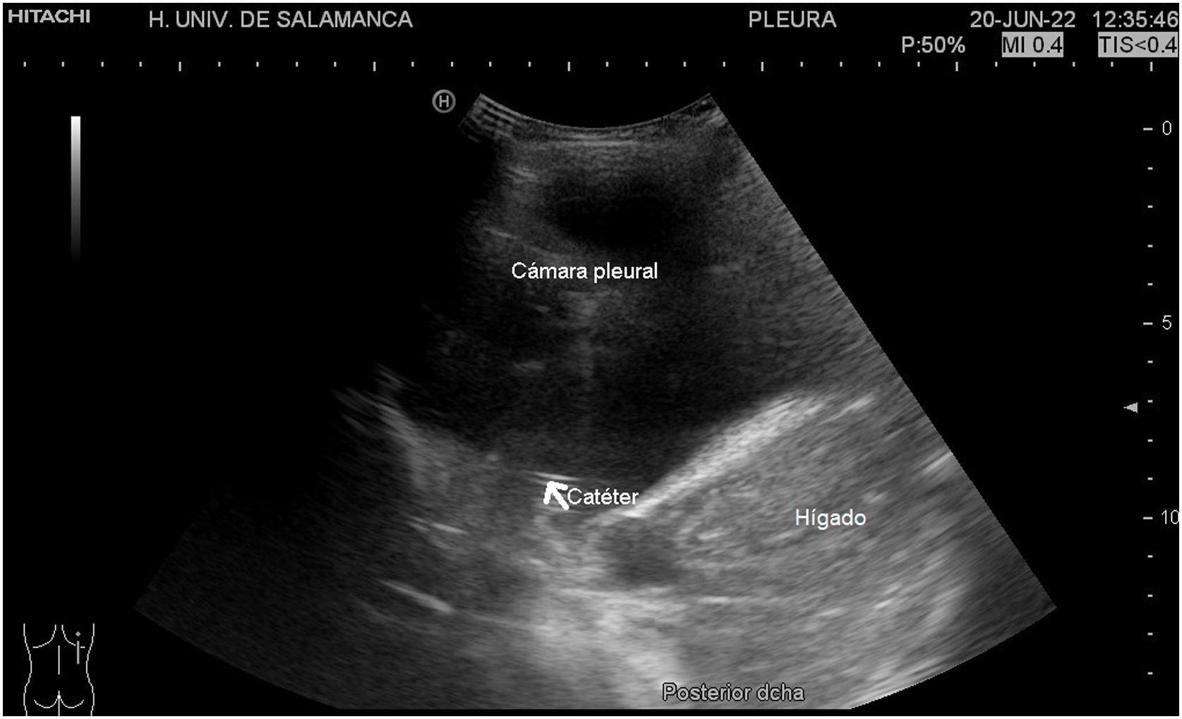
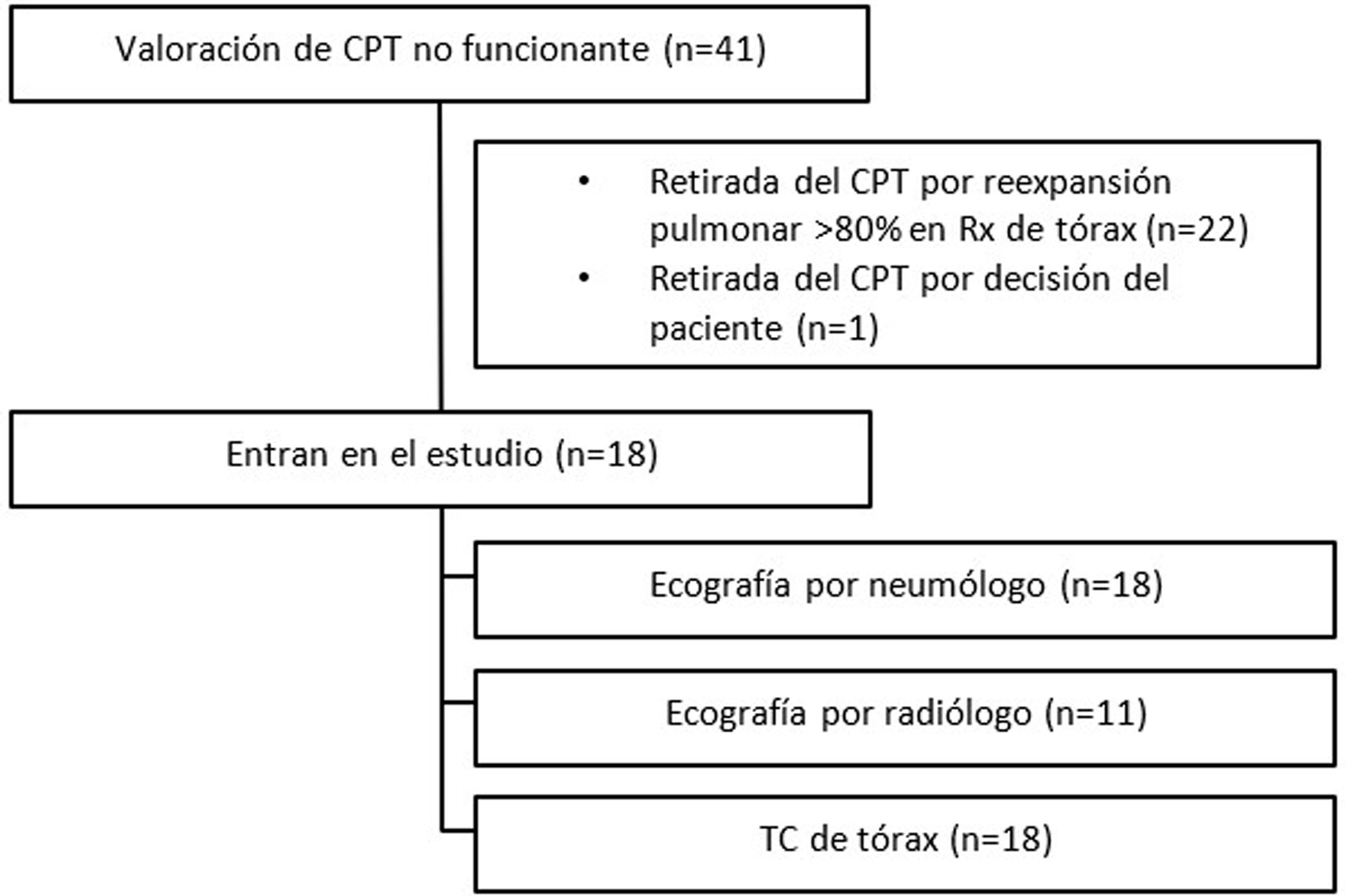
Pacientes y métodosEstudio prospectivo, descriptivo, multidisciplinar y multicéntrico de pacientes con derrame pleural maligno y CPT no funcionante sin reexpansión pulmonar. Se compararon las decisiones tomadas en base a la ecografía torácica realizada por un neumólogo y a la realizada por un radiólogo, con la TC de tórax como técnica de referencia.
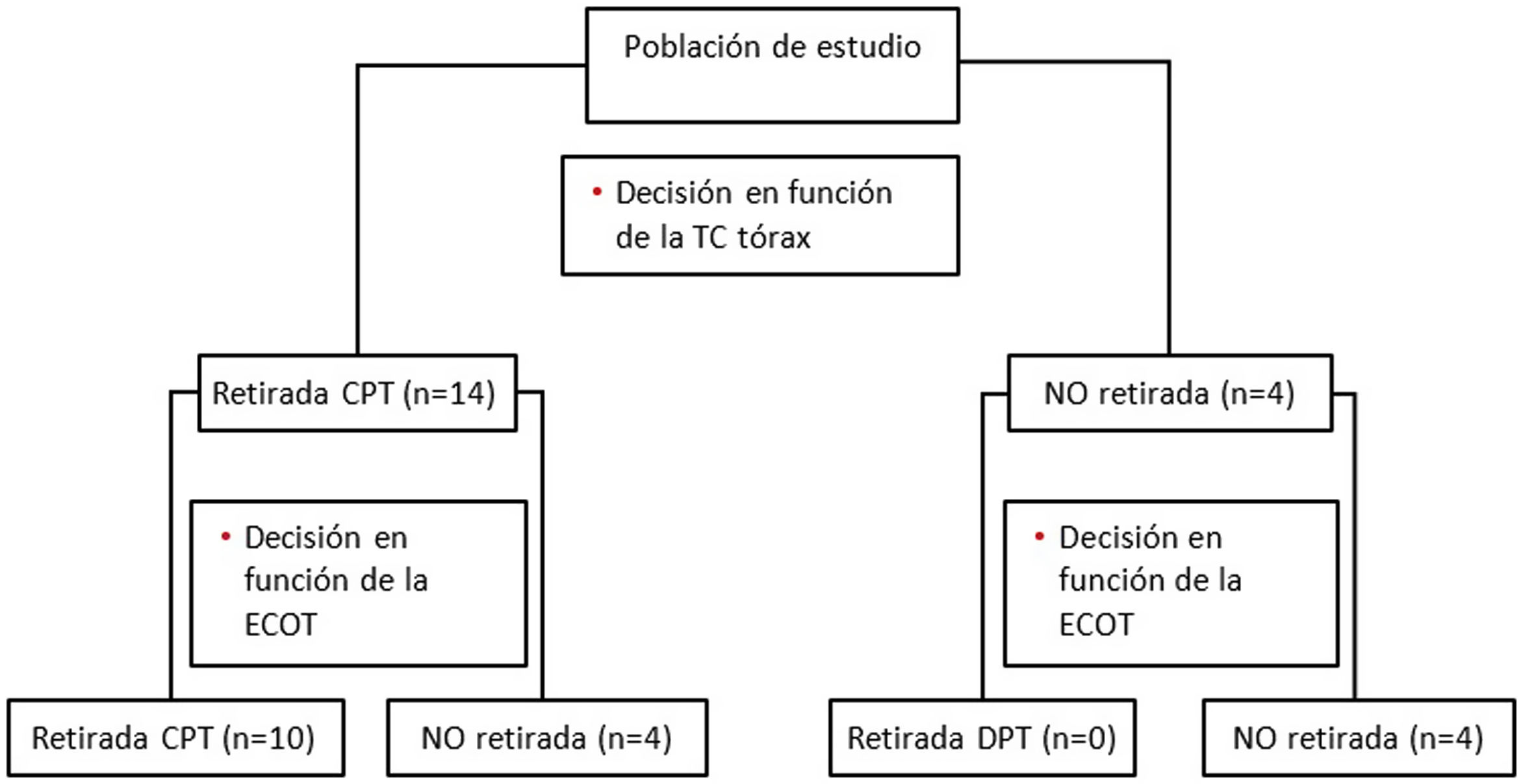
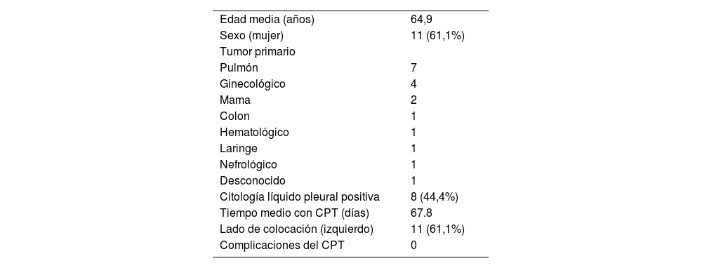
ResultadosSe analizan 18 pacientes, a todos ellos se les realiza ecografía por un neumólogo y TC de tórax, y en 11 de ellos también ecografía por un radiólogo. La ecografía realizada por el neumólogo presenta una sensibilidad del 60%, una especificidad del 100%, un VPP 100% y un VPN 66% en la decisión de la retirada correcta del CPT. La concordancia de ambas ecografías (del neumólogo y del radiólogo) fue del 100%, con un índice kappa de 1. Los 4 casos discordantes fueron aquellos en los que no se localizó el CPT en la ecografía.
ConclusionesLa ecografía torácica realizada por un neumólogo experto es una herramienta válida y sencilla para determinar pleurodesis espontánea y retirar un CPT no funcionante, lo que permitiría evitar una TC de tórax en aquellos casos en los que se objetive reexpansión pulmonar con la ultrasonografía.
There are no defined criteria for deciding to remove a non-functioning indwelling pleural catheter (IPC) when lung re-expansion on chest X-ray is incomplete. Chest computed tomography (chest CT) is usually used. The objective of this work is to validate the usefulness of chest ultrasound performed by a pulmonologist and by a radiologist compared to chest CT.
Patients and methodsProspective, descriptive, multidisciplinary and multicenter study including patients with malignant pleural effusion and non-functioning IPC without lung reexpansion. Decisions made on the basis of chest ultrasound performed by a pulmonologist, and performed by a radiologist, were compared with chest CT as the gold standard.
Results18 patients were analyzed, all of them underwent ultrasound by a pulmonologist and chest CT and in 11 of them also ultrasound by a radiologist. The ultrasound performed by the pulmonologist presents a sensitivity of 60%, specificity of 100%, PPV 100% and NPV 66% in the decision of the correct removal of the IPC. The concordance of both ultrasounds (pulmonologist and radiologist) was 100%, with a kappa index of 1. The 4 discordant cases were those in which the IPC was not located on the ultrasound.
ConclusionsThoracic ultrasound performed by an expert pulmonologist is a valid and simple tool to determine spontaneous pleurodesis and remove a non-functioning IPC, which would make it possible to avoid chest CT in those cases in which lung reexpansion is observed with ultrasonography.