metricas
covid
Buscar en
Radiología (English Edition)
Toda la web
Inicio Radiología (English Edition) Contrast-enhanced mammography and preoperative magnetic seed placement in breast...
Journal Information
Share
Share
Download PDF
More article options
Original articles
Available online 19 September 2024
Contrast-enhanced mammography and preoperative magnetic seed placement in breast cancer patients for the detection of residual disease following neoadjuvant systemic therapy
Mamografía con contraste y marcaje con semilla magnética para la detección de enfermedad residual en el cáncer de mama tras tratamiento neoadyuvante
A. Mariscal Martíneza,
Corresponding author
, E. Iglesias Bravob, H. Peris Alvàa, P. Rodríguez Martínezc, M. Luna Tomásd, I. Pascual Miguele, P. Puyalto de Pablof
a Unidad de Diagnóstico por la Imagen de la Mama, Servicio de Radiología, Hospital Universitario Germans Trias i Pujol, Badalona, Barcelona, Spain
b Servicio de Ginecología, Hospital Virgen de Valme, Sevilla, Spain
c Servicio de Anatomía Patológica, Hospital Universitario Germans Trias i Pujol, Badalona, Barcelona, Spain
d Servicio de Ginecología y Obstetricia, Hospital Universitario Germans Trias i Pujol, Badalona, Barcelona, Spain
e Servicio de Cirugía General, Hospital Universitario Germans Trias i Pujol, Badalona, Barcelona, Spain
f Servicio de Radiología, Hospital Universitario Germans Trias i Pujol, Badalona, Barcelona, Spain
Article information
Abstract
Full Text
Bibliography
Download PDF
Statistics
Figures (5)
Show moreShow less
Tables (3)
Table 1. Clinical/pathological findings.
Table 2. Mean differences, based on Student’s t-test parametric test, between residual tumour lesion size on CEM vs. pathological size.
Table 3. Assessment of residual tumour detection following NST by CEM.
Show moreShow less
Abstract
Purpose

Assess whether contrast-enhanced mammography (CEM) enables an evaluation of the residual size of breast tumours following neoadjuvant systemic therapy (NAST) in patients initially marked with magnetic seed.

Materials and methods

This single-centre prospective study was performed between March 2022 and April 2023 with patients with invasive breast carcinoma and lesional marking with magnetic seed. CEM was performed before and after NAST. The lesion size in CEM after NAST was compared to the pathological examination after surgery. Differences between sizes were evaluated and we determined the diagnostic capability indices.
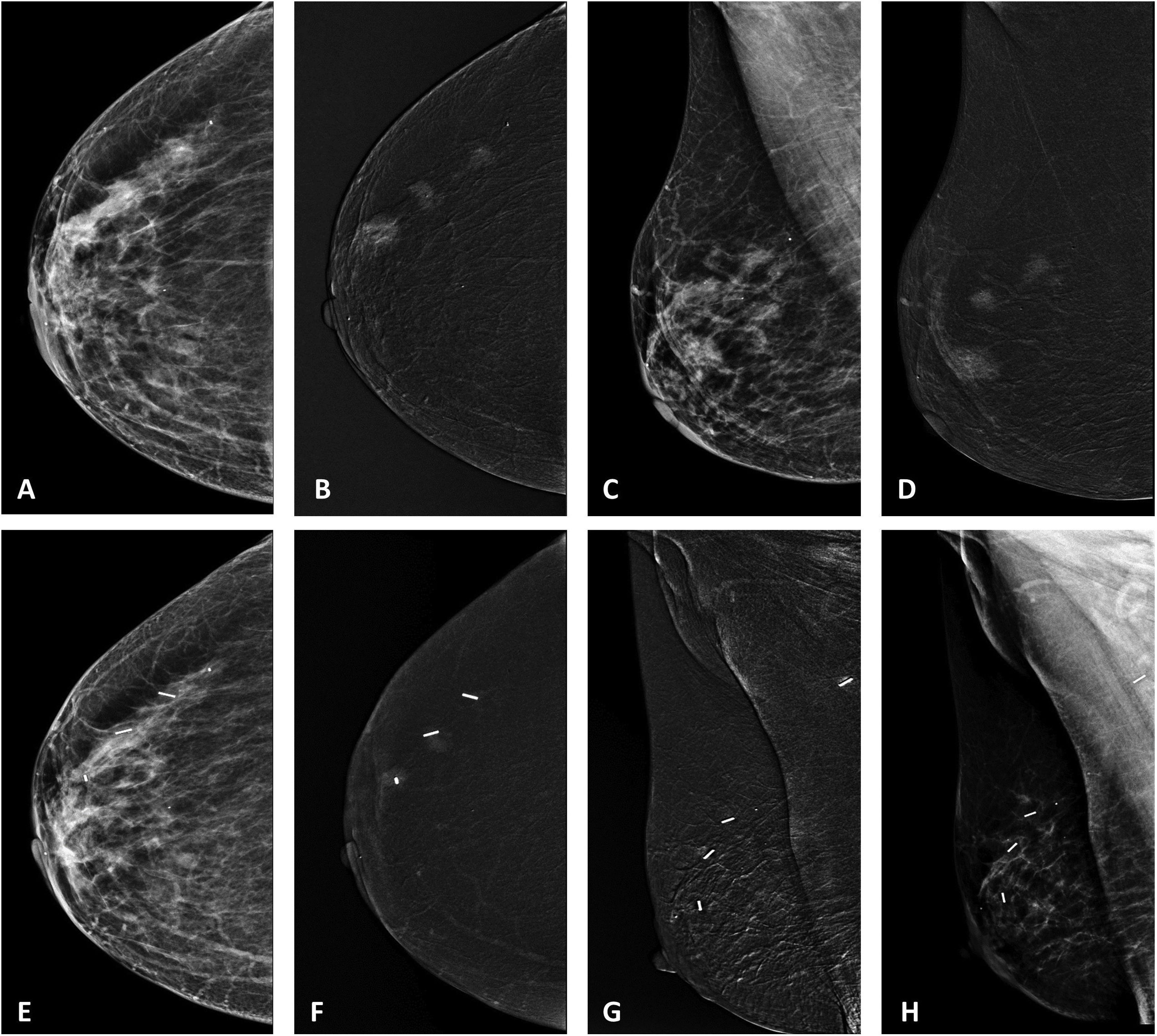
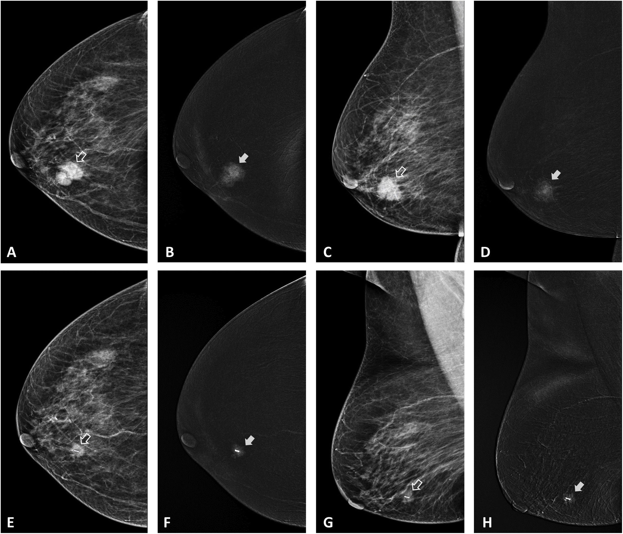
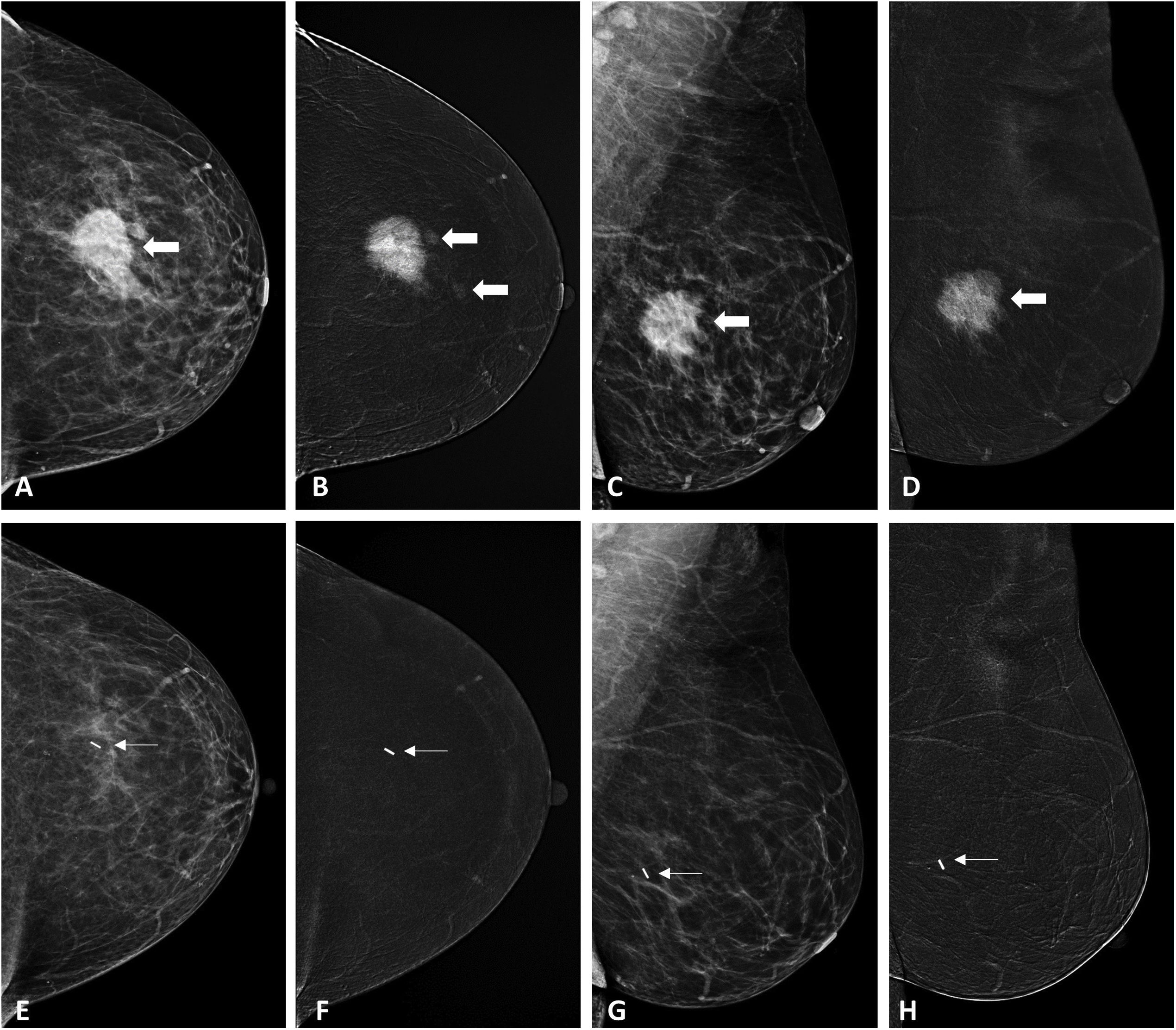
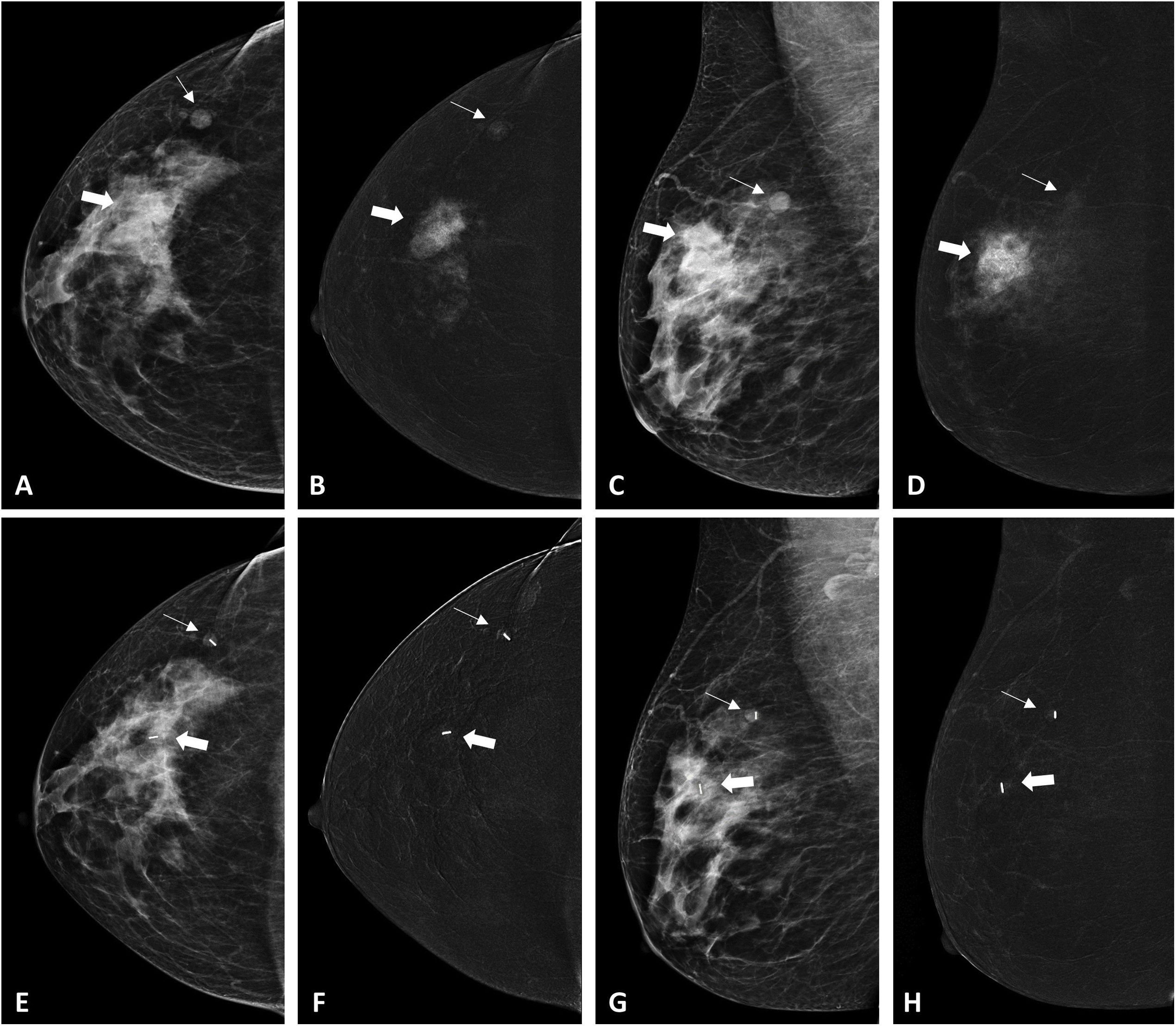
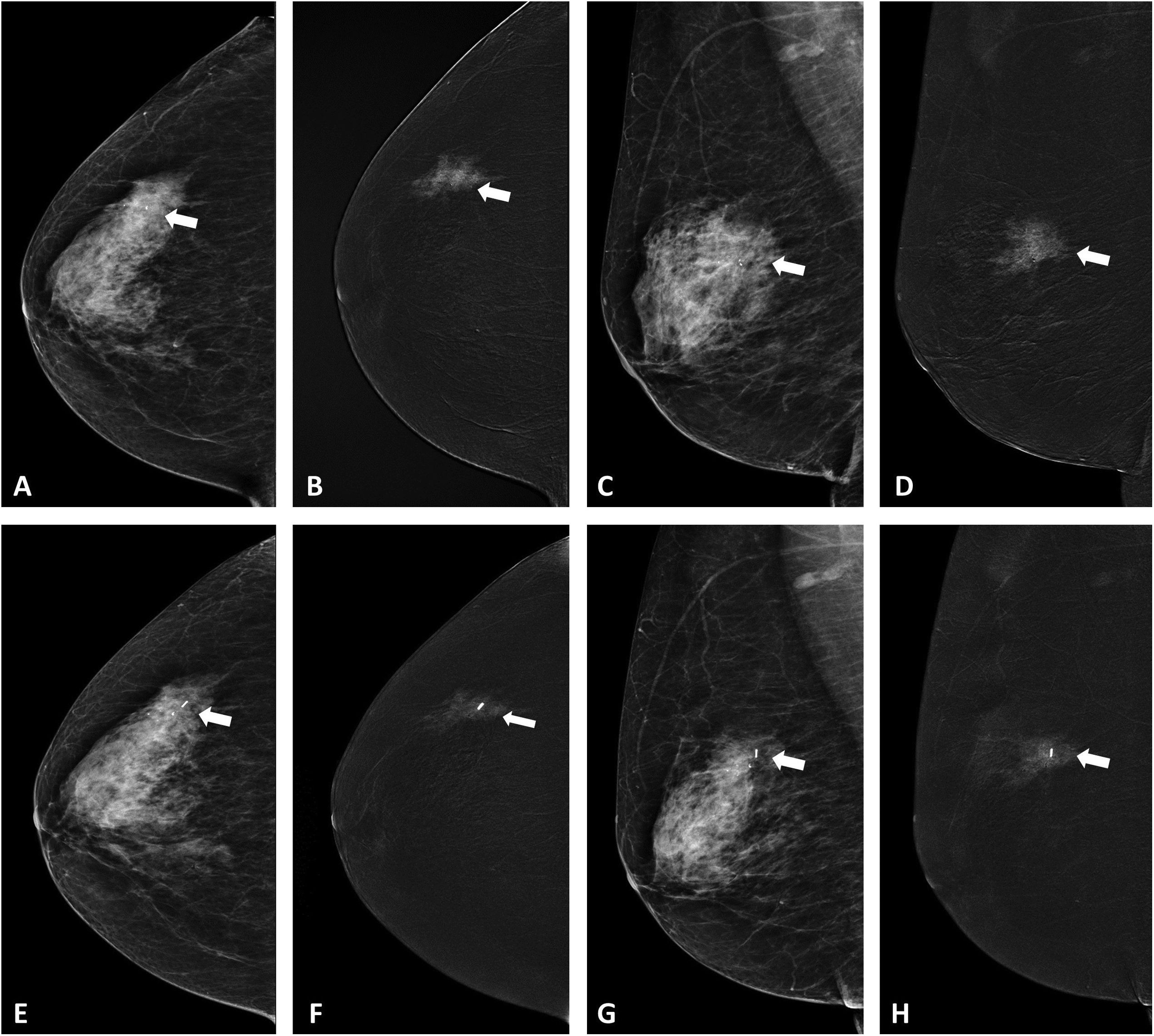
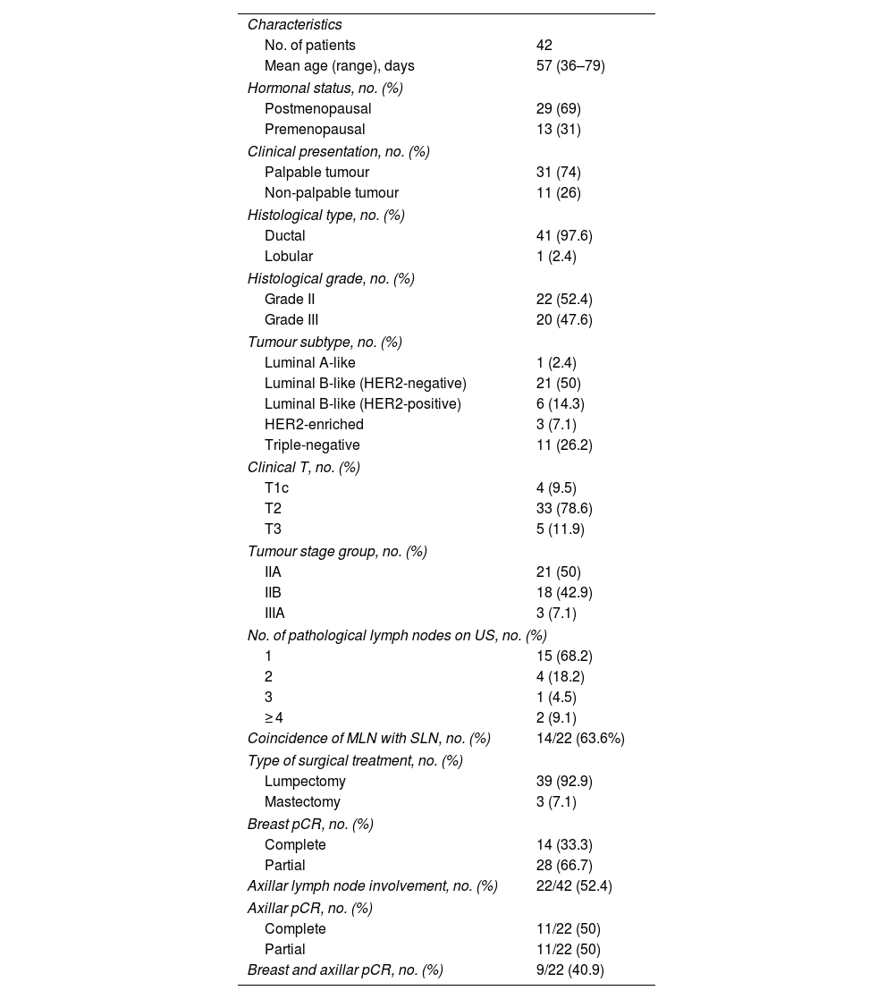
Results

The breast lesions marked with magnetic seed were successfully localised in the preoperative stage for the 42 patients included in the study and selective surgical excision was also achieved in all cases. Tumour diameter after NAST was determined by comparing enhancement on combined CEM images from before and after NAST. The mean diameter was 13.6 mm while post-surgical pathological examination determined the mean diameter to be 12.9 mm. There were therefore no statistically significant differences between the measurements.

Conclusions

There is a positive correlation and similarity between CEM and pathological examination with regards to the detection of residual disease after NAST, with high specificity and PPV.

Keywords:
Contrast-enhanced mammography
Magnetic seed
Neoadjuvant therapy
Breast neoplasms
Resumen
Objetivo

Valorar si la mamografía con contraste (MC) permite evaluar el tamaño tumoral residual en la mama tras realizar tratamiento sistémico neoadyuvante (TSN) en pacientes con cáncer de mama, marcado inicialmente con semilla magnética.

Materiales y métodos

Se realizó un estudio prospectivo unicéntrico entre marzo 2022 - abril 2023 con pacientes con cáncer de mama infiltrante y marcaje lesional con semilla; se realizó MC antes y después del TSN. Se comparó el tamaño lesional en MC posterior al TSN respecto al de la valoración anatomopatológica tras cirugía, se evaluaron las diferencias de los tamaños y se determinaron los índices de capacidad diagnóstica.

Resultados

En las 42 pacientes incluidas se consiguió con éxito la localización preoperatoria y la exéresis quirúrgica selectiva de las lesiones mamarias marcadas con semilla magnética. La media del diámetro mayor tumoral tras TSN, determinado por el realce en las imágenes recombinadas de la MC, fue de 13,6 mm vs 12,9 mm en el examen patológico postquirúrgico, no hallándose diferencias estadísticamente significativas entre las medias de las mediciones.

Conclusiones

La MC presenta una buena correlación y concordancia con la histopatología para detectar la enfermedad residual tras TSN, con elevada especificidad y VPP.

Palabras clave:
Mamografía con contraste
Semilla magnética
Tratamiento neoadyuvante
Cáncer de mama

Article

These are the options to access the full texts of the publication Radiología (English Edition)
Subscriber
Subscriber

If you already have your login data, please click here .

If you have forgotten your password you can you can recover it by clicking here and selecting the option “I have forgotten my password”
Purchase
Purchase article

Purchasing article the PDF version will be downloaded

Price 19.34 €

Purchase now
Contact
Phone for subscriptions and reporting of errors
From Monday to Friday from 9 a.m. to 6 p.m. (GMT + 1) except for the months of July and August which will be from 9 a.m. to 3 p.m.
Calls from Spain
932 415 960
Calls from outside Spain
+34 932 415 960
E-mail
Article options
es en pt

¿Es usted profesional sanitario apto para prescribir o dispensar medicamentos?

Are you a health professional able to prescribe or dispense drugs?

Você é um profissional de saúde habilitado a prescrever ou dispensar medicamentos