metricas
covid
Buscar en
Revista Colombiana de Reumatología (English Edition)
Toda la web
Inicio Revista Colombiana de Reumatología (English Edition) Interstitial lung disease in systemic lupus erythematosus: State of the art sear...
Journal Information
Visits
75
Review article
Full text access
Available online 21 June 2024
Interstitial lung disease in systemic lupus erythematosus: State of the art search
Enfermedad pulmonar intersticial en el lupus eritematoso sistémico: búsqueda avanzada
Visits
75
Juan Camilo Santacruz
Corresponding author
santa89@hotmail.com

Corresponding author.
, Marta Juliana Mantilla, Gustavo Rodríguez-Salas, Igor Rueda, Ana María Santos, John Londoño
Spondyloarthropathies Research Group, Universidad de La Sabana, Chía, Colombia
This item has received
Article information
Abstract
Full Text
Bibliography
Download PDF
Statistics
Figures (4)
Show moreShow less
Tables (1)
Table 1. Most representative articles to establish the lines of treatment in hierarchical order.
Abstract

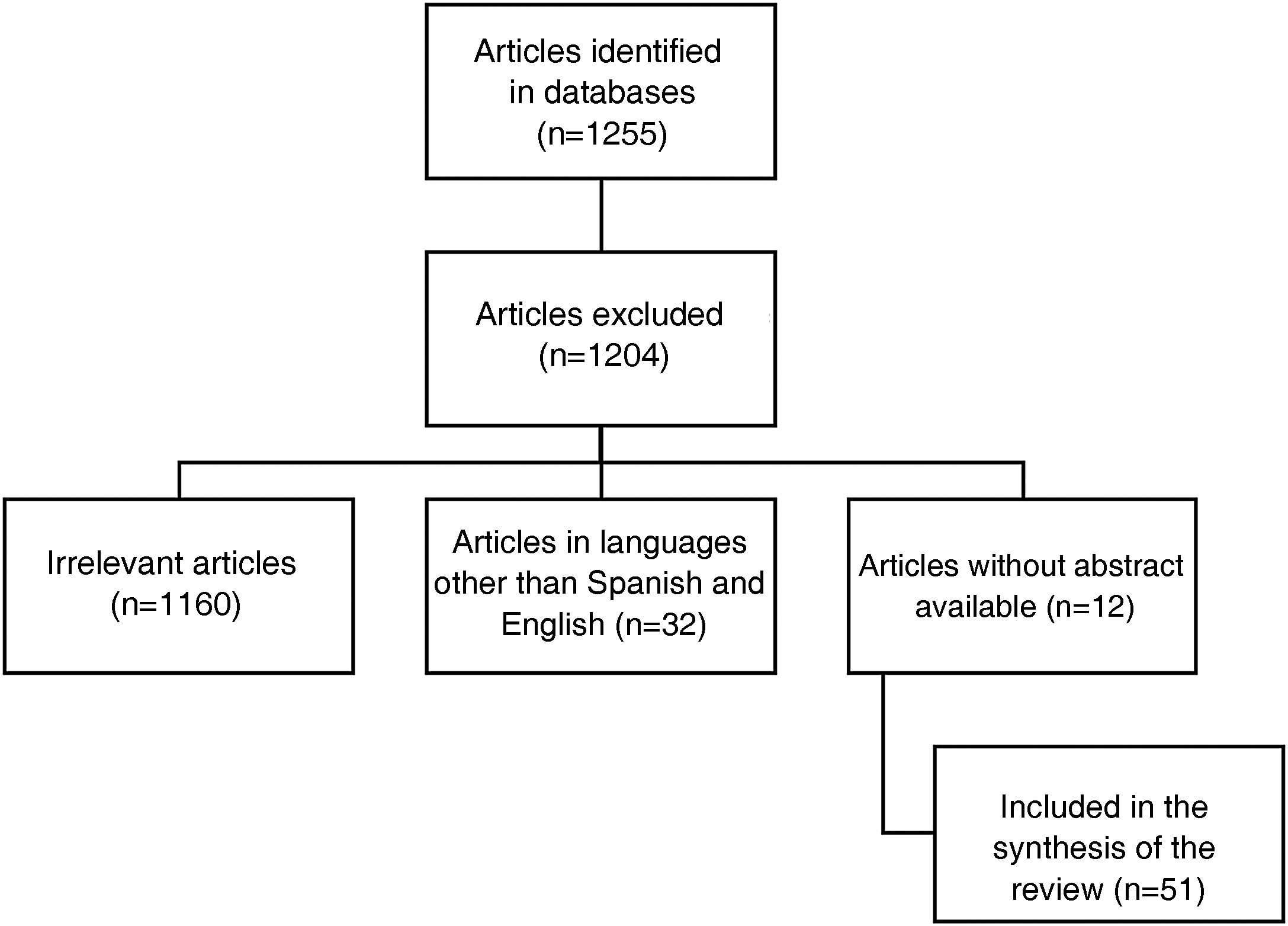
Interstitial lung disease in lupus is an entity that occurs infrequently and tends to progress slowly in most cases. Despite this, the therapeutic approach for moderate to severe cases is largely unknown because most of the evidence comes from case reports, many of which predate the advent of today’s known treatments for lupus. Additionally, little progress has been made in understanding its pathophysiology and current concepts come from other connective tissue diseases such as systemic sclerosis or are grouped within the group of interstitial pneumonias with autoimmune characteristics. This, to an extent, has been an obstacle for research in this field, and to date there is no unified diagnostic and therapeutic approach. Therefore we conducted a state-of-the-art search of the best evidence available to date, in terms of diagnostic methods and emerging therapies, to offer the clinician a practical vision for a comprehensive approach.

Keywords:
Lupus erythematosus systemic
Interstitial lung disease
Glucocorticoids
Nintedanib
Nonspecific interstitial pneumonia
Resumen

La enfermedad pulmonar intersticial en el lupus es una entidad que se presenta con poca frecuencia y en la mayoría de los casos tiende a ser de progresión lenta. A pesar de esto se desconoce en gran medida el enfoque terapéutico de los casos moderados a severos debido a que la mayor parte de la evidencia proviene de reportes de caso y muchos de ellos fueron anteriores al advenimiento de los nuevos tratamientos para el lupus que se conocen hoy en día. Adicionalmente, se ha avanzado poco en entender su fisiopatología, los conceptos actuales provienen de otras enfermedades del tejido conectivo como la esclerosis sistémica que en ocasiones son agrupadas dentro del grupo de neumonías intersticiales con características autoinmunes. Esto de cierta forma ha sido un obstáculo para la investigación en este campo, sin que se haya logrado un enfoque diagnóstico y terapéutico unificado. Por ello, se realiza una búsqueda avanzada con el objetivo de tener la mayor evidencia disponible hasta la fecha en cuanto a los métodos diagnósticos y terapias emergentes, ofreciendo al clínico una visión práctica para lograr su abordaje integral.

Palabras clave:
Lupus eritematoso sistémico
Enfermedad pulmonar intersticial
Glucocorticodes
Nintedanib
Neumonía intersticial no específica
Full Text
Introduction

Systemic lupus erythematosus (SLE) is a systemic autoimmune disease of variable severity with a tendency to present flare-ups over the course of its evolution. Immunological alterations, particularly the production of diverse antinuclear antibodies, are one of its determining characteristics. Both the innate and adaptive immune systems are involved in its pathophysiology, as well as the interaction between genes and environmental factors that cause sustained immunological alterations against autologous nucleic acids.1 At some point during the course of the disease, most patients with SLE show signs of involvement of the lung (either of the parenchyma or of its vasculature), the pleura, or the diaphragm.2,3 Pleuritic pain, cough, or dyspnea may be the first signs of lung involvement or may be due to the onset of the SLE.4

Despite interstitial lung disease (ILD) is a common manifestation in other connective tissue diseases, in SLE it is the exception, being an unusual finding that affects only 1%–15% of patients.5 Even though ILD in SLE is rarely severe and clinical progression is usually slow, two-thirds of patients will show asymptomatic abnormalities on pulmonary function tests and one-third will experience changes consistent with ILD on the chest high-resolution computed tomography (HRCT) scan.6,7 The reported prevalence of interstitial involvement varies between 3% and 8%, and an increase is observed as the time of evolution of the disease increases.8 ILD tends to be more common in older male adults with a late presentation of SLE.9

Like other autoimmune diseases, ILD in SLE can present the same histopathological findings as the different idiopathic interstitial pneumonias such as non-specific interstitial pneumonia (NSIP), usual interstitial pneumonia (UIP), organizing pneumonia (OP) and lymphocytic interstitial pneumonia (LIP). NSIP is the pattern most commonly seen in SLE, while UIP occurs infrequently.10–12 The severity of the interstitial lung disease does not correlate with lupus-specific serologic markers such as anti-dsDNA antibodies and complement consumption. However, it has been reported a weak association with anti-SSA/Ro antibodies.13

Methodology

A non-systematic narrative review of the literature in English and Spanish languages ​​was carried out, in accordance with the objective of having the most representative information available for the articles referenced in primary databases such as PubMed, Embase and Google Scholar. The medical subject headings (MeSH) terms used were: “Interstitial lung disease”, “Systemic lupus erythematosus”, “Autoimmune diseases” and “Pulmonary manifestations of lupus”, which were combined using the Boolean operators (AND, OR). A flowchart detailing the search strategy is presented in Fig. 1.

Fig. 1.

Search diagram.

(0.14MB).
Pathophysiology

There is little knowledge of the pathophysiology of ILD in SLE, but it is probably the result of an aberrant inflammatory response due to the imbalance between pro-inflammatory and anti-inflammatory cytokines.14 Approximately 15% of patients with ILD have an underlying connective tissue disease.15 It should be noted that almost all connective tissue diseases affect the lungs and produce pathological changes, although clinical symptoms may sometimes be absent. It is known that both T CD4+ cells and autoreactive B cells produce pathogenic autoantibodies. Several autoantibodies are identifiable in the different autoimmune diseases and vasculitis, but are of little use for the diagnosis and evaluation of the progression of the ILD.16

One study demonstrated that the chemokine ligand 1 (CXCL1) and its receptor CXCR2 may be involved in the development of interstitial pneumonia with autoimmune features (IPAF). CXCL1, through CXCR2, acts to recruit neutrophils, presumably exerting an inflammatory effect. CXCL1 levels were elevated in the plasma of patients with IPAF and were associated with exacerbations. In addition, CXCR2 was upregulated in the leukocytes and endothelial cells of the lungs of the patients with IPAF, compared with the patients with idiopathic pulmonary fibrosis.17

It has also been described that the baseline levels of the markers of fibrosis, Krebs von den Lungen-6 (KL-6) and surfactant protein A, increase in patients with IPAF who progress, negatively correlating with the results of the pulmonary function tests.18 It should be noted that these latter pathophysiological alterations have not been confirmed exclusively in patients with SLE, so further research is required to confirm their involvement in the progression of the interstitial lung disease in this context.

Clinical presentation and serological markers

The clinical manifestations of chronic ILD associated with SLE are chronic nonproductive cough, persistent exertional dyspnea, and pleuritic pain, although the majority of patients tend to be asymptomatic.19 Central cyanosis, bibasal rales, and digital hipoccratism may be observed on physical examination, although the latter finding is less common in SLE than in other idiopathic interstitial lung diseases.20

Patients with scleroderma-like features are at increased risk of developing ILD, as demonstrated by the association of sclerodactyly, abnormal nailfold capillaries, anti-RNP antibodies, and Raynaud's phenomenon. This suggests that the presence of an overlap syndrome or a connective tissue disease with mixed characteristics of scleroderma is related to the occurrence of ILD in SLE.21 High levels of C-reactive protein, hypocomplementemia, and the presence of cryoglobulins in serum have also been associated with its development.22 It is important to evaluate other serological markers, such as antibodies against extractable nuclear antigens, rheumatoid factor, antisynthetase antibodies, and creatine kinase in order to rule out overlap syndromes. Tests that indicate increased disease activity, such as C-reactive protein or hypocomplementemia, have been associated with progression of the ILD, but are not useful for its diagnosis.23

Pulmonary function tests and histopathologic findings

Pulmonary function tests (PFTs) are obtained to evaluate the pattern and severity of the respiratory failure. The most common finding in PFT in ILD associated with SLE is a decreased diffusing capacity of the lungs for carbon monoxide (DLCO); other findings may include a restrictive pattern on spirometry and oxygen desaturation during the 6-min walk test.24,25 A decreased DLCO, with normal lung volumes, may indicate the possibility of early-stage ILD, although the possibility of pulmonary vascular disease should be ruled out. Histopathological studies have reported the presence of lymphocytes and mononuclear interstitial and peribronchiolar infiltrates in biopsies taken from patients with SLE who had the NSIP pattern.26 Interstitial fibrosis may be present along with the deposits of IgG, IgM, C1q and C3 within the alveolar septa.27

Images

Chest X-rays may show some evidence of ILD, but HRCT is the gold standard to determine if there is interstitial involvement and define the radiological pattern and its specific histopathological correlation.28 A cross-sectional study of 34 patients with SLE conducted by Fenlon et al. correlated the HRCT findings with the clinical characteristics, chest X-rays and PFTs. 70% showed abnormalities on the HRCT, 24% had abnormal chest X-rays, 41% had abnormal PFTs, and 33% were considered to have ILD on the HRCT. No correlation was found between an abnormal HRCT and clinical symptoms, disease activity, smoking history, and treatment. The findings in the PFTs did not correlate with the severity of the ILD or the presence of bronchiectasis on the HRCT.29

Another study found a correlation between the extent of the disease on the HRCT, the longer duration of the disease and the decrease in DLCO, forced expiratory volume (FEV1) and forced vital capacity (FVC).30 In addition, the presence of honeycomb and ground glass areas on the HRCT has been correlated with a reduced DLCO.

Interstitial involvement tends to be more frequent in the lung bases, and is defined to a greater extent when the patient presents persistent respiratory symptoms.31 The most common radiological patterns of ILD in SLE include NSIP, OP, and LIP. LIP is characterized by diffuse infiltration of the interstitium by polyclonal lymphocytes and is often accompanied by lymphocytic alveolitis.

Ground-glass attenuation, centrilobular nodules, and septal thickening are frequently seen. Likewise, pulmonary cysts of variable size can be observed.32,33 OP has rarely been reported in SLE. It usually follows local lung damage, whether from infections, medications, irradiation, or chemical substances. It has been described as the initial manifestation of SLE and can occur regardless of the disease activity.34,35Fig. 2 presents a diagram with the classification characteristics of the idiopathic interstitial pneumonias.

Fig. 2.

Classification of idiopathic interstitial pneumonias.

BAL: bronchoscopy with alveolar lavage; LL: lower lobes; UL: upper lobes; LIP: lymphoid interstitial pneumonia; NSIP: non-specific interstitial pneumonia; UIP: usual interstitial pneumonia; OP: organizing pneumonia.

Source: Travis WD, Costabel U, Hansell DM, King TE Jr, Lynch DA, Nicholson AG, et al; ATS/ERS Committee on Idiopathic Interstitial Pneumonias. An official American Thoracic Society/European Respiratory Society statement: Update of the international multidisciplinary classification of the idiopathic interstitial pneumonias. Am J Respir Crit Care Med. 2013;188:733-48.

(0.59MB).
Bronchoalveolar lavage

Bronchoalveolar lavage (BAL) is considered an effective and relatively safe method to obtain inflammatory cell counts and secretion samples in patients with pulmonary disease. It has been reported that subclinical alveolitis occurs even in asymptomatic patients.36 One study analyzed the BAL fluid in untreated patients and with a recent diagnosis of SLE. The mean percentages of lymphocytes, neutrophils and macrophages were 23.6%, 6.2% and 70.5%, respectively. The patients with pulmonary symptoms had a predominance of lymphocytes, while those with radiographic changes consistent with ILD had a predominance of lymphocytes and neutrophils.37 Flexible fiberoptic bronchoscopy with BAL may be useful to rule out infections, hemorrhage, and malignancy, but it does not appear to be a useful tool for the diagnosis of interstitial lung disease.

Diagnosis

The diagnosis of ILD associated with SLE is fundamentally clinical, supported by the presence of extrapulmonary and serological manifestations in combination with a HRCT that confirms the findings related to interstitial disease with a specific radiological pattern. Other causes of interstitial involvement such as infections, drug toxicity, and acute heart failure should be excluded. Rarely, a lung biopsy is required if the diagnosis is unclear and the transbronchial biopsy results are not conclusive.38

Treatment

Just like with the treatment of other interstitial lung diseases, smoking cessation is strongly recommended. Supportive care with supplemental oxygen is indicated in patients with resting or exercise-induced hypoxemia. Vaccines against influenza, pneumococcus and COVID-19 should be administered according to current recommendation guidelines.39 The optimal therapy for ILD associated with SLE is not known due to the paucity of reported cases and the absence of controlled clinical trials. To define whether immunosuppressive treatment is warranted, the severity and rate of progression must be evaluated along with the indications for treatment of the extrapulmonary manifestations of SLE.

Treatment is generally reserved for patients who have a non-fibrosing radiological pattern with evidence of progression demonstrated by a drop in FVC or DLCO of at least 10%. Patients with a progressive fibrosing phenotype, such as UIP or fibrotic NSIP, are often less likely to benefit from immunosuppressive therapy, although they may achieve therapeutic responses with antifibrotic therapy if immunosuppressants fail to delay disease progression.40 For common radiological patterns, systemic glucocorticoids such as prednisone are initially indicated at doses of 0.5–1 mg/kg per day.

In an observational study of 14 patients with ILD associated with SLE it was described that three patients showed significant improvement with high doses of oral glucocorticoids (60 mg of prednisolone daily for at least 4 weeks).41 The choice of a glucocorticoid-sparing agent is based on the degree of respiratory failure and the comorbidities of the patient. In cases of mild to moderate ILD, mycophenolate or azathioprine are a reasonable option, although their evidence is still limited.42 In the case of patients with a flare of the disease or severe progressive ILD (marked hypoxemia and significant deterioration in the lung function tests), treatment is initiated with high doses of glucocorticoids (intravenous methylprednisolone 1 g daily for 3 days) and cyclophosphamide (usually intravenously) or rituximab with transition to azathioprine or mycophenolate after 6–12 months of treatment.43,44

No specific treatment has been established for fibrotic lung disease in patients with SLE. However, a clinical study of nintedanib, a tyrosine kinase inhibitor, that included patients with various progressive fibrosing ILDs, including ILD associated with autoimmune diseases, showed a reduction in the rate of FVC decline.45,46 The dose of nintedanib is 150 mg, twice a day, approximately 12 h apart. Liver function tests should be performed before starting it, since it is contraindicated in patients with moderate or severe liver failure (Child Pugh B or C). It is important to note that nintedanib may cause fetal damage. Therefore, there should be a negative pregnancy test before starting treatment in women of reproductive potential, and a highly effective method of contraception should be used during therapy.47

Lung transplantation may be the last treatment option for selected patients with advanced fibrosing ILD.48 Two diagrams that show the hierarchical order of the treatment of ILD associated with SLE are presented in Figs. 3 and 4. Likewise, the most relevant articles that support the recommendations of the treatment lines included in the aforementioned diagrams are listed in Table 1.

Fig. 3.

Suggested order of treatment according to the levels of evidence.

FVC: forced vital capacity; CYC: cyclophosphamide; DLCO: diffusing capacity of the lungs for carbon monoxide test; IV: intravenous; MMF: mycophenolate; RTX: rituximab.

(0.49MB).
Fig. 4.

Maintenance therapy according to the levels of evidence.

AZT: azathioprine; MMF: mycophenolate; NSIP: non-specific interstitial pneumonia; UIP: usual interstitial pneumonia.

(0.44MB).
Table 1.

Most representative articles to establish the lines of treatment in hierarchical order.

Author and year  Type of study  Drug  Comment  Reference 
Frankel et al., 2006Review articlePrednisoneIt describes a subjective improvement in respiratory symptoms  41
PFT values are not established 
Weinrib et al., 1990Clinical studyPrednisoneThe DLCO and the FVC improved or remained stable  23
A subjective improvement in respiratory symptoms was reported 
Schnabel et al., 1998Clinical studyCYC and prednisoneThe patients received 6−9 cycles of CYC (500 mg) and 50 mg of prednisone  51
Only 1 patient with SLE was included 
Lim et al., 2010Case reportRituximabImprovement in respiratory symptoms, SLAM score, and spirometry parameters  43
The findings in the CT scan corresponded to lupus pneumonitis 
Swigris et al., 2006Clinical studyMycophenolateThe dose of prednisolone was reduced by 5 mg with respect to the control  42
The FVC and the DLCO increased by 2.3% and 2.6%, respectively 
Only one patient with SLE was included 

FVC: forced vital capacity; CYC: cyclophosphamide; DLCO: diffusing capacity of the lungs for carbon monoxide; PFT: pulmonary function tests; SLAM: Systemic Lupus Activity Measure.

Follow-up

Follow-up should be performed at intervals of 3–6 months with PFT and evaluating if there are changes in symptoms or in physical examination. The frequency of PFTs can be reduced to every 2 years in patients who have a normal DLCO, do not present dyspnea, and PFTs remain unchanged for more than 3 years. A new HRCT may be considered if the patient develops new respiratory symptoms or if there is a decrease in lung volumes or in DLCO.49

Prognosis

The majority of the patients in the series described who have been treated with systemic glucocorticoids have achieved an improvement in DLCO and FVC or have remained stable with improvement in dyspnea, pleuritic pain, and chronic cough. Despite this, the clinical course of ILD in SLE is variable, although it usually progresses slowly with a tendency to improve or stabilize over time.50 However, the prognosis will also depend on the type of radiological pattern established, since a pattern of UIP will probably lead to further progression and a possibly irreversible course.

Conclusions

In ILD, despite being an infrequent finding in SLE, moderate to severe cases or those with a progressive fibrosing phenotype represent a great therapeutic challenge, due to the few cases reported in the literature, which is why many treatment strategies have been extrapolated from ILD associated with systemic sclerosis. Glucocorticoids remain the fundamental pillar of treatment and the consideration of co-initiating a glucocorticoid-sparing agent will depend on the comorbidities of the patient and the severity of the disease. In cases of flare-up or severe progressive disease, cyclophosphamide or rituximab should be used with subsequent transition to maintenance therapy. Antifibrotic therapy is indicated in any type of radiological pattern in the context of autoimmune diseases, although the optimal time for initiation and its duration are unknown.

It is probable that antifibrotic therapy will be more efficient in patients who have ILD with a progressive fibrosing phenotype to prevent the decline in FVC. However, the majority of studies have not evaluated mortality as the primary outcome. Even though it is true that the majority of patients with ILD associated with SLE have a better prognosis than cases of idiopathic pulmonary fibrosis, patients who present a progressive fibrosing phenotype and therapeutic failure require referral to a specialized center to consider lung transplantation.

Funding

This article has not received specific funding from the public sector, the commercial sector or non-profit entities.

Conflict of interest

The authors declare that they have no conflict of interest.

References
[1]
G.K. Bertsias, J.E. Salmon, D.T. Boumpas.
Therapeutic opportunities in systemic lupus erythematosus: state of the art and prospects for the new decade.
Ann Rheum Dis., 69 (2010), pp. 1603-1611
[2]
M.P.L.J. Keane 3rd.
Pleuropulmonary manifestations of systemic lupus erythematosus.
Thorax., 55 (2000), pp. 159-166
[3]
K. Tselios, M.B. Urowitz.
Cardiovascular and pulmonary manifestations of systemic lupus erythematosus.
[4]
D.B. Hellman, C.M. Kirsch, Q. Whiting-O’Keefe, J. Simonson, N.B. Schiller, M. Petri, et al.
Dyspnea in ambulatory patients with SLE: prevalence, severity, and correlation with incremental exercise testing.
J Rheumatol., 22 (1995), pp. 455-461
[5]
S.F.C. Mittoo.
Pulmonary manifestations of systemic lupus erythematosus.
[6]
T. Kozuka, T. Johkoh, O. Honda, N. Mihara, M. Koyama, N. Tomiyama, et al.
Pulmonary involvement in mixed connective tissue disease.
[7]
A.P. Andonopoulos, S.H. Constantopoulos, V. Galanopoulou, A.A. Drosos, N.C. Acritidis, H.M. Moutsopoulos.
Pulmonary function of nonsmoking patients with systemic lupus erythematosus.
Chest [Internet]., 94 (1988), pp. 312-315
[8]
O. Torre, S. Harari.
Pleural and pulmonary involvement in systemic lupus erythematosus.
[9]
G.S. Cheema.
QFJ. Interstitial lung disease in systemic lupus erythematosus.
Curr Opin Pulm Med., 6 (2000), pp. 424-429
[10]
D. Tansey, A.U. Wells, T.V. Colby, S. Ip, A. Nikolakoupolou, R.M. Du Bois, et al.
Variations in histological patterns of interstitial pneumonia between connective tissue disorders and their relationship to prognosis.
Histopathology., 44 (2004), pp. 585-596
[11]
R.A. Yood, D.M. Steigman, L.R. Gill.
Lymphocytic interstitial pneumonitis in a patient with systemic lupus erythematosus.
[12]
J.K. Min, Y.S. Hong, S.H. Park, J.H. Park, S.H. Lee, Y.S. Lee, et al.
Bronchiolitis obliterans organizing pneumonia as an initial manifestation in patients with systemic lupus erythematosus.
J Rheumatol., 24 (1997), pp. 2254-2257
[13]
M.T. Hedgpeth, D.W. Boulware.
Interstitial pneumonitis in antinuclear antibody-negative systemic lupus erythematosus: a new clinical manifestation and possible association with anti-Ro (SS-A) antibodies.
Arthritis Rheum., 31 (1988), pp. 545-548
[14]
F. Meloni, R. Caporali, A. Marone Bianco, E. Paschetto, M. Morosini, A.M. Fietta, et al.
BAL cytokine profile in different interstitial lung diseases: a focus on systemic sclerosis.
Sarcoidosis Vasc Diffus Lung Dis., 21 (2004), pp. 111-118
[15]
C. Strange, K.B. Highland.
Interstitial lung disease in the patient who has connective tissue disease.
Clin Chest Med., 25 (2004), pp. 549-559
[16]
S.K. Jindal, R. Agarwal.
Autoimmunity and interstitial lung disease.
Curr Opin Pulm Med., 11 (2005), pp. 438-446
[17]
E. Ramos-Martinez, R. Falfán-Valencia, G. Pérez-Rubio, M. Mejia, I. Buendía-Roldán, M.I. González-Pérez, et al.
Anti-aminoacyl transfer-RNA-synthetases (Anti-tRNA) autoantibodies associated with interstitial lung disease: pulmonary disease progression has a persistent elevation of the Th17 cytokine profile.
J Clin Med., 9 (2020), pp. 1356
[18]
S. Chartrand, J.J. Swigris, L. Peykova, J. Chung, A. Fischer.
A multidisciplinary evaluation helps identify the antisynthetase syndrome in patients presenting as idiopathic interstitial pneumonia.
J Rheumatol., 43 (2016), pp. 887-892
[19]
R.A. Matthay, M.I. Schwarz, T.L. Petty, R.E. Stanford, R.C. Gupta, S.A. Sahn, et al.
Pulmonary manifestations of systemic lupus erythematosus: review of twelve cases of acute lupus pneumonitis.
Medicine (Baltimore)., 54 (1975), pp. 397-409
[20]
E. Renzoni, P. Rottoli, G. Coviello, M.G. Perari, M. Galeazzi, M. Vagliasindi.
Clinical, laboratory and radiological findings in pulmonary fibrosis with and without connective tissue disease.
Clin Rheumatol., 16 (1997), pp. 570-577
[21]
E.J. Ter Borg, H. Groen, G. Horst, P.C. Limburg, A.A. Wouda, C.G.M. Kallenberg.
Clinical associations of antiribonucleoprotein antibodies in patients with systemic lupus erythematosus.
Semin Arthritis Rheum., 20 (1990), pp. 164-173
[22]
J.R. Hannah, D.P. D’Cruz.
Pulmonary complications of systemic lupus erythematosus.
Semin Respir Crit Care Med., 40 (2019), pp. 227-234
[23]
L. Weinrib, O.P. Sharma, F.P. Quismorio.
A long-term study of interstitial lung disease in systemic lupus erythematosus.
Semin Arthritis Rheum., 20 (1990), pp. 48-56
[24]
M. Nakano, H. Hasegawa, T. Takada, S. Ito, Y. Muramatsu, M. Satoh, et al.
Pulmonary diffusion capacity in patients with systemic lupus erythematosus.
[25]
H.A. Mohammad, A.A. Hassan, N.M.M. Osman, M.S. Mohamed.
Detection of pulmonary involvement in lupus patients with and without clinical pulmonary symptoms.
Egypt J Chest Dis Tuberc [Internet]., 63 (2014), pp. 463-469
[26]
M. Vivero, R.F. Padera.
Histopathology of lung disease in the connective tissue diseases.
Rheum Dis Clin North Am [Internet]., 41 (2015), pp. 197-211
[27]
H. Eisenberg, E.L. Dubois, R.P. Sherwin, O.J. Balchum.
Diffuse interstitial lung disease in systemic lupus erythematosus.
Ann Intern Med., 78 (1973), pp. 37-45
[28]
C.A. Ruano, R.N. Lucas, C.I. Leal, J. Lourenço, S. Pinheiro, O. Fernandes, et al.
Thoracic manifestations of connective tissue diseases.
Curr Probl Diagn Radiol., 44 (2015), pp. 47-59
[29]
H.M. Fenlon, M. Doran, S.M. Sant, É. Breatnach.
High-resolution chest CT in systemic lupus erythematosus.
Am J Roentgenol., 166 (1996), pp. 301-307
[30]
A.A. Bankier, H.P. Kiener, M.N. Wiesmayr, D. Fleischmann, M. Kontrus, C.J. Herold, et al.
Discrete lung involvement in systemic lupus erythematosus: CT assessment.
Radiology., 196 (1995), pp. 835-840
[31]
G.C. Ooi, H. Ngan, W.C.G. Peh, M.Y. Mok, M. Ip.
Systemic lupus erythematosus patients with respiratory symptoms: the value of HRCT.
Clin Radiol., 52 (1997), pp. 775-781
[32]
J.J. Swigris, A. Fischer, J. Gilles, R.T. Meehan, K.K. Brown.
Pulmonary and thrombotic manifestations of systemic lupus erythematosus.
Chest [Internet]., 133 (2008), pp. 271-280
[33]
T. Johkoh, N.L. Müller, H.A. Pickford, T.E. Hartman, K. Ichikado, M. Akira, et al.
Lymphocytic interstitial pneumonia: thin-section CT findings in 22 patients.
[34]
H. Takada, Y. Saito, A. Nomura, S. Ohga, K. Kuwano, N. Nakashima, et al.
Bronchiolitis obliterans organizing pneumonia as an initial manifestation in systemic lupus erythematosus.
Pediatr Pulmonol., 40 (2005), pp. 257-260
[35]
R.B. Gammon, T.A. Bridges, H. Al-Nezir, C.B. Alexander, J.I. Kennedy.
Bronchiolitis obliterans organizing pneumonia associated with systemic lupus erythematosus.
Chest., 102 (1992), pp. 1171-1174
[36]
P. Manganelli, F. Salaffi, A. Pesci.
Clinical and subclinical alveolitis in connective tissue diseases assessed by bronchoalveolar lavage.
Semin Arthritis Rheum., 26 (1997), pp. 740-754
[37]
P.N. Chhajed, K.P. Doshi, A.U. Athavale, L.S. Bichile.
SA. Bronchoalveolar lavage (BAL) in newly diagnosed patients with collagen vascular diseases.
Indian J Chest Dis Allied Sci., 40 (1988), pp. 243-250
[38]
G. Raghu, Y.N. Mageto, D. Lockhart, R.A. Schmidt, D.E. Wood, J.D. Godwin.
The accuracy of the clinical diagnosis of new-onset idiopathic pulmonary fibrosis and other interstitial lung disease: a prospective study.
Chest [Internet]., 116 (1999), pp. 1168-1174
[39]
G. Murdaca, A. Orsi, F. Spanò, F. Puppo, P. Durando, G. Icardi, et al.
Influenza and pneumococcal vaccinations of patients with systemic lupus erythematosus: current views upon safety and immunogenicity.
Autoimmun Rev [Internet]., 13 (2014), pp. 75-84
[40]
R. Amarnani, S.A. Yeoh, E.K. Denneny, C. Wincup.
Lupus and the lungs: the assessment and management of pulmonary manifestations of systemic lupus erythematosus.
[41]
S.K. Frankel, K.K. Brown.
Collagen vascular diseases of the lung.
Clin Pulm Med., 13 (2006), pp. 25-36
[42]
J.J. Swigris, A.L. Olson, A. Fischer, D.A. Lynch, G.P. Cosgrove, S.K. Frankel, et al.
Mycophenolate mofetil is safe, well tolerated, and preserves lung function in patients with connective tissue disease-related interstitial lung disease.
Chest [Internet]., 130 (2006), pp. 30-36
[43]
S.W. Lim, D. Gillis, W. Smith, P. Hissaria, H. Greville, C.A. Peh.
Rituximab use in systemic lupus erythematosus pneumonitis and a review of current reports.
Intern Med J., 36 (2006), pp. 260-262
[44]
M. Okada, K. Suzuki, M. Matsumoto, M. Nakashima, T. Nakanishi, K. Takada, et al.
Intermittent intravenous cyclophosphamide pulse therapy for the treatment of active interstitial lung disease associated with collagen vascular diseases.
Mod Rheumatol., 17 (2007), pp. 131-136
[45]
K.R. Flaherty, A.U. Wells, V. Cottin, A. Devaraj, S.L.F. Walsh, Y. Inoue, et al.
Nintedanib in progressive fibrosing interstitial lung diseases.
N Engl J Med., 381 (2019), pp. 1718-1727
[46]
A.U. Wells, K.R. Flaherty, K.K. Brown, Y. Inoue, A. Devaraj, L. Richeldi, et al.
Nintedanib in patients with progressive fibrosing interstitial lung diseases—subgroup analyses by interstitial lung disease diagnosis in the INBUILD trial: a randomised, double-blind, placebo-controlled, parallel-group trial.
Lancet Respir Med., 8 (2020), pp. 453-460
[47]
L. Richeldi, R.M. du Bois, G. Raghu, A. Azuma, K.K. Brown, U. Costabel, et al.
Efficacy and safety of nintedanib in idiopathic pulmonary fibrosis.
N Engl J Med., 370 (2014), pp. 2071-2082
[48]
M.M. Crespo, E.D. Lease, A. Sole, N. Sandorfi, L.D. Snyder, G.J. Berry, et al.
ISHLT consensus document on lung transplantation in patients with connective tissue disease: Part I: epidemiology, assessment of extrapulmonary conditions, candidate evaluation, selection criteria, and pathology statements.
J Hear Lung Transplant., 40 (2021), pp. 1251-1266
[49]
D. Khanna, H. Gladue, R. Channick, L. Chung, O. Distler, D.E. Furst, et al.
Recommendations for screening and detection of connective tissue disease-associated pulmonary arterial hypertension.
Arthritis Rheum., 65 (2013), pp. 3194-3201
[50]
C. Agusti, A. Xaubet, J. Roca, A.G.N. Agusti, R. Rodriguez-Roisin.
Interstitial pulmonary fibrosis with and without associated collagen vascular disease: results of a two year follow up.
Thorax., 47 (1992), pp. 1035-1040
[51]
A. Schnabel, M. Reuter, W.L. Gross.
Intravenous pulse cyclophosphamide in the treatment of interstitial lung disease due to collagen vascular diseases.
Copyright © 2022. Asociación Colombiana de Reumatología
Article options
Quizás le interese:
10.1016/j.rcreue.2023.12.002
No mostrar más