To evaluate the detection rate and therapeutic implication of the infiltration of the internal mammary chain (IMCI) by [18F]FDG PET/MRI for staging of patients with breast cancer.
MethodsProspective study including 41 women with breast cancer (stage ≥ IIB) staged by [18F]FDG PET/MR. Two-phase exam: breast imaging (prone), whole-body (supine).
TNM stage assessed by peer consensus with Nuclear Medicine and Radiology specialists. Study of the afferent vessel (AV) to IMC by breast MRI. IMCI was correlated with age, AV-IMC, T stage, breast quadrants, axillary and distant infiltration.
Therapeutic re-evaluation by a multidisciplinary committee.
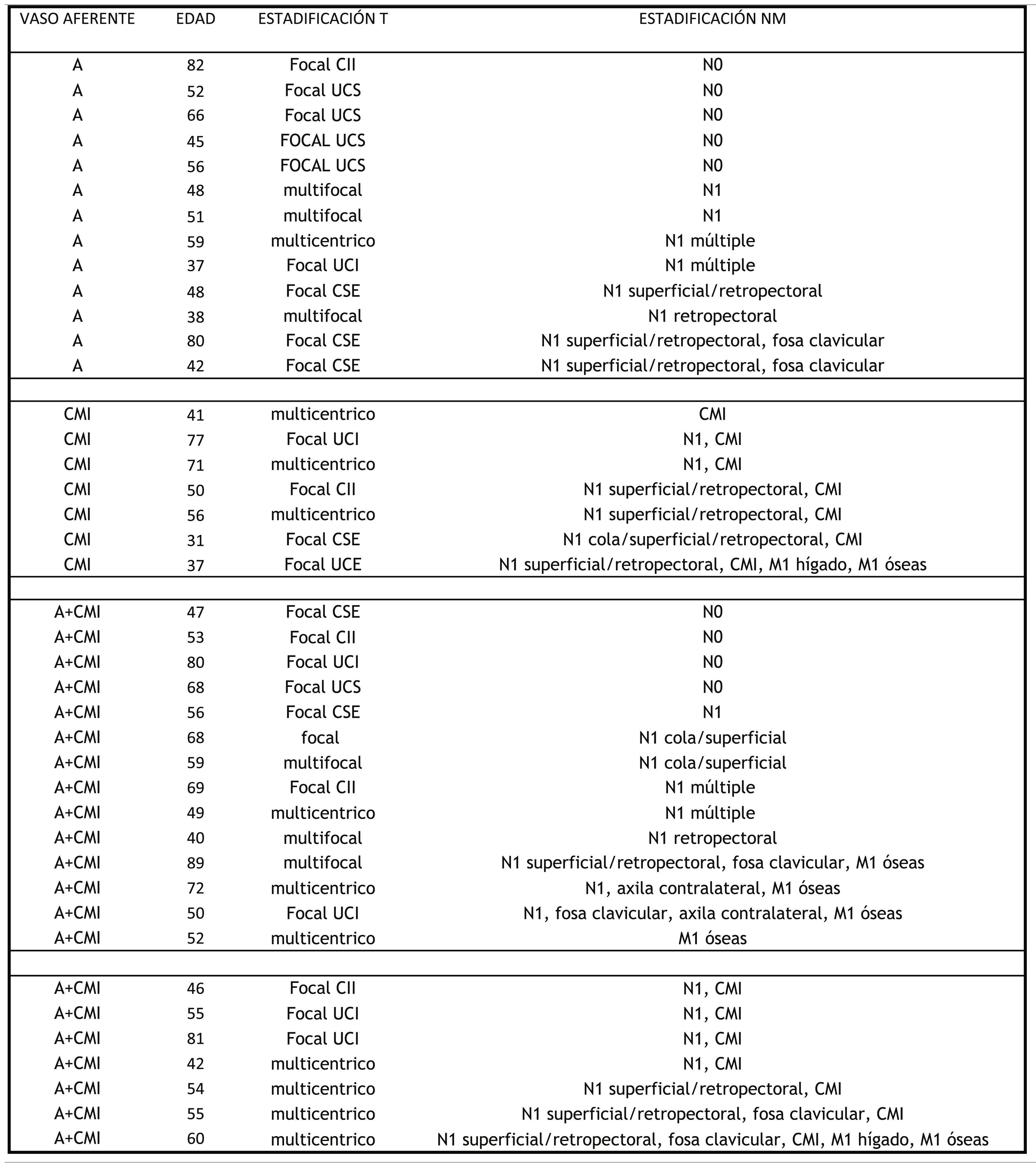
ResultsIMCI detection rate of 34% (14/41), with 8/14 patients under 55 years of age. All 14 patients with IMCI showed AV-IMC, 6 of them (43.9%) without VA-axillary. Of 27/41 patients without IMCI, in 13 (48.1%) only AV-axillary was found, in the remaining 14 (51.9%), AV-axillary and AV-IMC was found.
In 57% (8/14) tumours were multicentric and 42% (6/14) focal, in inner quadrants in 4/6 (66.7%).
In 1/14 patient (7.1%) only IMCI was found, in 9/14 (64.3%) axillary and IMC, in 4/14 patients (28.6%) distant lesions were detected.
Committee re-evaluation: no further treatment in 27/41 patients (65.8%), thoracic radiotherapy in 10/41 patients (24.4%), systemic therapy in 4/41 patients (9.7%).
ConclusionOur detection rate of IMCI in breast cancer staging by [18F]FDG PET/MR was 34%.
Related factors were age, multicentric tumours, inner quadrants, detection of AV-IMC, NM staging.
The evidence of IMCI allowed tailored therapy, with thoracic radiotherapy implementation in 24.4% of patients.
Evaluar la tasa de detección y la implicación terapéutica de la infiltración de la cadena mamaria interna (ICMI) mediante 18F-PET/RM en la estadificación de pacientes con cáncer de mama.
MétodoEstudio prospectivo, 41 mujeres con cáncer de mama (estadio ≥ IIB) estadificadas mediante 18F-FDG-PET/RM. Estudio en dos fases: Imágenes mamarias (decúbito prono), cuerpo completo (supino).
Estadificación TNM por consenso entre especialista en Medicina Nuclear y Radiología. Estudio vaso aferente (VA) a CMI por RM mamaria. Correlación ICMI con: edad, VA-CMI, estadificación T, cuadrante, infiltración axilar y a distancia.
Re-valoración terapéutica en comité multidisciplinar.
ResultadosTasa de detección de ICMN del 34% (14/41), siendo 8/14 < 55 años. Todas las 14 pacientes con ICMI muestran VA-CMI, en 6 de ellas (43,9%) sin VA-axilar. De 27/41 pacientes sin ICMI en 13 (48,1%) sólo VA-axilar, en los 14 restantes (51,9%) VA-axilar y VA-CMI.
El 57% (8/14) son multicéntricos y 42% (6/14) focales, en cuadrantes internos en 4/6 (66,7%).
En 1/14 paciente (7,1%) sólo ICMI, en 9/14 (64,3%) axilar y CMI, en 4/14 pacientes (28,6%) lesiones a distancia.
Decisión del comité: sin tratamiento adicionale en 27/41 pacientes (65,8%), radioterapia torácica en 10/41 pacientes (24,4%), terapia sistémica en 4/41 pacientes (9,7%).
ConclusiónLa tasa de detección de la ICMI en la estadificación del cáncer de mama mediante 18F-FDG PET/RM es del 34%.
Son factores asociados la edad, los tumores multicéntricos, los de cuadrantes internos, la existencia de VA- CMI, la estadificación NM.
La evidencia de ICMI permite la individualización de la terapia, indicando la radioterapia torácica en el 24,4%.
Article

Revista Española de Medicina Nuclear e Imagen Molecular (English Edition)


