Fibrous dysplasia is characterized by excessive proliferation of bone-forming mesenchymal cells. The maxilla is the most commonly affected facial bone, with facial asymmetry being the usual complaint. Surgery is the treatment of choice with two available options: conservative bone shaving or radical excision and reconstruction. We describe two cases of monofocal fibrous dysplasia of the midface causing facial asymmetry and treated by conservative surgery with good esthetic results.
A displasia fibrosa é caracterizada por uma proliferação excessiva células mesenquimais formadoras de osso. A maxila é o osso da face mais comumente afetado, sendo a assimetria facial o sinal mais comum. A cirurgia é o tratamento de escolha com duas opções possíveis: plastia óssea conservadora ou excisão radical seguida de reconstrução. Neste trabalho descrevemos dois casos de displasia fibrosa focal em terço médio da face, causando assimetria facial e tratada por cirurgia conservadora com bons resultados estéticos.
Fibrous dysplasia (FD) is a benign but chronic bone lesion characterized by the progressive replacement of normal bone with fibro-osseous connective tissue.1 Monostotic fibrous dysplasia is the most common form of this disease, characterized by the involvement of only one bone with no systemic manifestations. In the facial area, it is more frequently found in the maxillary bone of adolescents and young adults.1 Deformities leading to esthetic and functional disorders are observed in almost all cases.2 Plastic surgery is often recommended when the jaws are involved.3
Conservative management has been the standard of care, which involves removing the diseased bone via an intraoral approach.1,4 In aberrant cases, orthognathic surgery is indicated to restore the occlusion and facial deformity.3 This article describes the management of monostotic fibrous dysplasia affecting two adolescents. Esthetic and functional aspects are discussed.
Case reportCase report 1A 25-year-old male patient was referred to our unit with a complaint of severe swelling in the face that had been developing for the past two years. On clinical examination, the patient had some asymmetry in the left midface. The facial contour was compromised, and this caused severe esthetic discomfort to the patient (Figs. 1 and 2). A computed tomography (CT) scan showed a solid mass affecting a portion of the maxilla and the zygomatic bone on the left side (Figs. 5 and 6). Microscopic evaluation showed the typical histologic signs of FD including the presence of benign fibroblastic tissue with irregular spicules of woven bone and osteoblastic rimming embedded in fibrous tissue. Conservative surgical treatment was performed through intraoral access (Figs. 9 and 10). The contour of the midface was reestablished using chisels and drills. The esthetic result was satisfactory for the patient, who is in preparation for orthognathic surgery. The lesion did not show growth at a 26-month follow-up (Figs. 13 and 14).
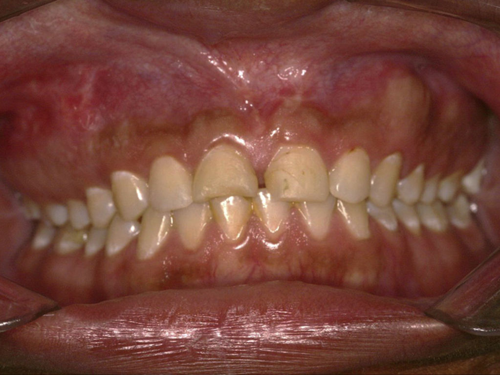
Case report 2A 22-year-old man had developed a swelling on the right side of the face that had persisted for one year. There was no history of pain, trauma, epistaxis, loosening of teeth, trismus, or diminished vision. Extraoral examination revealed a slight asymmetry on the right side of the maxilla that raised the nasal ala and modified the nasolabial fold. Intraoral examination revealed a hard swelling involving the right maxilla and zygoma (Figs. 3 and 4). A CT scan showed a radiodense mass involving the right zygomatic and maxilla, causing facial asymmetry (Figs. 7 and 8). An incisional biopsy showed irregular trabeculae of lamellar bone as well as woven bone with no definite arrangement lying in the compact stroma and confirms the diagnosis of fibrous dysplasia. The asymmetric area was surgically recontoured via an intraoral approach (Figs. 11 and 12). Postoperatively, the patient did well without complications. Preoperative occlusion was reestablished with relief of his pain. At the 14-month follow-up, an excellent esthetic profile of the midface was maintained (Figs. 15 and 16).
Craniofacial fibrous dysplasia can be classified as monostotic when it involves a single bone and polyostotic when it involves multiple bones. In the craniomaxillofacial skeleton, monostotic FD is more prevalent in the maxilla.5 The differential diagnoses of the condition include osteoma, osteosarcoma, cherubism and giant-cell granuloma.1,4 The progressive growth of fibrous dysplasia can lead to serious complications. When the lesion affects the midface, it can cause obstruction of the nostrils and difficulty opening the eyelids.6–8 The progression of the lesion into the oral cavity can compromise chewing and speaking. Periodontal and occlusal changes may also be present, and this may even result in tooth loss.2 Changes in the development of the jawbone usually affect dental occlusion. This condition may require orthodontic treatment and orthognathic surgery to correct the malposition of the teeth and jaw involved.7 Although occlusal changes were not present in the second patient, in the first patient, significant impairment of the position of the maxilla and mandible was observed and orthognathic surgery was indicated to restore occlusion and correct dentofacial deformity brought on by the disease process.
It is clear from clinical studies that the treatment necessary for this condition depends on its location in the craniofacial skeleton, its effect on function, and, ultimately, cosmetics. Skeletal deformities can require a surgical approach.1,8 The available options include two different approaches: conservative or radical. Conservative shaving or osseous contouring has been recommended by some authors5,8 who maintained that periodic contouring could be performed until a static phase was reached. Radical surgical therapy permits the complete removal of the lesion followed by immediate reconstruction.1,2
Some surgical access has been proposed for the treatment of fibrous dysplasia in the midface such as the midfacial degloving approach and the Weber-Ferguson incision.7,8 In both cases presented here, the intraoral approach offered appropriate conditions for the use of drills and chisel wear.
Although there was scope for radical excision of the fibrous dysplasia and reconstruction of the defect, poor patient compliance and the priority to relieve the symptoms and esthetic discomfort led us to adopt the conservative surgery, which had a fairly good postoperative result.
Ethical disclosuresProtection of human and animal subjectsThe authors declare that the procedures followed were in accordance with the regulations of the relevant clinical research ethics committee and with those of the Code of Ethics of the World Medical Association (Declaration of Helsinki).
Confidentiality of dataThe authors declare that they have followed the protocols of their work center on the publication of patient data.
Right to privacy and informed consentThe authors have obtained the written informed consent of the patients or subjects mentioned in the article. The corresponding author is in possession of this document.
Conflicts of interestThe authors have no conflicts of interest to declare.


