A 61-year-old male reported isolated dysphonia of 3 years’ evolution. Examination revealed bilateral paresis of the vocal folds in the paramedian position, with no dyspnoea or other accompanying symptoms.
Cervical CAT scan showed no alterations. A chest X-ray was performed which revealed bilateral hilar adenopathies. Since sarcoidosis was suspected, a thoracic CAT scan was requested which confirmed the presence of pretracheal, subcarinal and bilateral hilar enlarged lymph nodes.
Differential diagnosis of the adenopathies was started, a Mantoux test PPD was performed, which resulted negative, and calcaemia and angiotensin converting enzyme (ACE) levels were measured, which were normal. The patient underwent videomediastinoscopy and the adenopathies were biopsied. The pathology report showed non-caseating granulomas, therefore a diagnosis of sarcoidosis was made.
Treatment was started with prednisone 1mg/kg/day for 3 months, with complete recovery of left vocal fold (LVF) mobility, whereas the paralysis of the right vocal fold (RVF) persisted.
Case 2A 35-year-old woman reported isolated dysphonia of 2 years’ evolution. Examination revealed paralysis in the paramedian position of the LVF. Bilateral hilar adenopathies were revealed on cervicothoracic CAT scan, with no alterations at cervical level. Fibrobronchoscopy was performed with a biopsy, the histological study confirmed the presence of non-caseating granulomas, a diagnosis of sarcoidosis was made and treatment was started with prednisone 1mg/kg/day for 6 months.
After 6 months’ treatment, and despite the radiological disappearance of the adenopathies, the LVF paralysis persisted.
DiscussionAn aetiologic study must be started for patients with a history of dysphonia due to paralysis or paresis of the vocal folds (VF). Studies show that the most common causes of vocal fold paralysis in adults are endothoracic neoplasias and trauma (iatrogenic or otherwise) to the larynx, these 2 cases constitute more than half the cases of paralysis. However, between 10% and 42% of vocal fold paralyses are considered idiopathic; this is a percentage greater than 3%–17% of which represents the group of “rare” causes (e.g. granulomatous diseases). Paralysis of the left vocal fold is the most common.1
Therefore, aetiologic study of a case of vocal fold paralysis starts by ruling out the presence of endothoracic oncological processes and laryngeal trauma. Once these have been discounted, the rare causes of vocal fold paralysis should be considered, such as neurological, rheumatic, autoimmune or granulomatous disease.
Granulomatous diseases are divided into infectious, principally tuberculosis, and non-infectious, beriliosis and sarcoidosis, for example. We perform laboratory tests to make a differential diagnosis; PPD and ACE and calcaemia levels are useful. The diagnosis of sarcoidosis is based on the following criteria: (a) suggestive clinico-radiological features, (b) non-caseating granulomas shown in histology or a positive Kveim/Siltzbach test, (c) the exclusion of other granulomatous diseases and (d) compatible clinical evolution.2 However, the definitive diagnosis will be histopathological.
Sarcoidosis is a granulomatous disease of unknown aetiology which can affect any organ. The intra-thoracic region is most commonly affected (80%–90% of cases)3 with hilar, mediastinal lymph node and/or pulmonary infiltration.
Granulomas affecting the cervicofacial region in sarcoidosis occur in 9% of cases.4 Although laryngeal involvement can occur primarily due to granulomatous infiltration,4 several cases have been described of paralysis due to compression of the recurrent nerves caused by mediastinal adenopathies.5 Povedano et al.,6 based on the clinical and pathogenic characteristics of described cases of the disease, suggest 3 mechanisms of paralysis of the vocal folds in the context of sarcoidosis: (1) primary fixation due to granulomas, (2) neuropathy due to primary involvement of the vagus nerve, and (3) neuropathy due to compression of the recurrent nerve secondary to adenopathies. The distinction between these 3 possibilities will be made depending on the findings accompanying the vocal fold paralysis. We opt for the neuropathy group when other cranial nerves are involved, and for the group of primary fixation due to granulomas in the case of sarcoidosis of the larynx itself. Since the laryngeal CAT scans in the 2 cases which we present were normal, and there were no other neurological alterations, we assume that we can include them in the group of paralysis secondary to compressive adenopathy of the recurrent nerve.
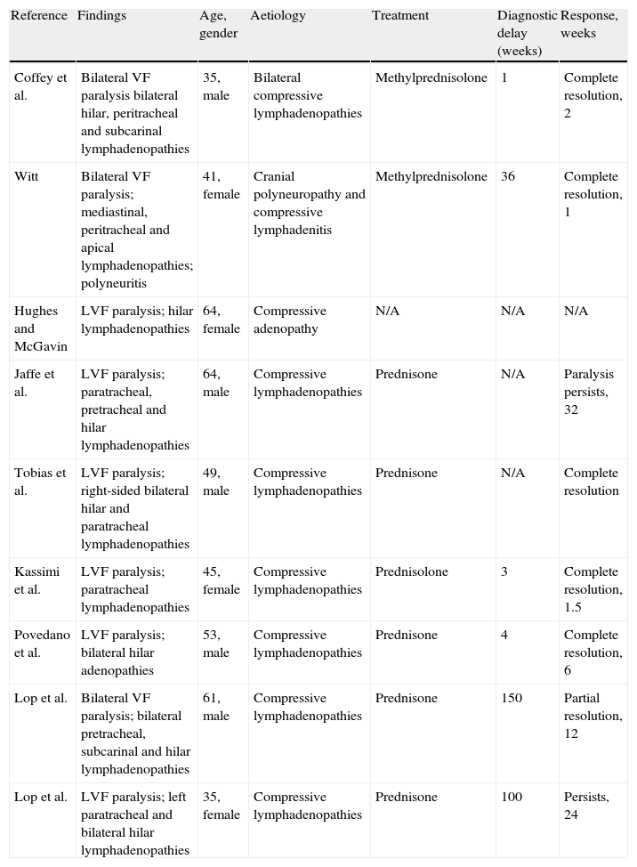
A great many of the cases published on vocal fold paralysis in the context of sarcoidosis fall into the compression neuropathy group. Coffey et al.5 revised most of the published cases (Table 1) of recurring paralysis in the context of sarcoidosis and found that these were largely left-sided recurrent paralyses; this data is to be expected given the long intra-thoracic route of the left recurrent nerve.7 However, the same authors describe a case of bilateral recurrent paralysis which is justified by the existence of adenopathies at paratracheal level and of the right subclavian artery, where the lower right laryngeal nerve recurs.5 Witt8 and Alon9 also presented cases of bilateral or right-sided recurrent paralysis in sarcoidosis, but these were associated with other cranial neuropathies, and constitute examples of neurosarcoidosis and therefore could be included in group 2 of those proposed by Povedano et al.6
Published Cases of Vocal Fold Paralysis Associated With Sarcoidosis.
| Reference | Findings | Age, gender | Aetiology | Treatment | Diagnostic delay (weeks) | Response, weeks |
| Coffey et al. | Bilateral VF paralysis bilateral hilar, peritracheal and subcarinal lymphadenopathies | 35, male | Bilateral compressive lymphadenopathies | Methylprednisolone | 1 | Complete resolution, 2 |
| Witt | Bilateral VF paralysis; mediastinal, peritracheal and apical lymphadenopathies; polyneuritis | 41, female | Cranial polyneuropathy and compressive lymphadenitis | Methylprednisolone | 36 | Complete resolution, 1 |
| Hughes and McGavin | LVF paralysis; hilar lymphadenopathies | 64, female | Compressive adenopathy | N/A | N/A | N/A |
| Jaffe et al. | LVF paralysis; paratracheal, pretracheal and hilar lymphadenopathies | 64, male | Compressive lymphadenopathies | Prednisone | N/A | Paralysis persists, 32 |
| Tobias et al. | LVF paralysis; right-sided bilateral hilar and paratracheal lymphadenopathies | 49, male | Compressive lymphadenopathies | Prednisone | N/A | Complete resolution |
| Kassimi et al. | LVF paralysis; paratracheal lymphadenopathies | 45, female | Compressive lymphadenopathies | Prednisolone | 3 | Complete resolution, 1.5 |
| Povedano et al. | LVF paralysis; bilateral hilar adenopathies | 53, male | Compressive lymphadenopathies | Prednisone | 4 | Complete resolution, 6 |
| Lop et al. | Bilateral VF paralysis; bilateral pretracheal, subcarinal and hilar lymphadenopathies | 61, male | Compressive lymphadenopathies | Prednisone | 150 | Partial resolution, 12 |
| Lop et al. | LVF paralysis; left paratracheal and bilateral hilar lymphadenopathies | 35, female | Compressive lymphadenopathies | Prednisone | 100 | Persists, 24 |
Despite the disparity in laterality, both the unilateral and the bilateral cases showed an excellent response to treatment with corticosteroids, given the reversion of inflammation and lymph node oedema. However, in the cases where introduction of treatment was delayed, the response to corticosteroids was reduced.8 This observation is reinforced in the two cases which we present, both share a long evolution time prior to diagnosis (2 years).
In conclusion, it is important to include granulomatous diseases in the differential diagnosis of recurrent paralysis.
Conflict of InterestsThe authors have no conflict of interest to declare.
Please cite this article as: Lop Gros J, García Lorenzo J, Quer Agustí M, Bothe González C. Sarcoidosis y parálisis de cuerdas vocales: a propósito de 2 casos y revisión de la literatura. Acta Otorrinolaringol Esp. 2014;65:378–380.


