INTRODUCTION
Tuberculosis (TB) is still an important worldwide public health problem affecting 16 million of people; 1.8 million of them die yearly, and 1.9 billion are estimated to be infected by Mycobacterium tuberculosis (Mt) 1. The incidence of TB has been increasing and 8.2 million of new cases are registered yearly, 1.3 million of them in children and adolescents under the age of 15 years 2.
Despite of being very well established in adults, TB diagnosis and treatment in children are sometimes difficult to achieve, because of their particularly aspects 4. In general, TB is diagnosed by the identification of Mt in about 30 % to 40 % of cases 4. However, in children the pauci-bacilar form is more common and Mycobacteria isolation and identification are even less frequent. Therefore, TB diagnosis in children is done mainly by a positive history of contact with a baciliferous adult, by a positive Mantoux tuberculin skin test (MTST) or by an abnormal CXR 3,5. Gastric contents aspired for Mt research and culture has low indexes of positivity and requires children hospitalization 6. Routine bacillus of Calmete-Guérin (BCG) vaccination, usually in the first month of life, is another point that difficults TB diagnosis in children. Although BCG protects against severe forms of TB, it becomes harder to interpret a positive purified protein derivative (PPD) test 7,8.
The clinical picture of TB is very wide and can simulate several illnesses from a common cold to a chronic pneumopathy like asthma. Almost 17 % of children with TB who are admitted in hospitals present wheezing, and can be misdiagnosed as asthmatics during an acute attack 3. In this article we report a patient with pulmonary TB, initially classified as moderate-to-severe asthmatic with recurrent pneumonia.
CASE REPORT
J.C.S., second son of three offspring, was followed as an outpatient at the Allergy Clinic, since he was 3 years and 8 month-old. At that time, he had a history of wheezing in the last 12 months. His first wheezing episode was due to climate changes (cough, and dyspnea, without fever) and was treated with short-acting β2-agonists with partial clinical improvement. Afterwards, he presented wheezing episodes bi-weekly. When he was three years old a pneumonia (CXR, right pulmonary base) was treated with amoxicillin and short-acting β2-agonists with clinical improvement. Other 4 episodes of pneumonia, confirmed by CXR and treated as previously, had occurred in a 6 month period. There was not reference of nocturnal perspiration, productive cough, fever or lose weight.
His younger brother (two years and five month-old) had a positive history of fever of undetermined origin lasting 3 months, associated to sinusal tachycardia, anemia and energy-protein malnutrition. As inpatient he was evaluated and all lab tests done (toxoplasmosis, cytomegalovirus, HIV) and PPD were negative. During hospitalization he had pericarditis and was treated as TB pericarditis (Isoniazid, Pyrazinamide and Rifampicine). He improved and was free of symptoms after 6 months of treatment. All contactants had been evaluated and were healthful. The primary focus of TB contact was not identified.
As JCS was admitted by recurrent pneumonia associated to wheezing he was evaluated for TB. He was well-nourished (98,6 % adequacy of weight for stature and 96.5 % of stature for age), heart rate 100 beats/min, without changes in pulmonary and cardiac auscultation as well as in abdominal examination. Skin prick tests to inhalant allergens were negative, WBC was normal, and the hemosedimentation rate was 14 mm. Parasites in stool (3 samples) and gastroesophageal reflux evaluation (scintillography and upper gastrointestinal series) were negative. Normal levels of sweat electrolytes and serum immunoglobulins A, G and M (244 mg/dl, 1430 mg/dl and 125 md/dl, respectively) were observed. There were no antibodies to Toxocara cannis, PPD, and acid alcohol resistant bacilli in gastric aspirates were negative.
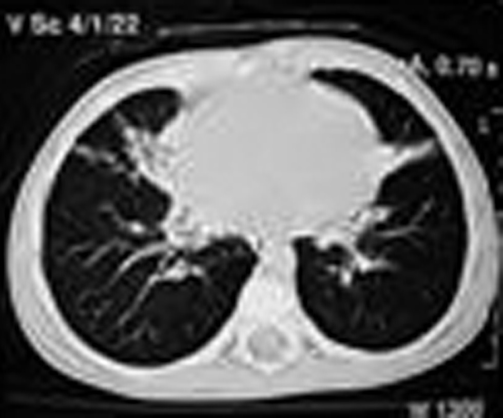
Hyperinsulflation, image of persistent condensation in right base, and hilar adenopathy were observed at several CXR. CCT showed condensation in right base, hypodensity, and atelectasis in left hemithorax, compatible with pneunomitis (fig. 1). At this time, JCS initiated nocturnal productive cough and perspiration, without fever.
Figure 1.--CCT before treatment. Area of heterogeneous increase in pulmonary transparency, condensation in medium lobe. Fibro-atelectatic band in lingula.
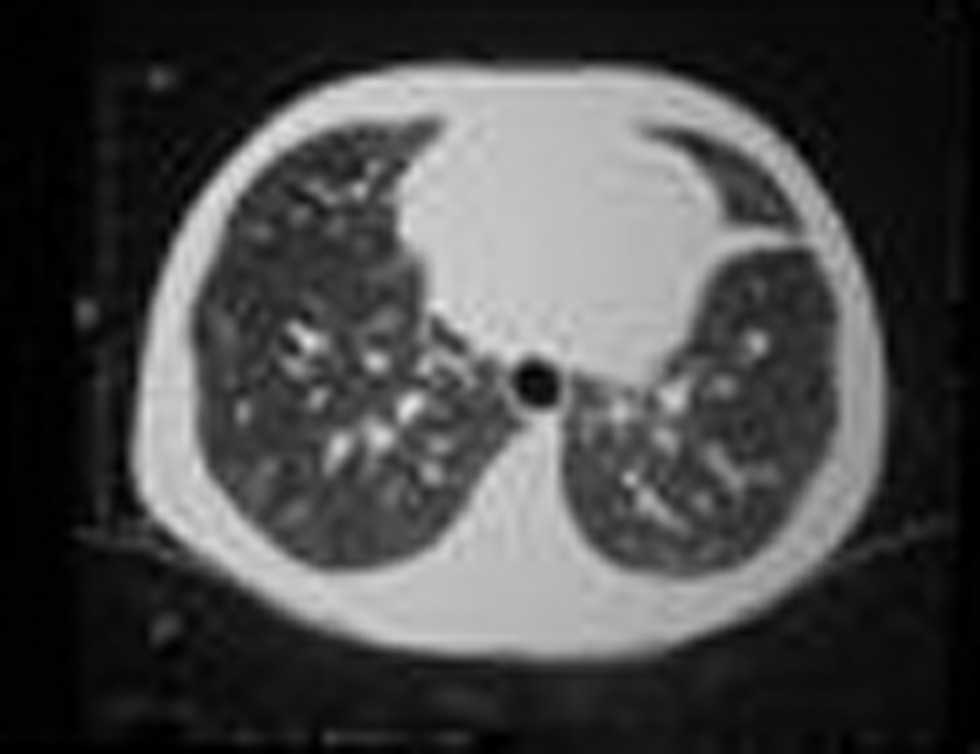
Due to difficulty in establishing TB diagnosis, we decided to start proof treatment (Isoniazid [10 mg/kg/ day], Pyrazinamide [35mg/kg/day] and Rifampicine [10 mg/kg/day]). Significant improvement in symptoms was observed after four months of treatment. Anteroposterior CXR, after treatment, showed absence of condensation in right base and CCT evidenced fibrosis or atelectatic band in left base, probably a scar (fig. 2).
Figure 2.--CCT after treatment. It shows disappearance of pulmonary area condensation in medium lobe, remaining the fibro-atelectatic band in lingula.
DISCUSSION
The decision to institute TB treatment, in our patient, was based on clinical data supported by lab tests and strengthened by positive epidemiological history. According to the Center for Disease Control and Prevention (CDC) due to the high risk of TB spread in children above four years of age, treatment must be initiated, as soon as TB is suspected.
TB diagnosis in younger children, especially in the vaccinated ones, is a difficult task. OMS standardized some criteria to facilitate its diagnosis. According to these criteria, TB should be: a) suspected when there is history of confirmed TB contact; loss of weight, cough and wheezing that does not improve with appropriate therapy or painless lymphadenopathy; b) probable - in children with one of the following: PPD higher than 10 mm; suggestive CXR; biopsy of granuloma lesion with suggestive histology; good response to antituberculosis therapy; and c) confirmed - bacillus detected by microscopy or culture in secretion or tissue, or with the identification of Mt by specific cultures 1.
TST has been the most utilized exam to evaluate patients suspected to be with TB. In Brazil, PPD-RT23 (2UT) is the standard reagent applied in TST and according to the induced induration diameter individuals are classified as: non reactor (0-4 mm), weak reactor (5-9 mm) and strong reactor (> 10 mm). Children who have been vaccinated or who had had contact with another atypical mycobacterium generally have a weak reaction. The strong reactors are those individuals who had had contact with Mt (rarely the vaccinated ones present this type of reaction). It is reported that the possibility of being sick is bigger when TST is greater than 15 mm 9,10.
The American Academy of Pediatrics utilizes three other cutoffs to classify a positive TST: ≥ 5 mm, ≥ 10 mm, ≥ 15 mm, depending on history and inherent risk factors. For example, TST ≥ 5 mm is considered positive if a child had contact with a possible baciliferous adult, or if he is imunossupressed 7.
Some studies demonstrated that the majority of children vaccinated with BCG has TST ≥ 10 mm that diminishes with age, except if the child has contact with a baciliferous patient, suggesting cumulative effect of vaccine and exposition to TB disease 7. Similar picture occurs with children who receive more than one dose of BCG 7, because vaccination stimulates delayed hypersensitivity response to TST 10. However, this higher hypersensivity cannot be differentiated from TB disease, increasing the difficulty in the diagnosis of TB in children. The accomplishment routine to TST is of great value for aid in TB diagnosis in countries where BCG vaccination is not a routine.
Our patient had negative TST. According to Shingadia & Novelli 11, around 10 % of children with a positive culture for TB do not react initially to TST. TST can become reactive in many children during treatment. It suggests that TB itself can contribute for immunossupression. Other factors can be associated with a false negative TST such as: virus infection (human immunodeficiency virus, measles and chickenpox), bacterial infections (typhoid fever and brucellosis), been vaccinated with alive virus (polio, measles and chickenpox), concomitant disease (chronic renal insufficiency, lymphoma and leukemia), use of corticosteroids, inadequate technique (inappropriate material or incorrect administration, error in measuring, and problems with the extract) and finally error in interpreting the results 7.
In children the best way to isolate Mt is from gastric aspired culture although its positivity is only around 30 % 6. TB CXR pattern can show hilar adenopathy, hyperinsulflation, atelectasis, bronchiectasis, alveolar consolidation, pleural fluid with or without empyema and rarely cavitation. In miliary TB, pulmonary injure is diffuse, and it is common to see hilar adenopathy and diffuse bilateral reticular infiltrate 11. CCT shows more details than CXR, distribution of thoracic lymph nodes and its characteristics can help to discard other diseases like sarcoidosis (without low density center) or lymphoma (anterior lymphadenopathy) 12.
The CXR of our patient showed adenopathy and persistent condensation in the middle lobe, independently from treatment. CCT showed signals of active pneumonia, however the patient had persistent cough without fever.
Bronchoscopy followed by isolation of Mt in bronchial secretion obtained from patients suspected to have TB is less sensitive than gastric contents. However, bronchoscopy is an important method for diagnosing endobronchial TB and/or opportunistic infection, in patients with immunodeficiency, and foreign body aspiration 11.
Wheezing in TB patients might be secondary to extrinsic compression due to lymphadenopathy or even though the blockage due to endobronchial granuloma in those with endobronchial TB 3,11. Our patient had recurrent wheezing that improved with inhaled short-acting β2-agonists. This could not be explained by adenopathy compression but probably to pulmonary parenchyma injury, in association with atelectasis and bronchial hyperreactivity.
Recently, other methods have been used to improve TB diagnosis. Amplification of small amounts of bacterial nucleic acid by polymerase chain reaction (PCR) has a 40 % to 60 % of sensitivity 11. Antibodies to Mt antigens, including PPD, can be detected by enzyme-linked assay (ELISA). However, it has low sensitivity and specificity, as well as, inadequate reproducibility that reinforces its inadequate use for children with TB 11.
TB continues causing considerable morbidity and mortality in children, adolescents and adults worldwide. Childhood TB represents an alert signal suggesting recent transmission from an infective adult. Early diagnosis and suspicious is very important, because it would represent a decrease in morbidity. Sometimes, TB diagnosis is difficult and dependent on large investigations. We present a clinical case of persistent wheezing due to TB initially treated as asthma. Patients with persistent wheezing whose clinical history differs from usual asthma picture, especially in children with recurrent pneumonia, must be investigated for other diseases such as TB before starting prophylactic asthma treatment.
Correspondence:
C.S.K. La Scala
Rua dos Otonis, 725
Vila Clementino.
São Paulo, SP CEP 04025 - 002. Brazil
E-mail: cintialascala@uol.com.br


