To describe the characteristics of type 1 choroidal neovascularisation (CNV) in age-related macular degeneration (ARMD) using two different optical coherence tomography angiography (OCT-A) devices sequentially during a standard protocol of three intravitreal injections of an anti-vascular endothelial growth factor (anti-VEGF).
MethodsThe study included 6 eyes with naïve neovascular ARMD. Macular OCT-A images were acquired using AngioPlex Cirrus HD-OCT 5000 (Carl Zeiss Meditec, Inc., Dublin, USA) and DRI OCT Triton SS-OCT Angio (Topcon, Medical Systems, Inc., Oakland, NJ, USA). The macular OCT-A scan covered an area of 3mm×3mm. Distinct morphological patterns and quantifiable features of the neovascular membranes were studied on en face projection images, which were taken at different stages of the follow-up.
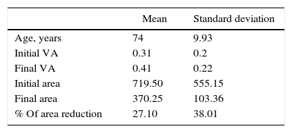
ResultsTreatment response could be estimated using the OCT-A criteria of CNV activity. Higher activity scores before treatment resulted in a greater decrease in the membrane area. The estimated net decline in area ranged from 83.5% to 1.4%. The OCT-A performed one-week after treatment revealed the greatest area reductions.
ConclusionsOCT-A provides new possibilities for the non-invasive assessment of features of neovascular networks and CNV structural morphology. Newly described activity criteria can also guide therapeutic decisions, and help in evaluating responses. Quantitative and qualitative information can be provided with this technique. However, further software development and future investigation are essential to define the role of this tool on a daily basis.
Describir las características de la neovascularización coroidea (NVC) tipo 1 en pacientes con degeneración macular asociada a la edad (DMAE), utilizando la angiografía por tomografía de coherencia óptica (A-OCT) secuencialmente durante el transcurso de un protocolo estándar de 3 inyecciones intravítreas de fármaco anti-VEGF.
MétodosSeis ojos con DMAE neovascular no tratados previamente fueron incluidos. Se obtuvieron imágenes por A-OCT empleando AngioPlex Cirrus HD-OCT 5000 (Carl Zeiss Meditec, Inc., Dublin, EE. UU.) y DRI OCT Triton SS-OCT Angio (Topcon, Medical Systems, Inc. Oakland, NJ, EE. UU.). El área estudiada comprende un escáner macular de 3×3mm. Diferentes patrones morfológicos y aspectos cuantificables de las membranas neovasculares han sido evaluados con imágenes en proyección en face, que fueron tomadas en distintos tiempos del seguimiento de los pacientes.
ResultadosEl grado de respuesta al tratamiento fue estimado empleando criterios de actividad de NVC para A-OCT. Puntuaciones más altas en los ítems de actividad antes del tratamiento resultaron en mayores reducciones del área de las membranas. Los resultados finales de reducción de área oscilaron entre el 83,5 y el 1,4%. Las A-OCT realizadas a la semana de tratamiento revelaron los mayores porcentajes de reducción.
ConclusionesLa A-OCT ofrece la posibilidad de analizar en profundidad las características morfológicas y estructurales en NVC de tipo 1. Los criterios de actividad permiten guiar decisiones terapéuticas y evaluar la respuesta al tratamiento. Con esta técnica puede obtenerse información útil tanto cualitativa como cuantitativa. Sin embargo, son necesarios avances en el desarrollo del software y en investigación para poder definir el papel de esta herramienta en la práctica diaria.
Artículo
Comprando el artículo el PDF del mismo podrá ser descargado
Precio 19,34 €
Comprar ahora


