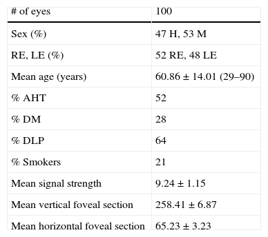
To establish normal values of macular thickness and volume obtained by the Cirrus SD-OCT (Carl ZeissMeditec, Dublin, CA, USA). Secondly, to assess the association between macular thickness and volume, sex and age.
Material and methodsA prospective study was conducted on patients who were seen in a hospital Retina Unit, and who only had retinal disease in one eye. All the Macular Cube 512×128 scan protocols were performed by the same operator. Only the healthy eye was scanned in each patient.
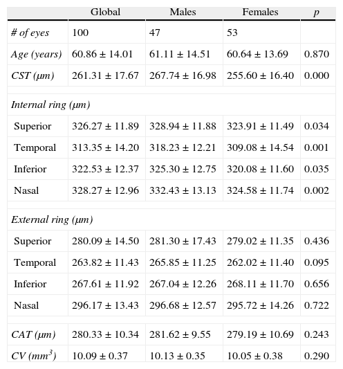
ResultsA total of 100 eyes of 100 patients were analysed. The mean central foveal thickness was 261.31±17.67μm, and was significantly (p<.05) higher in males (267.74±16.98μm) than in females (255.60±16.40μm). The mean obtained for the volume of the cube was 10.09±0.37mm 3, and the mean thickness of 280.33±10.34 cube um, with no statistically significant differences between gender being found (p<.05). The mean macular thickness is less at central level, increases in the inner perifoveal ring, and then decreases in the outer perifoveal ring. Furthermore, of all quadrants the greatest thickness was the nasal (328.27±12.96μm), followed by the upper (326.27±11.89μm), lower (322.53±12.37mm) sectors, with the temporal sector being the thinnest (313.35±14.20μm). The mean age of the patients was 60.86±14 years.
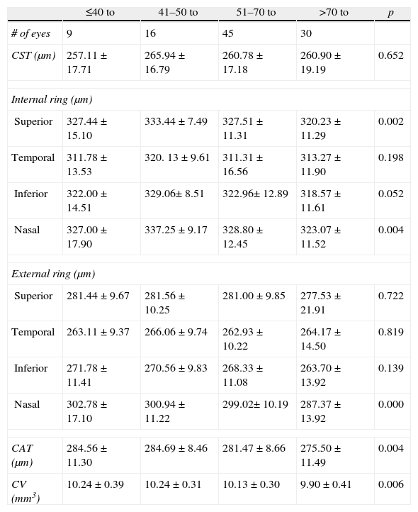
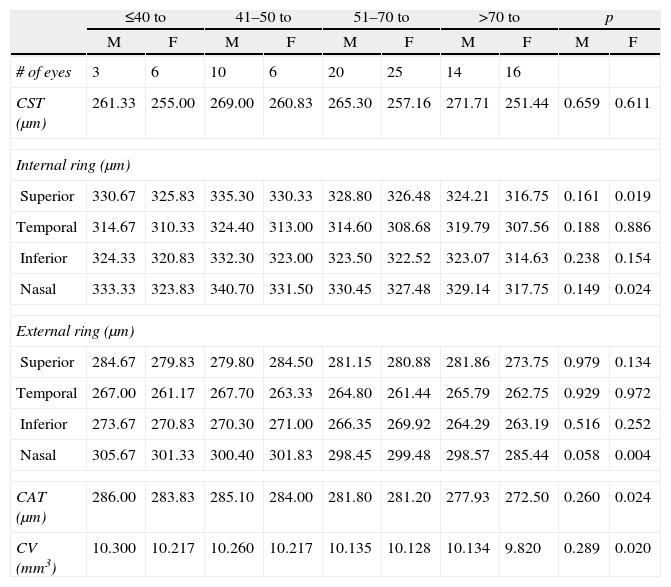
ConclusionThe mean central foveal thickness and the thickness of the inner perifoveal ring are significantly higher in men than in women. Both the mean volume and thickness of the cube, as well as nasal and inner superior sectors decrease with age, being significant only in women.
Establecer los valores de normalidad de espesor y volumen macular obtenidos mediante SD-OCT Cirrus (Carl ZeissMeditec, Dublín, CA, EE. UU.). Secundariamente, evaluar la asociación entre espesor y volumen macular, sexo y edad.
Material y métodoEstudio prospectivo, con pacientes que fueron visitados en la unidad de retina del HUNSC y solo presentaban enfermedad retiniana en uno de los ojos. Todos los protocolos Macular Cube 512×128 fueron realizados por un mismo operador. De cada paciente, solo se analizó el ojo sano.
ResultadosSe analizaron 100 ojos de 100 pacientes. El espesor foveal central medio fue 261,31±17,67μm, siendo significativamente (p<0,05) mayor en hombres (267,74±16,98μm) que en mujeres (255,60±16,40μm). La media obtenida para el volumen del cubo fue de 10.09±0.37mm3 y para el espesor medio del cubo de 280.33±10.34μm; no encontrando diferencias estadísticamente significativas entre sexos (p<0,05). El espesor macular medio es menor a nivel central, aumenta en el anillo perifoveal interno y disminuye a continuación en el anillo perifoveal externo. Asimismo, de todos los cuadrantes el de mayor espesor es el nasal (328,27±12,96μm), seguido del superior (326,27±11,89μm), inferior (322,53±12,37μm) y por último, el de menor espesor es el temporal (313,35±14,20μm). La edad media de los pacientes incluidos es de 60,86±14 años.
ConclusiónEl espesor foveal central medio y el espesor del anillo perifoveal interno son significativamente mayores en hombres que en mujeres. Tanto el volumen como el espesor medio del cubo, así como los sectores nasales y el sector supero interno, decrecen con la edad, únicamente de forma significativa en las mujeres.
Artículo
Comprando el artículo el PDF del mismo podrá ser descargado
Precio 19,34 €
Comprar ahora


