We aimed to evaluate the usefulness of magnetic resonance imaging (MRI) in the diagnosis of occult fractures of the scaphoid and to determine the advantages and cost in comparison with the traditional follow-up protocol.
Materials and methodsThe traditional approach at our center consisted of immobilization and periodic clinical and radiological follow-up (plain-film X-rays and computed tomography in the final phase of the process). The new protocol called for a limited MRI study consisting of coronal T1- and T2-weighted fat suppression sequences if the findings at plain-film X-rays continued to be negative at the first follow-up examination with the traumatologist (10 days after trauma). We evaluated the MRI findings, the time the patient was immobilized, the cost of each protocol, and the dose of radiation received.
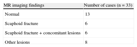
ResultsWe included 33 cases of patients with clinically suspected fractures of the scaphoid and negative findings on plain-film X-rays. In 13 patients, the MRI findings were negative. In 12 patients, the MRI findings confirmed the diagnosis of a fracture of the scaphoid, which was associated with other pathology in 6 cases. In 8 patients, another pathological process was diagnosed. The cost of the new protocol was €131.06 per patient; the cost of the traditional protocol was €114.41 or €151.06 per patient, depending on the follow-up studies required. The new protocol reduced the dose of radiation by eliminating successive radiologic studies.
ConclusionsThe new protocol improved the management of these patients, reducing the time of immobilization, improving joint rigidity, and reducing the time off work. The limited MRI study makes it possible to diagnose other radiologically occult lesions. The cost of the new protocol is similar to that of the traditional protocol and even lower in some cases. The new protocol results in a reduction in the dose of radiation.
Nuestro objetivo es valorar la utilidad de la resonancia magnética (RM) en el diagnóstico de las fracturas ocultas de escafoides, mostrando las ventajas y el coste comparativo frente al protocolo de seguimiento tradicional.
Material y métodoEl protocolo de actuación tradicional en nuestro centro, consistía en inmovilización y revisiones clínico-radiológicas periódicas (radiología convencional y tomografía computarizada en la fase final del proceso). En el nuevo protocolo, si en el primer control del traumatólogo (10 días post- traumatismo) la radiología convencional seguía siendo negativa se realizaba un protocolo limitado de RM de muñeca (coronal T1 y T2-supresión grasa). Se valoraron los hallazgos visualizados en RM, tiempo de inmovilización del paciente, coste económico de ambos protocolos y dosis de radiación recibida.
ResultadosSe incluyeron 33 casos de pacientes con sospecha clínica de fractura de escafoides y radiología negativa. En 13 pacientes la RM fue negativa. En 12 se confirmó el diagnóstico de fractura de escafoides, 6 asociadas a otra patología. En 8 se diagnosticó otro proceso. El coste del nuevo protocolo fue de 131.06€ por paciente y de 114.41€ ó151.06€ para el tradicional, según las revisiones necesarias. Se redujo la dosis de radiación al eliminar la realización de sucesivas exploraciones radiológicas.
ConclusionesEl nuevo protocolo mejora el manejo de estos pacientes, reduciendo el tiempo de inmovilización, mejorando la rigidez articular y disminuyendo el periodo de baja laboral. Permite el diagnóstico de otras lesiones radiológicamente ocultas. El coste es similar, e incluso inferior en algunos casos, y la irradiación es menor.
Artículo
Comprando el artículo el PDF del mismo podrá ser descargado
Precio 19,34 €
Comprar ahora


