


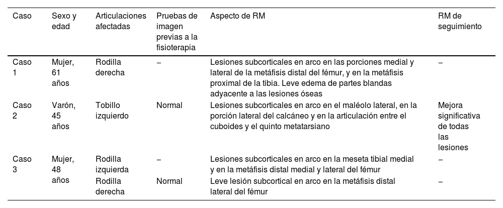
La diatermia terapéutica por ultrasonidos es un método no invasivo ampliamente disponible, utilizado para ablandar el tejido cicatricial, resolver el edema y acelerar la cicatrización de heridas. Sin embargo, no existe consenso sobre la eficacia de este método y se carece de información sobre complicaciones específicas. Presentamos tres casos de lesiones óseas focales relacionadas temporalmente con fisioterapia mediante diatermia por ultrasonidos. El primer paciente presentaba lesiones en las porciones medial y lateral de la metáfisis distal del fémur derecho, así como en la metáfisis proximal de la tibia. El segundo paciente presentaba lesiones en el tobillo izquierdo. El tercer paciente presentaba lesiones bilaterales en la rodilla. Las lesiones se caracterizaron como subcorticales lineales o en arco con señal hipointensa en las imágenes ponderadas en T1 y señal hiperintensa en las imágenes ponderadas en T2 y por densidad de protones, similares a la osteonecrosis. Sin embargo, estas lesiones se distribuyeron en zonas superficiales, en las que se aplica directamente el ultrasonido. Todos los pacientes refirieron mejoría o desaparición del dolor tras la interrupción de la diatermia por ultrasonidos. Por lo tanto, la diatermia terapéutica por ultrasonidos puede asociarse con lesiones óseas focales. Este diagnóstico debe tenerse en cuenta cuando aparecen lesiones óseas superficiales relacionadas con el tiempo de tratamiento y en zonas que no suelen estar afectadas por la osteonecrosis.
Therapeutic ultrasound diathermy is a widely available noninvasive method, used to softening scar tissue, resolving edema, and accelerating wound healing. However, there is no consensus regarding the efficacy of this method and there is a lack of information on specific complications. We report three cases of focal bone lesions time-related with physiotherapy using ultrasound diathermy. The first patient presented with lesions in the medial and lateral portions of the distal metaphysis of the right femur, as well as in the proximal metaphysis of the tibia. The second patient presented with lesions in the left ankle. The third patient presented with lesions in the knee, bilaterally. The lesions were characterized by subcortical linear or arc lesions with hypointense signal on T1-weighted imaging and hyperintense signal on T2- and proton-density weighted imaging, similar to osteonecrosis. However theses lesions were distributed in superficial areas, in which the ultrasound is directly applied. All patients reported improvement or resolution of pain after discontinuation of ultrasound diathermy. Therefore, therapeutic ultrasound diathermy may be associated with focal bone lesions. This diagnosis should be considered when superficial bone lesions appear time-related to therapy and in areas not usually affected by osteonecrosis.

