To assess the importance of false-negative and false-positive findings in computed tomography (CT) and 18F-FDG positron emission tomography (PET) in mediastinal lymph node staging in patients undergoing surgery for non-small cell lung cancer (NSCLC).
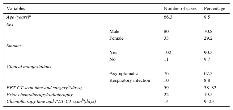
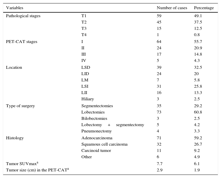
Material and methodsThis retrospective study included 113 consecutive patients and 120 resected NSCLCs; 22 patients received neoadjuvant treatment. We compared the findings on preoperative 18F-FDG PET-CT studies with the postoperative pathology findings. Lymph node size and primary tumor size were measured with CT, and lymph nodes and primary tumors were evaluated qualitatively and semiquantitatively (using standardized uptake values (SUVmax)) with PET.
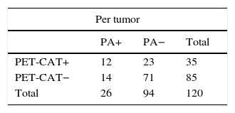
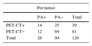
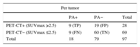
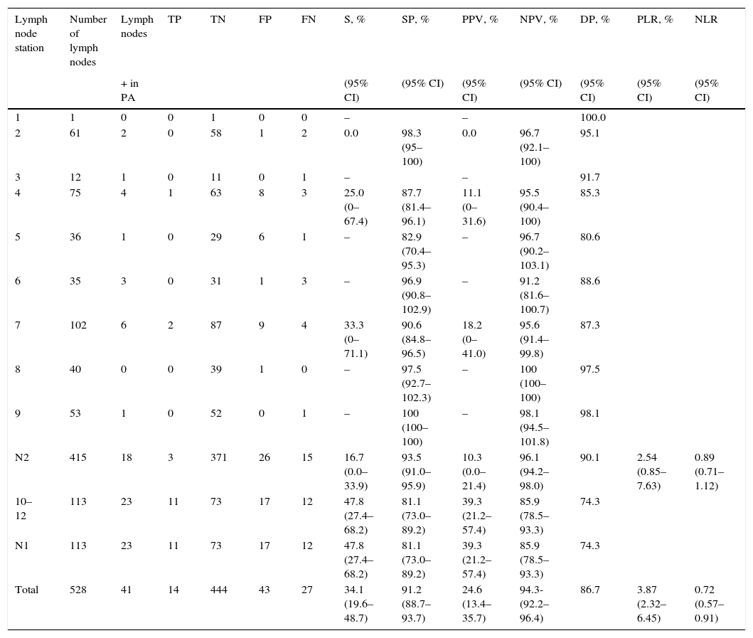
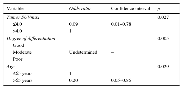
ResultsMetastatic lymph nodes were found in 26 (21.7%) of the 120 tumors and in 41 (7.7%) of the 528 lymph node stations analyzed. 18F-FDG PET-CT yielded 53.8% sensitivity, 76.6% specificity, 38.9% positive predictive value, 85.7% negative predictive value, and 71.7% diagnostic accuracy. The false-negative rate was 14.2%. Multivariable analysis found that the factors associated with false-negative findings were a moderate degree of differentiation in the primary tumor (p=0.005) and an SUVmax of the primary tumor >4 (p=0.027). The false-positive rate was 61.1%, and the multivariable analysis found that lymph node size >1cm was associated with false-positive findings (p<0.001).
ConclusionsIn mediastinal lymph node staging in patients with NSCLC, 18F-FDG PET-CT improves the specificity and negative predictive value and helps clinicians to select the patients that will benefit from surgery. Given the high rate of false positives, histological confirmation of positive cases is recommendable.
Valorar las implicaciones de los falsos negativos (FN) y de los falsos positivos (FP) de la tomografía computarizada (TC) y la tomografía por emisión de positrones (PET) con fluorodesoxiglucosa (18F-FDG) en nuestro medio en la estadificación ganglionar mediastínica de pacientes operados de carcinoma de pulmón de células no pequeñas (CPCNP).
Material y métodosEstudio retrospectivo de 113 pacientes consecutivos con 120 CPCNP operados; 22 pacientes recibieron tratamiento neoadyuvante. Se compararon los resultados obtenidos en la 18F-FDG PET-TC prequirúrgica con los patológicos. Se analizaron el tamaño ganglionar y del tumor primario en la TC, y su valoración cualitativa y semicuantitativa (SUVmáx) en la PET.
ResultadosSe encontraron ganglios metastásicos en el 21,7% de los 120 tumores y en el 7,7% de las 528 estaciones ganglionares analizadas. La 18F-FDG PET-TC en el estudio por tumor mostró una sensibilidad del 53,8%, una especificidad del 76,6%, un valor predictivo positivo del 38,9%, un valor predictivo negativo del 85,7% y una precisión diagnóstica del 71,7%. La tasa de FN fue del 14,2%. El análisis multivariable mostró que un grado de diferenciación moderado del tumor primario (p = 0,005) y una SUVmáx del tumor primario >4 (p = 0,027) eran los factores asociados con los FN. La tasa de FP fue del 61,1% y el tamaño ganglionar >1cm era el factor asociado con los FP (p < 0,001).
ConclusionesLa 18F-FDG PET-TC en la estadificación ganglionar mediastínica de pacientes con CPCNP mejora la especificidad y el valor predictivo negativo, y ayuda al clínico a seleccionar los pacientes que se beneficiarán de la cirugía. Dada la elevada tasa de FP, es recomendable que, antes de excluir a pacientes de la cirugía, se confirmen histológicamente los casos positivos.
Artículo
Comprando el artículo el PDF del mismo podrá ser descargado
Precio 19,34 €
Comprar ahora


