Existe una carencia de métricas cuantitativas de la calidad del hueso trabecular alveolar, factor determinante en implantología. El objetivo de este estudio es desarrollar una metodología con tomografía computarizada multidetector para objetivar la calidad del hueso trabecular y establecer diferencias entre los distintos tipos y el estado de las piezas dentarias mediante procesado de imágenes y análisis estructural.
Materiales y métodosSe analizan 20 pacientes con exploración de tomografía computarizada multidetector dental para la valoración del hueso mandibular y posiciones dentales. El análisis de las imágenes incluyó la segmentación automática de la mandíbula, obtención de secciones perpendiculares a la arcada dentaria y análisis estructural del hueso trabecular de cada sección. Se obtuvieron la ratio entre volumen de hueso y volumen total de la sección, el grosor, la separación y el número trabecular, y la atenuación promedio en unidades Hounsfield. Se analizaron diferencias entre tipos de diente (incisivos, caninos, premolares y molares) y entre estados de las piezas dentarias (ausente o presente).
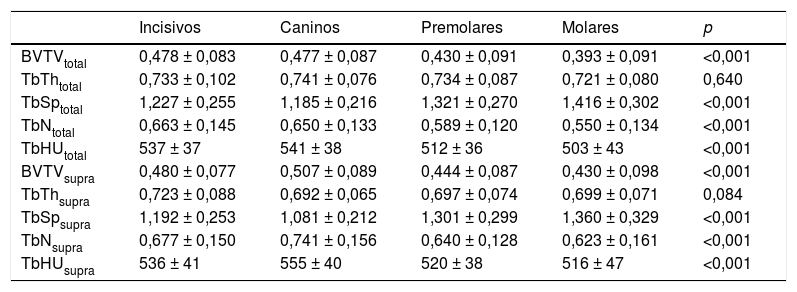
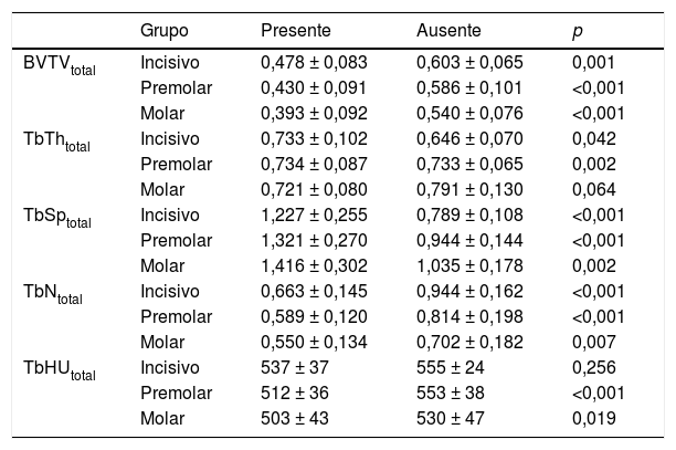
ResultadosSe obtuvieron diferencias estadísticamente significativas entre los tipos y estados de las piezas. Por tipo, los incisivos mostraron mayor ratio de hueso trabecular, con disminución progresiva para caninos, premolares y molares. Por estado, las secciones pertenecientes a dientes ausentes presentaron mayor ratio de hueso que con el diente presente.
ConclusionesLa metodología desarrollada permite cuantificar las propiedades estructurales del hueso alveolar a partir de imágenes de tomografía computarizada multidetector. Los resultados obtenidos objetivan el estado del sustrato óseo de cara a la planificación y seguimiento de la colocación de implantes dentales.
There is a lack of quantitative measures of the quality of alveolar trabecular bone, an important factor in implantology. This study aimed to develop a method of objectively assessing the quality of trabecular bone by means of image processing and structural analysis of multidetector computed tomography images and to establish differences between tooth types and tooth presence/absence.
Materials and methodsWe analyzed 20 patients who underwent multidetector computed tomography to evaluate mandibular bone and tooth positioning. Image analysis included automatic segmentation of the mandible, obtainment of sections perpendicular to the dental arch, and structural analysis of the trabecular bone in each section. We calculated the ratio between the volume of bone and the total volume of the section, the thickness, the trabecular number, and the mean attenuation in Hounsfield units. We analyzed the differences among different tooth types (incisors, canines, premolars, and molars) and between present and absent teeth.
ResultsWe found statistically significant differences between different tooth types and between sections in which teeth were present or absent. Incisors had a greater ratio of trabecular bone; the ratio of trabecular bone progressively decreased from the incisors to the canines, premolars, and molars. The ratio of trabecular bone was greater in sections in which teeth were absent than in those in which teeth were present.
ConclusionsThe method allows to quantify the structural properties of alveolar bone from multidetector computed tomography images. Our results provide an objective picture of the bone substrate that can be useful for planning and following up dental implant procedures.
Artículo
Comprando el artículo el PDF del mismo podrá ser descargado
Precio 19,34 €
Comprar ahora


