La lumbalgia y la lumbociatalgia crónica son una de las causas más frecuentes de consulta médica. La mayor parte de los pacientes se diagnostican con criterios clínicos, no necesitando pruebas de imagen y siendo episodios autolimitados. No existe consenso actualmente sobre el mejor manejo de estos pacientes ni cuál es la medida más costo-eficaz. Sin embargo, generalmente las pruebas de imagen están indicadas en el caso de que sea crónico (> 6 semanas). El gold standard es la resonancia magnética (RM), lo que supone una sobrecarga en los servicios de radiología debido a la alta prevalencia de esta patología y la generación de importantes listas de espera.
ObjetivosEvaluar si la tomografía computerizada lumbar (TC) presenta la misma validez que la RM lumbar en el estudio de la lumbalgia/lumbociatalgia crónica. Valorar si existe concordancia de los resultados obtenidos entre dos radiólogos con distinto grado de experiencia.
Materiales y métodosEstudio retrospectivo de pacientes con lumbalgia/lumbociatalgia crónica, a los que se les realizó tanto RM como TC lumbar. La lectura de las imágenes la ha realizado un radiólogo experto con más de 25 años de experiencia y un adjunto de primer año de manera independiente. Se estiman los índices de validez de sensibilidad y especificidad y se analizaron la concordancia intra e interobservadores mediante el cálculo del índice Kappa de concordancia y del test de asimetría de McNemar.
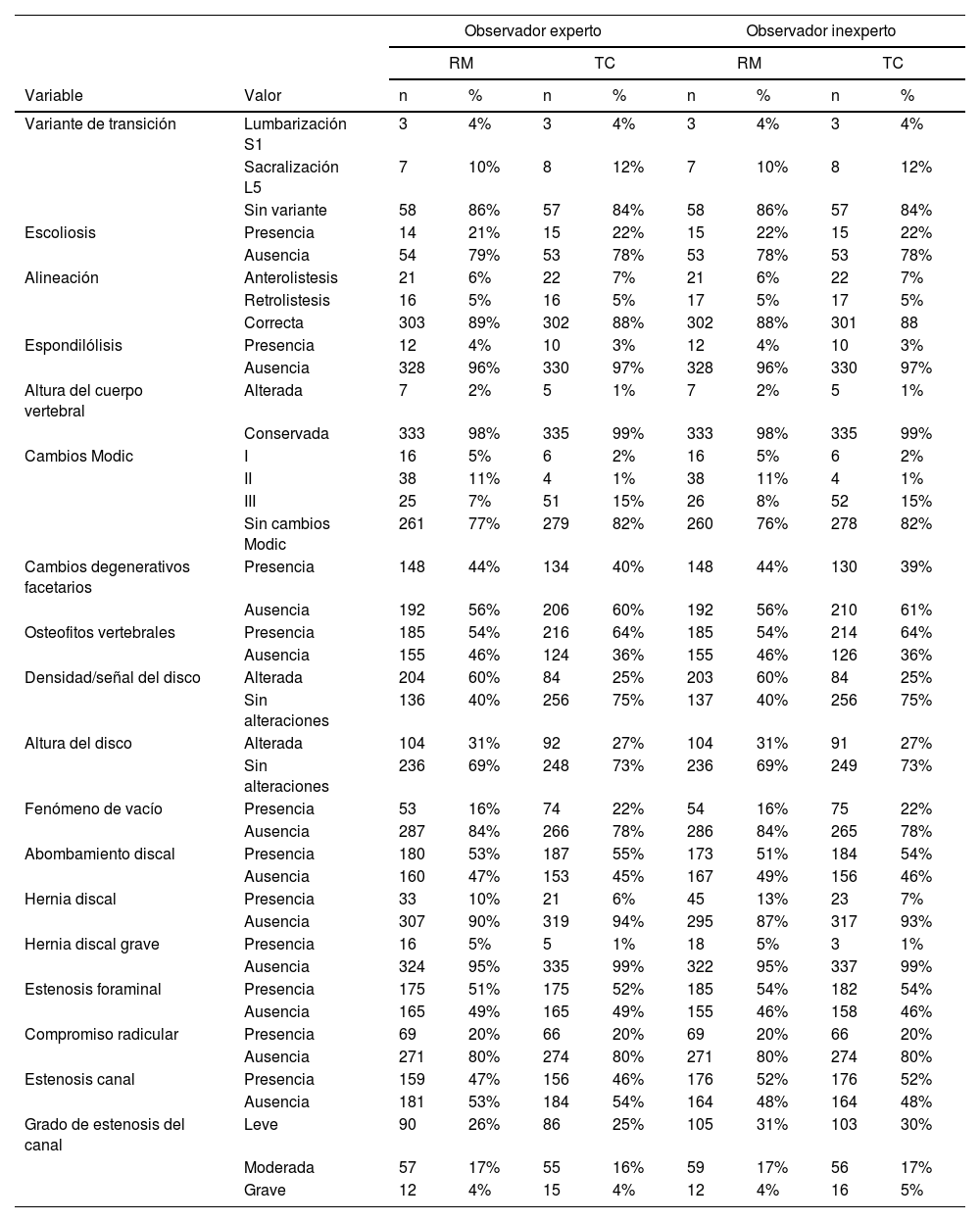
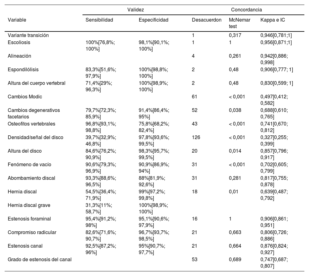
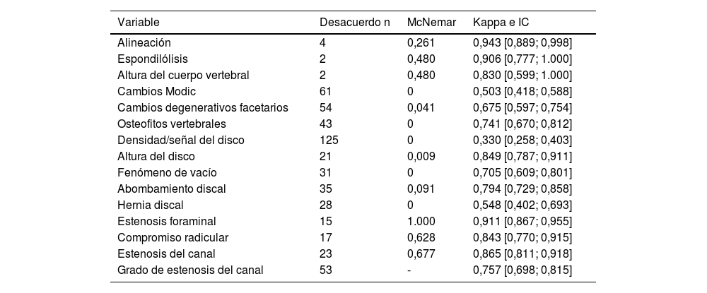
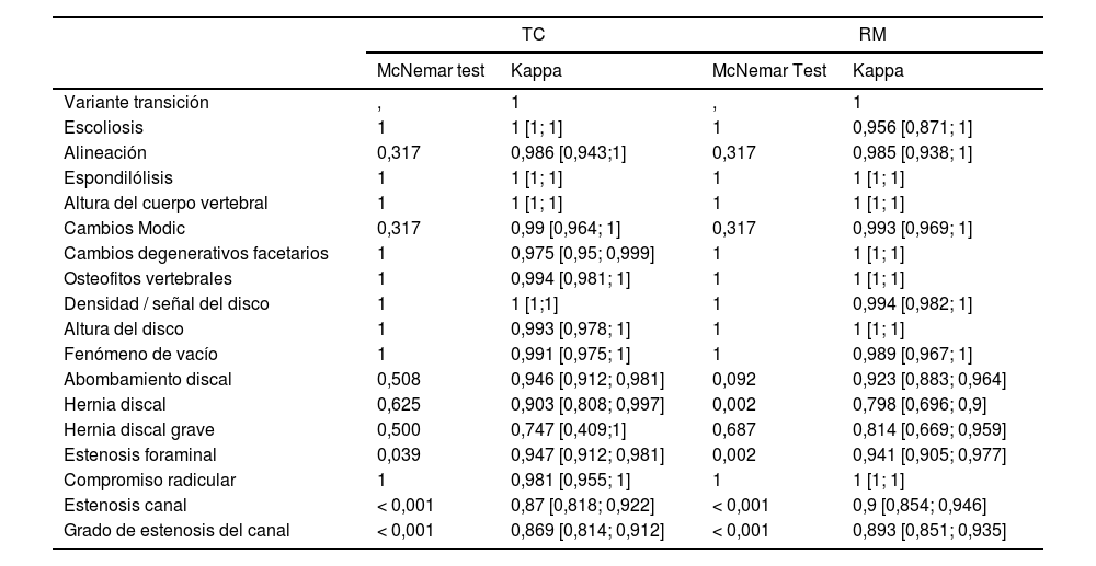
ResultadosSe han evaluado un total de 340 niveles vertebrales en 68 pacientes adultos con lumbalgia o lumbociatalgia crónica, mujeres en un 63.2% y con una edad media de 60.3 años (DE 14.7). La TC presenta índices de sensibilidad y especificidad altos (>80%) en todas las variables, pero la sensibilidad es muy baja en la señal de disco (40%) y en el diagnóstico de hernia (55%). La concordancia entre ambas técnicas es buena (kappa>0.7), salvo para los cambios Modic (kappa=0.497), los cambios degenerativos (kappa=0.688), la señal de disco (kappa=0.327) y la hernia discal (kappa=0.639). En cuanto a la concordancia entre observadores, en general es alta con kappa>0.8. En TC, las discrepancias asimétricas se presentan en la valoración de la estenosis foraminal, la estenosis de canal y del grado de estenosis de canal, sobrediagnosticadas por el observador inexperto.
ConclusionesLa TC lumbar permite valorar la mayoría de las variables con una sensibilidad y especificidad similar a la RM excepto en los cambios de la densidad del disco, la presencia de hernia y los cambios Modic tipo I y II. También estos datos se obtienen independientemente del grado de experiencia del radiólogo. Así, se concluye que la información que aporta cualquiera de las dos técnicas es igual de válida para el correcto manejo de los pacientes.
Low back pain (LBP) is one of the most frequent reasons for medical consultation. Most of the patients will have nonspecific LBP, which usually are self-limited episodes. It is unclear which of the diagnostic imaging pathways is most effective and costeffective and how the imaging impacts on patient treatment. Imaging techniques are usually indicated if symptoms remain after 6 weeks. Magnetic resonance imaging (MRI) is the diagnostic imaging examination of choice in lumbar spine evaluation of low back pain; however, availability of MRI is limited.
ObjectivesTo evaluate the diagnostic accuracy of computed tomography (CT) with MRI (as standard of reference) in the evaluation of chronic low back pain (LBP) without red flags symptoms. To compare the results obtained by two radiologists with different grades of experience.
Materials and methodsPatients with chronic low back pain without red flags symptoms were retrospectively reviewed by two observers with different level of experience. Patients included had undergone a lumbar or abdominal CT and an MRI within a year. Once the radiological information was collected, it was then statistically reviewed. The aim of the statistical analysis is to identify the equivalence between both diagnostic techniques. To this end, sensitivity, specificity and validity index were calculated. In addition, intra and inter-observer reliability were measured by Cohen's kappa values and also using the McNemar test.
Results340 lumbar levels were evaluated from 68 adult patients with chronic low back pain or sciatica. 63.2% of them were women, with an average age of 60.3 years (SD 14.7). CT shows high values of sensitivity and specificity (>80%) in most of the items evaluated, but sensitivity was low for the evaluation of density of the disc (40%) and for the detection of disc herniation (55%). Moreover, agreement between MRI and CT in most of these items was substantial or almost perfect (Cohen's kappa-coefficient > 0’8), excluding Modic changes (kappa=0.497), degenerative changes (kappa0.688), signal of the disc (kappa=0.327) and disc herniation (kappa=0.639). Finally, agreement between both observers is mostly high (kappa>0.8). Foraminal stenosis, canal stenosis and the grade of the canal stenosis were overdiagnosed by the inexperienced observer in the evaluation of CT images.
Conclusions and significanceCT is as sensitive as lumbar MRI in the evaluation of most of the items analysed, excluding Modic changes, degenerative changes, signal of the disc and disc herniation. In addition, these results are obtained regardless the experience of the radiologist. The rising use of diagnostic medical imaging and the improvement of image quality brings the opportunity of making a second look of abdominal CT in search of causes of LBP. Thereby, inappropriate medical imaging could be avoided (2). In addition, it would allow to reduce MRI waiting list and prioritize other patients with more severe pathology than LBP.
Artículo
Comprando el artículo el PDF del mismo podrá ser descargado
Precio 19,34 €
Comprar ahora


