


Scaphocapitate, or Fenton syndrome, is a rare injury. This article presents three new cases that were treated by open reduction and internal fixation with miniscrews, obtaining good results at 16 months follow-up.
El síndrome de la fractura-luxación transescafo-hueso grande o síndrome de Fenton, constituye una lesión muy poco frecuente. En este artículo se presentan 3 casos que fueron tratados mediante reducción abierta y fijación interna con minitornillos, obteniendo buenos resultados a los 16 meses del seguimiento medio.
Trans-scaphocapitate fracture-dislocation, scaphocapitate or Fenton syndrome, is a special form of trans-scaphoid–perilunate carpal fracture in which there is a combined fracture of the scaphoid and capitate bones and in which the proximal fragment of the latter is rotated over 90°, generally 180°.1–3 Very few cases of this syndrome have been published and most of them are unique.4–15 The problem is that this lesion, mainly the capitate bone fracture, is often overlooked in the initial radiographic examination, thus leading to functional sequelae with difficult solution.
This work is based on 3 new cases and also conducts an extensive review of the literature on this particular lesion, commenting on its aetiology, diagnosis and treatment.
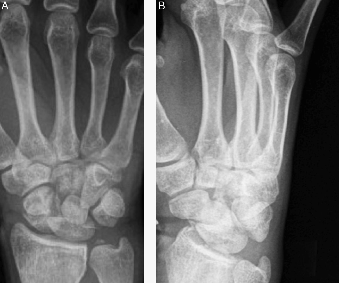
Material and methodBetween 2007 and 2010 we treated 3 patients (2 males and 1 female) for Fenton syndrome at our centre. Their mean age was 26 years (19, 21 and 32 years) and the affected wrist was the right wrist in all cases. Two of the 3 patients reported suffering the lesion due to a motorcycle accident and the third had an accidental fall. All cases reported significant pain and swelling of the dorsum of the wrist. The radiographic study by anteroposterior, lateral and oblique projections showed an association of both fractures, as well as a rotation over 90° of the head of the capitate bone with respect to the rest of the bone (Fig. 1).
 Anteroposterior projection. (B) Oblique projection. Note the association of a scaphoid fracture with another, located in the neck of the capitate, with the proximal fragment rotated 180°.")
In addition, computed tomography (CT) studies revealed a rotation of the proximal fragment of the capitate bone (Fig. 2).

CT study of the wrist of the same patient as in Fig. 1. Note the rotation of the head of the capitate bone.
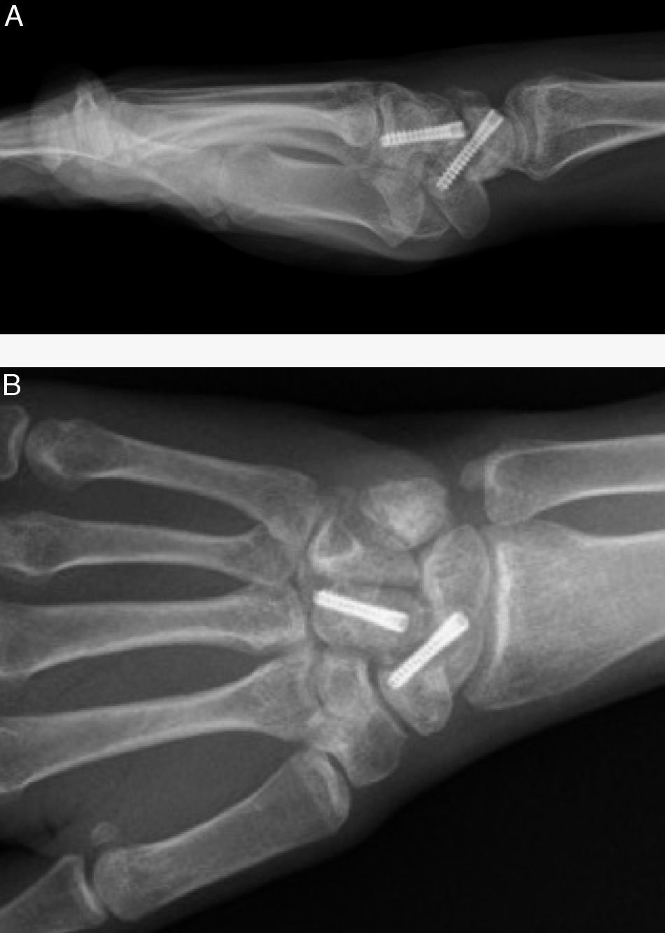
The treatment established for all 3 patients (which was not delayed more than 3 days) consisted of open reduction of both fractures and internal fixation, using Acutrak® miniscrews (Acumed, USA) in 2 cases and Herbert-Whipple® miniscrews (Zimmer, USA) in the remaining case (Fig. 3).
 Anteroposterior projection. (B) Lateral projection.")
Moreover, we also temporarily stabilised the 2 carpal rows using Kirschner wires. Following surgery, the wrist was immobilised for 6 weeks, after which the cast and Kirschner wires were withdrawn and rehabilitation with the assistance of a physiotherapist was started.
The mean follow-up period was 16 months, with a minimum of 12 and a maximum of 24 months. At the end of this period we assessed the presence of pain, balance of the wrist joint, fracture consolidation and the presence of osteoarthritis and/or necrosis of the head of the capitate and/or scaphoid bones.
ResultsNone of the patients reported pain at the end of the follow-up period, although 1 of the 3 patients reported mild discomfort after strenuous activity. The mean wrist joint balance was 55° extension, 50° flexion, 22° ulnar tilt and 8° radial tilt. The consolidation of both fractures was achieved at 8 weeks in all 3 cases. The postoperative CT scan performed at 6 months enabled us to rule out the development of degenerative changes and avascular necrosis of the proximal pole of the capitate and scaphoid bones.
All patients were satisfied with the outcome.
DiscussionAccording to Adler and Shaftan,11 capitate bone fractures are classified into 3 types: (1) isolated fractures, both transverse at the neck level or oblique affecting the distal dorsal cortex; (2) scaphocapitate or Fenton syndrome; and (3) capitate bone fractures associated with other fractures of the carpal bones.
Fenton syndrome is a special form of trans-scaphoid–perilunate fracture-dislocation.1 This severe carpal injury is extremely rare, which is why there are very few publications on the topic and most of them deal with a single case.4–15 In fact, the most extensive series consists of 5 cases.16 The injury mechanism involved is controversial, although most authors agree that it would consist of a hand trauma with forced hyperextension associated with axial compression. According to Stein and Siegel,14 the lesion would go through 3 stages during the sequence of trauma: in the first stage, the neck of the capitate bone would break on impact with the dorsal edge of the radius; in the second stage, the force would continue and would fracture the scaphoid, while the proximal fragment of the capitate bone would rotate 90°; and in the third stage, the hand would return to its initial position, thus rotating the proximal fragment of the capitate bone to 180° (Fig. 4).
. (A) Position of the wrist in hyperextension at the time of impact. Note that the capitate bone is situated at 90° with respect to the radius. (B) The posterior edge of the radius fractures the capitate bone. (C) Fracture of the scaphoid and rotation of the proximal pole of the capitate to 90°. (D) As the wrist returns to the neutral position, the proximal pole of the capitate continues to rotate up to 180°.")
Mechanism of injury in Fenton syndrome (extract from an article by Stein and Siegel,14 with permission from the publisher, JBJS). (A) Position of the wrist in hyperextension at the time of impact. Note that the capitate bone is situated at 90° with respect to the radius. (B) The posterior edge of the radius fractures the capitate bone. (C) Fracture of the scaphoid and rotation of the proximal pole of the capitate to 90°. (D) As the wrist returns to the neutral position, the proximal pole of the capitate continues to rotate up to 180°.
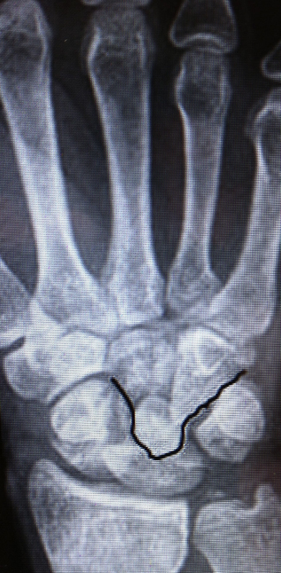
Fenton syndrome may be associated with other complex fractures-dislocations of the carpus17 or of the distal end of the ulna and radius,18 may also occur in children19 and may even be bilateral.13 In any case, the diagnosis of Fenton syndrome is difficult, mainly due to the extreme rotation undergone by the proximal fragment of the capitate bone. For this reason, it is important to pay very close attention when assessing radiographs. This assessment involves identifying the 3 carpal arcs of Gilula.20 Arc I is formed by the margins of the proximal surfaces of the scaphoid, lunate and triquetrum bones, arc II is formed by the edges of the distal surfaces of these same bones, and arc III is formed by the edges of the proximal surfaces of the capitate and hamate bones. In Fenton syndrome, arc III becomes completely altered, indicating the presence of a lesion in the midcarpal joint (Fig. 5).

Disruption of arc III of Gilula20 in the anteroposterior projection in a patient with Fenton syndrome.
However, we believe that obtaining a complementary CT scan is almost essential, not only to confirm both fractures, but also to assess the degree of rotation of the capitate head. Moreover, it enables planning of the most appropriate surgical technique. In this sense, although some studies report satisfactory results without reduction of the capitate bone fracture, we believe the only way to prevent possible complications, including capitate head necrosis,21 is to carry out open reduction of both fractures and to stabilise them with internal fixation. In this regard, the synthesis of fractures can be performed with Kirschner wires and/or either Herbert®, Herbert-Wipple® and/or Acutrak® miniscrews, as in our cases. As for the approach route, this may be volar, dorsal or combined. In our 3 patients, we performed surgery via a dorsal approach to initially synthesise the scaphoid fracture. This procedure is done in any trans-scaphoid-perilunate fracture-dislocation, as the scaphoid bone integrates the 2 carpal rows, so its stabilisation facilitates reduction of the capitate bone. Next, we reduced the dislocation of the proximal fragment of the capitate bone and also synthesised it with a screw. Since the lesion was accompanied by severe involvement of the intercarpal ligament, we supplemented the stabilisation with temporary fixation of both carpal rows using Kirschner wires, although some authors do not consider this step necessary. The immobilisation period varies from 6 to 9 weeks and assisted rehabilitation is required since there is significant postoperative stiffness, as was the case in our patients. With regard to prognosis, it will depend on whether the reduction obtained in both fractures is optimal and stable, or else has a deficit in reduction and/or poor stabilisation. However, given the severity of the lesion, sequelae are common regardless of whether a perfect reduction has been achieved. These sequelae include necrosis of the capitate head fragment and/or radiocarpal and midcarpal osteoarthritis. Patients should be informed of these events.
Level of evidenceLevel of evidence IV.
Ethical responsibilitiesProtection of people and animalsThe authors declare that this investigation did not require experiments on humans or animals.
Confidentiality of dataThe authors declare that this work does not reflect any patient data.
Right to privacy and informed consentThe authors declare that this work does not reflect any patient data.
Conflict of interestsThe authors have no conflict of interests to declare.
Please cite this article as: Natera Cisneros L, et al. Síndrome de Fenton. Rev Esp Cir Ortop Traumatol. 2012;56:369–73.
