


The fifth metacarpal fracture is a frequent pathology that can cause deformity and functional impairment for the adequate grip of the hand. Reintegration into daily or working activities is related to the treatment received and rehabilitation. In fractures of the neck of the fifth metacarpal, internal fixation with a Kirschner's wire is a conventional treatment method with variants that affect its outcome.
Aim of the studyTo compare the functional and clinical results of the treatment of fifth metacarpal fracture with the use of retrograde vs. antegrade Kirschner wires.
Material and methodsComparative, longitudinal, prospective study at a third-level trauma centre in patients with a fifth metacarpal neck fracture, with clinical, radiographic and Quick DASH scale follow-up at the 3rd, 6th, and 8th postoperative week.
ResultsSixty patients were included (58 men, 2 women), age of 29.63±10.15 years, with a fifth metacarpal fracture, treated by closed reduction and stabilisation with a Kirschner wire. The antegrade approach showed a metacarpophalangeal flexion range at 8 weeks of 89.11° (p<0.001; 95% CI [−26.81; −11.42]), a DASH scale value of 18.17 (p<0.001; 95% CI [23.45; 39.12]), and an average of 27.35 days to return to work (p=0.002; 95% CI [16.22; 62.14]), compared with the retrograde approach.
ConclusionStabilisation with antegrade Kirschner wire showed superior functional results, and metacarpophalangeal range of motion, compared to those operated via retrograde approach.
La fractura del quinto metacarpiano es una dolencia muy frecuente que puede ocasionar deformidad y afectación funcional para la prensión adecuada de la mano. La reinserción a las actividades cotidianas o laborales se relaciona con el tratamiento recibido y la rehabilitación. En las fracturas de cuello del quinto metacarpiano la fijación interna con aguja Kirschner es un método de tratamiento convencional con variantes que afectan su desenlace.
ObjetivoComparar los resultados funcionales y clínicos del tratamiento de las fracturas del quinto metacarpiano con el uso de agujas Kirschner vía retrógrada versus anterógrada.
Material y métodosEstudio comparativo, longitudinal, prospectivo, realizado en un hospital de tercer nivel de traumatología en pacientes con fractura de cuello del quinto metacarpiano, con seguimiento clínico, radiográfico y con escala Quick DASH a la tercera, sexta y octava semana postoperatoria.
ResultadosSe incluyeron 60 pacientes (58 varones, 2 mujeres), con un promedio de edad de 29,63±10,15 años, con fractura del quinto metacarpiano, tratados mediante reducción cerrada y estabilización con aguja Kirschner. La vía anterógrada mostró un rango de flexión metacarpofalángica a las 8 semanas de 89,11 grados (p<0,001; IC 95%; −26.81; −11,42), un valor de la escala DASH de 18,17 (p<0,001; IC 95%: 23,45; 39,12) y un promedio de 27,35 días de incapacidad laboral (p=0,002; IC 95%: 16,22; 62,14), comparada con la vía retrógrada.
ConclusiónLa estabilización con aguja Kirschner vía anterógrada mostró superioridad en los resultados funcionales y en la amplitud de movimiento metacarpofalángica comparados con los operados por vía retrógrada a la octava semana del postoperatorio.
Fractures of the fifth metacarpal neck (FFMN) account for 51–68% of all metacarpal fractures. They are typically accompanied by volar angulation and malrotation of the distal segment.1,2 A volar angulation of up to 70° is deemed acceptable and can be managed conservatively2; however, in fractures with angulation and malrotation, better outcomes are achieved by surgical stabilisation.3 There are a number of surgical strategies that have been compared, including various configurations of percutaneous K-wire (KWS): intermetacarpal, crossover,4 intramedullary, and interlocking K-wire from the proximal end.5
Similarly, several biomechanical studies have compared fixation of FFMN: closed reduction and intramedullary internal fixation (CRIMF) with AK vs. low-profile miniplates6–8 and comparative studies of CRIMF with KW via antegrade with low-profile miniplate, revealing better outcomes in terms of range of motion in treatment with antegrade intramedullary fixation compared to those treated with miniplate at three months.6 Treatment by CRIMF with KW exhibits advantages over other treatments due to its simplicity, the fact that it is less invasive, and that it is quicker. It also has disadvantages, such as lack of absolute stability, possible migration of KWs, soft tissue injury, infection, and the need to remove the synthesis material.9 This treatment can be performed either via antegrade or retrograde approach and each of these types of treatment requires different surgical skills.
Anterograde treatment of FFMNs has been reported to be possible to perform in the operating room by axillary block, with pneumatic tourniquet, and under radiographic control (C-arm), with 2 divergent 1.4-mm KWs, with the tip bent anteriorly to 20° from the base of the fifth MTN.7,8 Nevertheless, outpatient treatment increases the efficiency of the procedure,10 and the use of local anaesthesia allows for faster recovery and discharge.11–13 The use of a single KW has also proven to suffice to produce satisfactory clinical outcomes without causing rotational misalignment of the distal fragment.14,15
The objective of the present study was to compare two CRIMF techniques using AK (antegrade compared to retrograde), performed in the outpatient setting to manage FFMN, and to determine their short-term (8 weeks) functional, mobility, and pain outcomes, given that there is literature that reports that both techniques are equal after six months of treatment.7
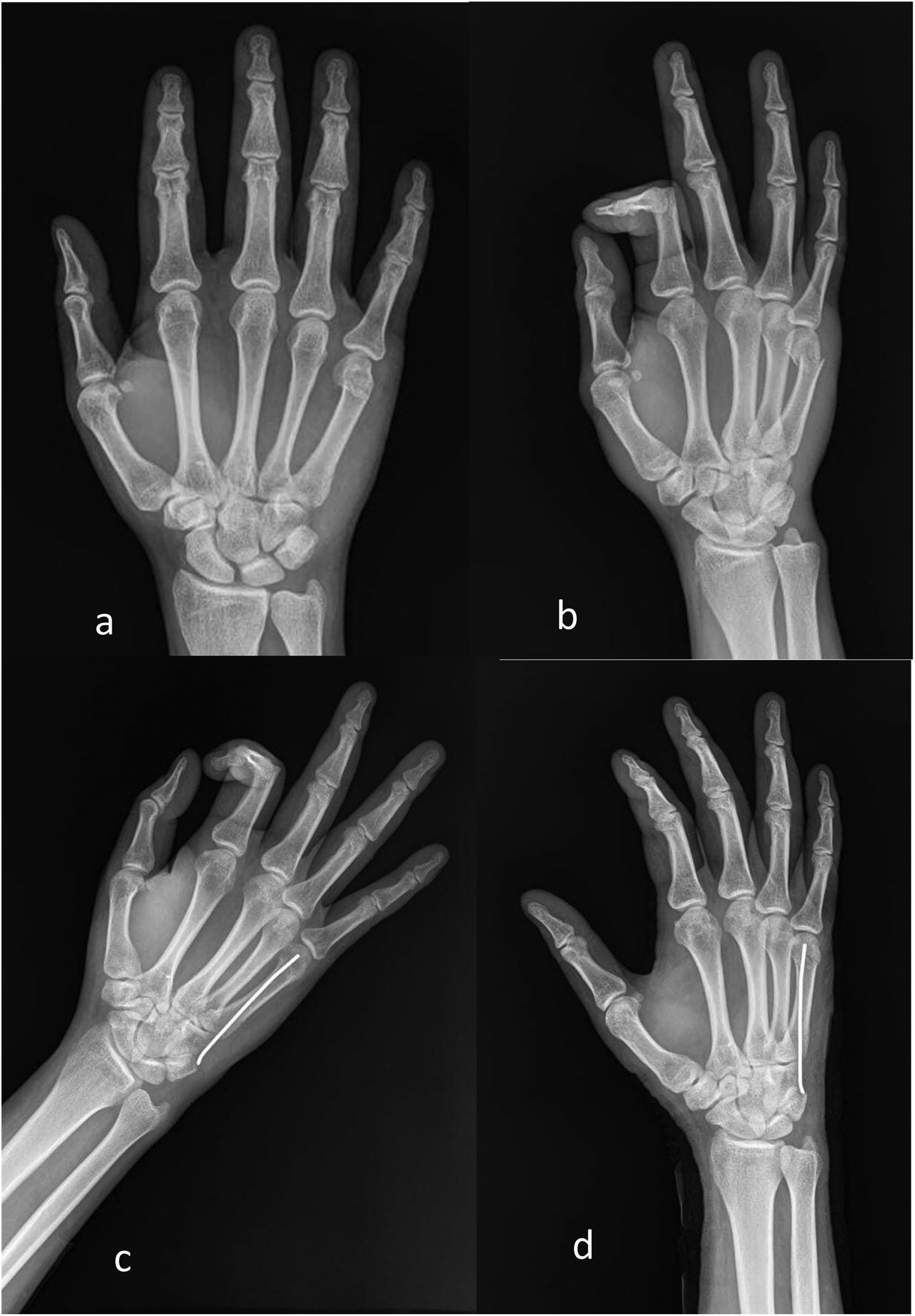
Material and methodsAn observational, prospective study was carried out in a tertiary hospital, after being approved by the local Ethics Committee of the Mexican Social Security Institute (R-2017-2105-27), including patients with FFMN with an evolution time of less than one week and treated by CRIMF via retrograde (Fig. 1) or antegrade (Fig. 2) with AK. Both are standard outpatient treatments in this hospital unit and the choice is at the surgeons’ discretion. Patients treated with other types of osteosynthesis material, open fractures, and patients stabilised for more than one week were excluded.
 and postoperative X-rays (c and d) of the retrograde Kirschner wire procedure in patients with a fracture of the head of the fifth metacarpal.")
 and postoperative X-rays (c and d) of the antegrade Kirschner wire procedure in patients with a fracture of the head of the fifth metacarpal.")
The surgical site was prepared and isolated with sterile fields in the ambulatory surgical theatre, under local aneasthesia, with the patient in dorsal decubitus and under fluoroscopic control. Again, under fluoroscopic visualisation, closed reduction using flexion–extension of the fifth finger was performed until anatomical reduction of the FFMN was achieved.
Retrograde technique- •
The MCP joint was flexed to 90° to reduce the fracture and to expose the articular surface of the MCP.
- •
Reduction of the fracture was verified, and the entrance point of the KW was determined by fluoroscopy, taking care to avoid excessive injury to the articular cartilage and not to perforate the extensor tendons of the fifth toe.
- •
Zx using a power instrument, the KW was inserted 1.4mm retrograde, directly to the intramedullary canal.
- •
The reduction was checked; the KW was bent and cut and was left outside the skin.
- •
The exit point of the KW was covered with sterile gauze and an antebrachidigital splint was placed in the intrinsic plus position.
- •
A dorsal incision of approximately 5mm was made in the base of the fifth MCP and blunt disection was performed, taking care to avoid injuring the doral sensitive branch of the ulnar nerve and the common extensor tendons of the fifth finger, close to the entrance point.
- •
Closed reduction was then performed by means of traction of the fifth finger with respect to the MCP axis with the MCP joint flexed to 90°. Reduction was verified by fluoroscopy.
- •
An entrance orifice as performed at the base of the fifth MCP using a motorised drill and 2.0bit.
- •
The tip of the KW was bent (1.4mm) approximately 20° and was mounted on a T-handle.
- •
Through the already created orifice in the bone, the tip of the KW was inserted percutaneously towards the ulnar edge.
- •
The KW was directed to the cortical bone of the border radial del MCP using semicircular movements.
- •
The KW was advanced to the facture line where it was pinned into the head of the MCP.
- •
The reduction was verified and the KW was bent and cut and left outside of the skin.
- •
The incision was sutured with 3-0 nylon and the KW exit was covered with sterile gauze and an antebrachydigital splint was placed in the intrinsic plus position.
Outpatient follow-up was performed at the third, sixth, and eighth postoperative weeks. The home therapy exercises were explained both verbally and in writing to all participants and were performed starting in the second postoperative week and for a period of six weeks. The exercise programme consisted of 3 daily series (morning, afternoon, and evening) for 20–30min and 4–6 exercises per series.16 The KW was removed in the presence of clinical and radiographic data of consolidation, as evaluated by two independent and blinded observers. The KWs were removed in an outpatient operating theatre, under analgesia, asepsis of the region, and axial traction of the KW.
Demographic data (age, sex, occupation, lateral dominance, affected side, and time of KW removal) and clinical data were collected from the participants in the study: pain as rated on a visual analogue scale (VAS)17; the Quick DASH questionnaire18 was completed, and passive flexion and extension of the MCP joint was measured by goniometer from the 0 position, presence-absence of angulation correction of the fracture line, and the presence of complications. The presence of fracture angulation correction was regarded when an angle of less than 30° was found between the initial radiographs and the radiographs at the end of the follow-up period. The complications observed were recorded.
Calculation of sample size and statistical analysisThe sample size was calculated with a prevalence of patients with FFMN treated with CRIMF with K-wires of 18% and the level of statistical significance (α) was set at 0.05 to detect an effect size of 20% of the functional outcome using the Quick DASH questionnaire (based on the effect size found at six months in the work of Kim and Kim),7 leading to a total of 52 patients. The sample size was set at 60 patients, in a 1:1 ratio between those treated with CRIMF with antegrade and retrograde K-wires.
A descriptive analysis was conducted using frequencies and percentages for qualitative variables, and mean and standard deviation for quantitative variables. Inferential analyses were performed using the Mann–Whitney U test and Wilcoxon rank sum for paired samples for the quantitative values obtained from the Quick DASH scale, pain (VAS), and range of mobility (flexion and extension of the MCP joint) for the third, sixth, and eighth postoperative weeks. Fisher's exact test was used to determine the presence of correction and complications in each treatment group. Analysis of concordance between the injured hand and lateral dominance was carried out using weighted kappa. An α value=0.05 was assumed for statistical significance testing. The results were analysed using SPSS® 21.0 statistical software (demo version).
ResultsSixty subjects were included, 96.7% of whom were male, with a mean age of 29.63±10.15 years. The retrograde technique group consisted of 26 patients and the retrograde [sic] technique group of 34 patients (1:1.3). Both groups were comparable in age (p=0.257) and sex (p=0.184) (Table 1). As for the participants’ occupation, 16.7% were machine operators (n=10), followed by human resource professionals, students, and labourers, each accounting for 13.3% (n=8); 10% were engineers (n=6), 6.7% were warehouse workers (n=4), while there were 3.3% (n=2) each of programmers, logistics, drivers, labellers, supervisors, maintenance, housekeeping, and nurses. Right hand lateral dominance was present in 93.3% (n=56) and the remaining 6.7% (n=4) were left hand dominant, which were related to the fractured hand in 92.9% (n=52) for the right-handed cohort and 100% for the left-handed subgroup, exhibiting a level of concordance (dominant hand=fractured hand) of 0.634 (kappa; p<0.001). The average time at which the K-wires were removed for the retrograde procedure was 45.38±21.16 (29–112 days) compared to 42.05±8.8 (33–63 days) for the antegrade procedure, exhibiting no statistically significant differences between them (Mann–Whitney U, p=0.742; 95% CI: [−5.60, 11.48]).
Statistically significant differences were found in the variables of pain (p<0.001), Quick DASH questionnaire (p<0.001), and flexion–extension of the MCP joint (p<0.001) between the treatment groups from the third to the eighth week of follow-up in favour of the antegrade technique (Table 2). Analysis of the dependent variables at postoperative follow-up was performed and the change in value at the various stages of postoperative follow-up was confirmed. Statistically significant differences were observed in pain assessment (p<0.001), functional assessment by means of the Quick DASH questionnaire (p<0.001), and flexion degrees (p<0.001) between the third, sixth, and eighth weeks of treatment in both treatment groups. MCP joint extension demonstrated no difference between weeks of follow-up in the retrograde pin group (p=0.46) (Table 3). The initial angulation of the head of the fifth MCP was fully corrected in 18 patients (69.23%) treated with retrograde pinning and in 32 patients (94.11%) treated with antegrade pinning, showing statistically significant differences between both treatment groups for the presence-absence of anatomical correction (Fisher's exact test; p=0.014). Four patients with complications were detected in the retrograde pin group (extensor tendon injury), with no complications found in the antegrade group (Fisher's exact test; p=0.094). Neither group experienced complications of infectious aetiology, and as the follow-up was short term, there was no loss to follow-up of patients.
Comparative analysis of the range of movement, pain, and functionality between each treatment group.
| Week | Technique | Mean | SD | pa | |
|---|---|---|---|---|---|
| Flexion (degrees) | 3 | Retrograde | 46.93 | 4.8 | <.001 |
| Antegrade | 77.35 | 7.52 | |||
| 6 | Retrograde | 53.07 | 7.51 | <.001 | |
| Antegrade | 83.82 | 7.81 | |||
| 8 | Retrograde | 68.46 | 14.05 | <.001 | |
| Antegrade | 89.11 | 2.64 | |||
| Extension (degrees) | 3 | Retrograde | 1.15 | 2.19 | <.001 |
| Antegrade | 8.82 | 4.85 | |||
| 6 | Retrograde | 1.53 | 3.15 | <.001 | |
| Antegrade | 10.58 | 4.28 | |||
| 8 | Retrograde | 3.07 | 5.96 | <.001 | |
| Antegrade | 14.11 | 5.07 | |||
| VAS | 3 | Retrograde | 5.76 | .83 | <.001 |
| Antegrade | 4.94 | .74 | |||
| 6 | Retrograde | 4.53 | .66 | <.001 | |
| Antegrade | 3.23 | .75 | |||
| 8 | Retrograde | 3.69 | 1.03 | <.001 | |
| Antegrade | 1.88 | 1.11 | |||
| Quick DASH | 3 | Retrograde | 73.59 | 9.57 | <.001 |
| Antegrade | 46.38 | 18.72 | |||
| 6 | Retrograde | 60.13 | 9.89 | <.001 | |
| Antegrade | 31.68 | 13.09 | |||
| 8 | Retrograde | 49.47 | 10.50 | <.001 | |
| Antegrade | 18.17 | 10.29 | |||
SD: standard deviation; VAS: visual analogue scale; Quick DASH: Quick Disability of Arm Shoulder and Hand.
Change in movility, pain, and functionality at weeks three, six, and eight between both treatment group.
| Retrograde | Antegrade | ||||||||||
|---|---|---|---|---|---|---|---|---|---|---|---|
| Differences | Differences | ||||||||||
| Week | Mean | SD | 3rd–6th | 6–8th | pa | Mean | SD | 3rd–6th | 6–8th | pa | |
| Flexion (degrees) | 3 | 49.6 | 12.6 | 6.2 | 77.35 | 7.41 | 6.47 | ||||
| 6 | 55.8 | 12.5 | 14.2 | .001 | 83.82 | 7.7 | 5.91 | <.001 | |||
| 8 | 70.0 | 14.9 | <.001 | 89.11 | 2.6 | <.001 | |||||
| Extension (degrees) | 3 | 1.92 | 3.18 | 0.38 | 8.82 | 4.78 | 1.76 | ||||
| 6 | 2.30 | 3.80 | 1.54 | .157 | 10.58 | 4.22 | 3.53 | .058 | |||
| 8 | 3.84 | 6.05 | .46 | 14.11 | 4.99 | .001 | |||||
| VAS | 3 | 5.76 | .81 | −1.26 | 4.94 | .74 | −1.01 | ||||
| 6 | 4.5 | .64 | −0.8 | <0.001 | 3.23 | .74 | −1.35 | <.001 | |||
| 8 | 3.7 | 1.01 | <0.001 | 1.88 | 1.1 | <.001 | |||||
| Quick DASH | 3 | 73.6 | 9.38 | −13.5 | 46.38 | 18.44 | −14.7 | ||||
| 6 | 60.1 | 9.7 | −10.7 | <0.001 | 31.68 | 12.9 | −13.5 | <.001 | |||
| 8 | 49.4 | 10.3 | <0.001 | 18.18 | 10.13 | <.001 | |||||
SD: standard deviation; VAS: visual analogue scale; Quick DASH: Quick Disability of Arm Shoulder and Hand.
In the present study, MCPFs treated by CRIMF with K-wire via the antegrade approach displayed superior results with respect to function, pain, range of motion, and the presence of complications when compared to retrograde approaches. In the literature, surgical treatment of MCP fractures plus rehabilitation therapy have proven favourable functional outcomes for both the MCP joint and hand performance as a whole.16,19–21 Our sample was predominantly male, which is in line with the literature in epidemiological series,22 with a greater ratio of males:females than those reported in epidemiological series (5.08:1 vs. 29:1).
The fractured hand was significantly related to the individual's handedness (kappa<0.001) in more than 90% of the cases. This finding justifies the need for rapid, effective, and easily performed management to restore hand function for productive activities or activities of daily living for each individual patient. It should be noted that most of the patients in the sample with MCPF are of working age1–3; in the case of the present study, the mean age was 29.63 years, with a minimum of 18 and a maximum of 62 years.
In a clinical trial from the literature, 46 patients were included who had displaced MCPFs with a dorsal apical angulation greater than 30°, treated by CRIMF with percutaneous antegrade or retrograde K-wire. Radiographic assessments of dorsal apical angulation and axial shortening were performed preoperatively and 6 months postoperatively. More favourable results using the antegrade approach were reported over the retrograde approach group from the first check-up at three months. At six months, there was no significant difference with respect to VAS, Quick DASH, and range of motion.7 In the present study, the follow-up period was short and accompanied by early and targeted rehabilitation exercises, with an average of 42–45 days at the time of KW removal. In the latter case, the K-wires were removed under analgesia, asepsis, and in an outpatient setting, using direct traction. Unlike Kim and Kim's study, the K-wires were not left under the skin, thereby sparing the patients another anaesthetic procedure, and the removal time in the present review was halved, with no complications as far as consolidation is concerned.
In the present study, when comparing both procedures (retrograde vs. antegrade) at the same weeks of evolution, differences were observed among the variables evaluated: VAS, Quick DASH, and flexion and extension of the MCP joint. Both approaches and placement of the K-wire had acceptable results in each of the variables analysed; however, the antegrade approach exhibited superiority over the retrograde approach as early as the third week. These results can be attributed to early mobilisation and rehabilitation, as with the antegrade route, the K-wire does not block any joints. In this study, mobilisation was allowed at the end of the second week, with no displacement, loss of reduction, migration of the synthetic material, or impairment of consolidation, as has been reported in the literature.5 The retrograde approach yielded acceptable functional and pain reduction results during follow-up, albeit these were not superior to the results of the antegrade approach. When it came to removal of the retrograde KWs, two extreme results of 110 and 112 days were observed, which widened the dispersion of the data in the sample. These two results were due to the fact that no previous radiographic data of consolidation were detected; it was therefore decided to postpone the removal of the synthetic material.
In a comparative study of MCP and phalangeal fractures with plates vs. antegrade CRIMF, greater benefit was observed with the latter, with no difference between the two procedures at six months. Plating involved fewer complications, although perioperative costs and resources were greater compared to the antegrade K-wires.6 The outcomes found with CRIMF using antegrade KW were superior in the short term, as reported in other similar series.23–25 Statistically, flexion and extension were better in the antegrade group compared to the retrograde group during the third, sixth, and eighth weeks of follow-up. Clinically speaking, the ranges of motion changed sufficiently in the anterograde group as to manage to close the fist of the affected hand and to extend the fifth MCP actively to at least the 0 position, which was not possible in the retrograde group. In each treatment group, both antegrade and retrograde, improvements were found in the clinical assessment, but the extension of the MCP joint in the retrograde treatment group failed to demonstrate short-term differences when comparing this variable at the third, sixth, and eighth weeks of treatment (repeated measures). Extension in the retrograde group increased only up until the time of K-wire withdrawal. We believe this is attributable to the joint locking caused by leaving the AK exposed distally, which prevents mobility and early full extension and maintains the joint in flexion as a result of the impingement of the base of the proximal phalanx on the KW.
The bibliography indicates that regardless of the type of treatment, open reduction plus internal fixation by means of an implant (locking or low-profile plate, or with the use of screws),6,18 intramedullary KW (retrograde or anterograde),7,8,24,25 or even with conservative management,26–28 at 3- and 6-month or 2-year follow-ups, the VAS, range of motion, and Quick DASH variables become uniform and achieve adequate results, albeit at different time points, which can be attributed to each type of treatment. This effect was achieved in our series at postoperative week 8, where functional and pain perception differences between treatment groups were similar.
The retrograde approach is a simpler technique to perform and in experienced hands can be performed on an outpatient basis and even without the need for fluoroscopy, and offers acceptable ranges of motion and function. However, these results take longer compared to the antegrade approach. In both surgical procedures there is always a risk of injury to the extensor tendon and the sensory branch of the ulnar nerve.23
We have considered several limitations to the present study, which is observational and had a short-term follow-up (8 weeks). Nevertheless, a relatively brief follow-up demonstrated that it is possible to observe favourable results in this period of time. This is relevant, inasmuch as this is a condition that mostly affects young patients of working age who need to return to work soon.
Another limitation is the difficulty of performing full MCP joint movements in the retrograde KW group by leaving it outside the skin and thus blocking the joint. This can be overcome by leaving the synthesis material passing through the skin at the base of the fifth MCP without blocking the joint, and avoiding the bias of the start of rehabilitation in the retrograde group.
ConclusionsCRIMF of MCPF with KW via the antegrade approach demonstrated better functional and range-of-motion (flexion–extension) results compared to the retrograde approach at three, six, and eight weeks postoperatively; however, both techniques are appropriate options with good outcomes in the short term. The complications encountered (extensor tendon injury) occurred in patients who underwent retrograde approach.
Level of evidenceLevel of evidence ii.
FundingThe authors state that the research was conducted with their own resources and the ascribed Hospital Unit.
Conflict of interestsThe authors have no conflict of interests to declare.
Right to privacy and informed consentThe authors have obtained informed consent from the patients and/or subjects referred to in the article. This document is held by the corresponding author.
Ethics committee approvalLocal Ethics Committee of the Mexican Institute of Social Security (R-2017-2105-27).
