To evaluate the utility of positron emission tomography-computed tomography (PET/CT) as an imaging tool for the characterization of immune-mediated inner ear disease (IMIED), providing measurements of the inner ear region activity as well as detecting possible involvement of other organs.
Material and methodsThe study included 28 patients with IMIED and 4 sex-matched and age-matched control subjects with no history of ear disease.
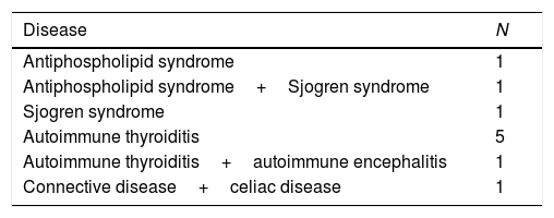
Eighteen patients were considered to be suffering from primary IMIED and 10 patients from secondary. PET/CT scans with 18F-FDG were performed to assess systemic involvement as well as inner ear region activity.
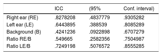
Interpretation of PET/CT scans was performed independently by two nuclear medicine physicians blinded to clinical history. In order to assess inter-rater agreement before performing the analysis of the inner ear, different Bland & Altman plots and the intraclass correlation coefficients were estimated.
ResultsDifferent metabolically active foci findings were reported in 13 patients. Four patients diagnosed as primary IMIED showed thyroid and aorta activity. Regarding the inner-ear semiquantitative analysis, the inter-rater agreement was not sufficiently high. Comparisons between groups, performed using Mann–Whitney test or Kruskal–Wallis tests, showed no differences.
ConclusionsThe study showed 18F-FDG PET/CT could be an important tool in the evaluation of IMIED as it can support the characterization of this entity providing the diagnosis of unknown or underestimated secondary IMIED. Nevertheless, we consider PET is not an adequate tool to approach the inner ear because of the small size and volume of the cochlea which makes the assessment very difficult.
Evaluar la utilidad de la PET/TC con 18F-FDG en la caracterización de la enfermedad inmunomediada del oído interno primaria (EIOI) aportando datos que ayuden a valorar la actividad inflamatoria y la existencia o ausencia de enfermedad sistémica asociada.
Material y métodosEstudio prospectivo sobre 28 pacientes con sospecha de EIOI o EIOI primaria diagnosticada y controles sin enfermedad ótica conocida con PET FDG realizado por otro motivo.
Dieciocho pacientes presentaban EIOI primaria y diez EIOI secundaria. A todos se les realizó un PET/TC con 18F-FDG para valorar la actividad metabólica en el oído interno y la presencia de afectación sistémica.
La interpretación del resultado del PET fue realizada por dos médicos nucleares sin conocimiento de la clínica del paciente. Para valorar la reproductibilidad de las medidas se realizaron análisis Bland&Altman y correlación de coeficientes interclase.
ResultadosSe encontraron hallazgos sospechosos de afectación sistémica en 13 pacientes. Cuatro de ellos correspondieron a pacientes diagnosticados previamente de EIOI primaria que mostraron actividad inflamatoria (tiroidea y aórtica). En cuanto al análisis semicuantitativo de la actividad metabólica en el oído interno, la variabilidad interobservador fue muy alta y no fue posible establecer diferencias adecuadas entre grupos.
ConclusionesEste estudio demuestra que el PET-TC con 18F-FDG puede tener un papel en la evaluación de pacientes con sospecha de EIOI primaria descartando la presencia de datos que sugieran inflamación sistémica. Consideramos que no es adecuado tratar de cuantificar la actividad metabólica del oído interno probablemente por el pequeño tamaño del mismo.
Artículo

Revista Española de Medicina Nuclear e Imagen Molecular (English Edition)
Comprando el artículo el PDF del mismo podrá ser descargado
Precio 19,34 €
Comprar ahora


