metricas
covid
Buscar en
Revista Española de Medicina Nuclear e Imagen Molecular
Toda la web
Inicio Revista Española de Medicina Nuclear e Imagen Molecular Positron emission tomography/computed tomography (PET/CT) with 2-deoxy-2-[18F]-f...
Información de la revista
Vol. 26. Núm. 5.
Páginas 306 (septiembre 2007)
Compartir
Compartir
Descargar PDF
Más opciones de artículo
Vol. 26. Núm. 5.
Páginas 306 (septiembre 2007)
Acceso a texto completo
Positron emission tomography/computed tomography (PET/CT) with 2-deoxy-2-[18F]-fluoro-D-glucose ( 18FDG) in a patient with cutaneous T-cell lymphoma
Tomografía por emisión de positrones/tomografía axial computarizada (PET//TAC) con 2-deoxi-2-[18F]-fluoro-D-glucosa ( 18FDG) en un paciente con linfoma cutáneo de células T
Visitas
2046
S. Mahmooda, SR. Martínez de Llanob
a Nuclear Medicine Service. Department of Radiology. Memorial Sloan-Kettering Cancer Center. New York. USA. MRC Clinical Sciences Centre and Division of Neuroscience, Faculty of Medicine, Imperial College, Hammersmith Hospital-Cyclotron Building. London UK.
b Nuclear Medicine Service. Department of Radiology. Memorial Sloan-Kettering Cancer Center. New York. USA. Montefiore Medical Center / Albert Einstein College of Medicine. Bronx, New York, USA.
Este artículo ha recibido
Información del artículo
Texto completo
Bibliografía
Descargar PDF
Estadísticas
Figuras (1)
Texto completo

CASE REPORT

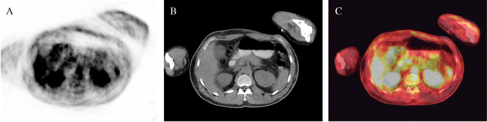
A 65-year-old man with a history of lymphoma was referred for a positron emission tomography/ computed tomography (PET/CT) with 2-deoxy-2-[18F]-fluoro-D-glucose F-(18FDG) study to evaluate a collection in the subcutaneous fat of the anterolateral upper abdomen reported on a CT scan. PET/CT scan (fig. 1) revealed mildly increased tracer activity localizing to multifocal discrete areas of haziness and infiltration in the subcutaneous tissues of the abdomen. Subsequent skin biopsy demonstrated subcutaneous panniculitis-like T-cell lymphoma.

Fig. 1.--Transaxial (A) positron emission tomography (PET) (B) computed tomography (CT) and (C) PET/CT fused images (LSO Biograph PET/CT scanner) at 70 minutes post injection of 16.5 mCi of 18F-FDG, demonstrating increased FDG uptake in multiple ill-defined subcutaneous nodules in the abdominal wall, with a maximum standardized uptake value (SUVmax) of 1.2 over the left anterolateral abdominal wall.

Cutaneous T-cell lymphoma (CTCL) is a rare disease, characterized by indolent patches and plaques in early stages, and extracutaneous involvement in advanced stages. Mycosis fungoides is the most common type of CTCL1.

18F-FDG PET/CT scan might add valuable clinical information and guide biopsy of lesions with the highest SUV, thus facilitating patient management and follow up1-3.


Correspondencia:

S. Mahmood.

Department of Nuclear Medicine.

Montefiore Medical Center / Albert Einstein College of Medicine.

1695 A Eastchester Road.

Bronx, NY 10461.

E-mail: smahmood@yahoo.com

Bibliography
[1]
Valencak J, Becherer A, Der-Petrossian M, Trautinger F, Raderer M, Hoffmann M..
Positron emission tomography with [18F] 2-fluoro-D-2- deoxyglucose in primary cutaneous T-cell lymphomas..
Haematologica, 89 (2004), pp. 115-6
[2]
Von Nida J, Randell P, Heenan P..
Granulomatous mycosis fungoides with extensive chest wall involvement..
Australas J Dermatol, 45 (2004), pp. 42-6
[3]
Elstrom R, Guan L, Baker G, Nakhoda K, Vergilio JA, Zhuang H, et al..
Utility of FDG-PET scanning in lymphoma by WHO classification..
Opciones de artículo
es en pt

¿Es usted profesional sanitario apto para prescribir o dispensar medicamentos?

Are you a health professional able to prescribe or dispense drugs?

Você é um profissional de saúde habilitado a prescrever ou dispensar medicamentos