metricas
covid
Buscar en
Revista Española de Medicina Nuclear e Imagen Molecular
Toda la web
Inicio Revista Española de Medicina Nuclear e Imagen Molecular Posterior scapular lymph nodes in a patient with follicular lymphoma on whole-bo...
Información de la revista
Vol. 27. Núm. 3.
Páginas 202-203 (mayo 2008)
Compartir
Compartir
Descargar PDF
Más opciones de artículo
Vol. 27. Núm. 3.
Páginas 202-203 (mayo 2008)
Acceso a texto completo
Posterior scapular lymph nodes in a patient with follicular lymphoma on whole-body 2-deoxy-2-[ 18F]-fluoro-D-glucose ( 18F-FDG) positron emission tomography/computed tomography (PET/CT)
Posterior scapular lymph nodes in a patient with follicular lymphoma on whole-body 2-deoxy-2-[ 18F]-fluoro-D-glucose ( 18F-FDG) positron emission tomography/ computed tomography (PET/ CT)
Visitas
2458
S. Mahmooda, SR. Martínez de Llanob
a Nuclear Medicine Service. Department of Radiology. Memorial Sloan-Kettering Cancer Center. New York. NY. USA. Montefiore Medical Center/Albert Einstein College of Medicine. Bronx. NY. USA.
b Nuclear Medicine Service. Department of Radiology. Memorial Sloan-Kettering Cancer Center. New York. NY. USA. MRC Clinical Sciences Centre and Division of Neuroscience. Faculty of Medicine. Imperial College. Hammersmith Hospital-Cyclotron Building. London. UK.
Este artículo ha recibido
Información del artículo
Texto completo
Bibliografía
Descargar PDF
Estadísticas
Figuras (1)
Texto completo

CASE REPORT

A 58-year-old man with a history of follicular lymphoma complained of progressively enlarging non-tender adenopathy, increasing fatigue, new-onset peripheral edema, progressive anemia, and demonstrated a monoclonal kappa restricted population on the flow-cytometry. Patient was referred for a re-staging 18F-FDG PET/CT study, with a clinical suspicion of progression to a leukemic phase of his lymphoma, to base therapy specific to his disease. PET/CT images demonstrated extensive hypermetabolic supradiaphragmatic and infradiaphragmatic lymphadenopathy, including symmetric nodes posterior to the scapulae bilaterally (fig. 1).

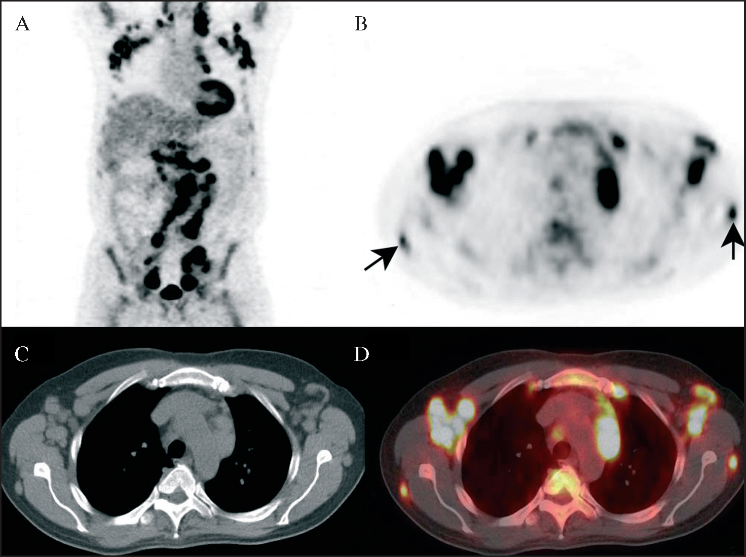
Fig. 1.--Coronal PET images (A), transaxial PET (B), CT (C) and PET/CT fused images (D), (Biograph PET/CT scanner, Siemens Medical Solutions, Erlangen, Federal Republic of Germany) at 75 minutes post injection of 14.2 mCi of 18F-FDG demonstrating extensive hypermetabolic lymphadenopathy in cervical, supraclavicular, mediastinal with a maximum standardized uptake value (SUVmax) of 9.4, axillary with a SUVmax of 8.5 on the left and 8.8 on the right side, periceliac, paraaortic with a SUVmax of 11.2, aortocaval, bilateral external iliac, bilateral inguinal, and bilateral lymph nodes posterior to the scapulae, with a SUVmax of 4.6 on the left and 3.6 on the right side, respectively (arrows). Mildly increased tracer activity seen within the bone marrow is likely reactive in nature.

Follicular lymphoma, a type of non-Hodgkin's lymphoma, is one of the most common lymphoma entities in the United States. Upon presentation, most patients have disseminated disease affecting peripheral lymph nodes and/or bone marrow1. 18F-FDG PET has proved useful in the diagnosis, staging, follow-up and assessment of treatment response of Hodgkin's disease and non-Hodgkin's lymphoma (especially more aggressive types). Further, the widespread use of combined PET/CT has also increased the sensitivity and specificity of FDG PET scans2-6.

We present a case of follicular lymphoma with involvement of lymph nodes posterior to the scapulae bilaterally. To our knowledge, posterior scapular nodes have not been described previously in the literature. Due to the multiple foci of markedly increased FDG avidity, more aggressive treatment was employed in our patient with Cyclophosphamide, Doxrubicin, Oncovin (Vincristine), Prednisone (CHOP) and Rituximab (CHOP-R), with complete remission.


Correspondence:

S. Mahmood, MD.

Attending Physician/Assistant Professor of Nuclear Medicine.

Department of Nuclear Medicine.

Montefiore Medical Center/Albert Einstein College of Medicine.

1695A Eastchester Road.

Bronx, NY 10461,

E-mail: smahmood@yahoo.com

Recibido: 09-10-2007.

Aceptado: 14-01-2008.

Bibliography
[1]
Wohrer S, Jaeger U, Kletter K, Becherer A, Hauswirth A, Turetschek K, et al..
18F-fluoro-deoxy-glucose positron emission tomography (18F-FDG-PET) visualizes follicular lymphoma irrespective of grading..
Ann Oncol, 17 (2006), pp. 780-4
[2]
Imaging: staging and evaluation of lymphoma using nuclear medicine. Semin Oncol. 2005;321 Suppl 1:S11-8.
[3]
Jhanwar YS, Straus DJ..
The role of PET in lymphoma..
J Nucl Med, 47 (2006), pp. 1326-34
[4]
Raanani P, Shasha Y, Perry C, Metser U, Naparstek E, Apter S, et al..
Is CT scan still necessary for staging in Hodgkin and non-Hodgkin lymphoma patients in the PET/CT era? Ann Oncol, 17 (2006), pp. 117-22
[5]
Reinhardt MJ, Herkel C, Altehoefer C, Finke J, Moser E..
F-FDG positron emission tomography for therapy control of Hodgkin's and non-Hodgkin's lymphoma patients:when do we really need FDG-PET? Ann Oncol, 16 (2005), pp. 1524-9
[6]
Schoder H, Noy A, Gonen M, Weng L, Green D, Erdi YE, et al..
Intensity of 18Fluorodeoxyglucose uptake in positron emission tomography distinguishes between indolent and aggressive non-Hodgkin's lymphoma..
J Clin Oncol, 23 (2005), pp. 4643-51
Opciones de artículo
es en pt

¿Es usted profesional sanitario apto para prescribir o dispensar medicamentos?

Are you a health professional able to prescribe or dispense drugs?

Você é um profissional de saúde habilitado a prescrever ou dispensar medicamentos

Quizás le interese:
10.1016/j.remn.2022.03.001
No mostrar más