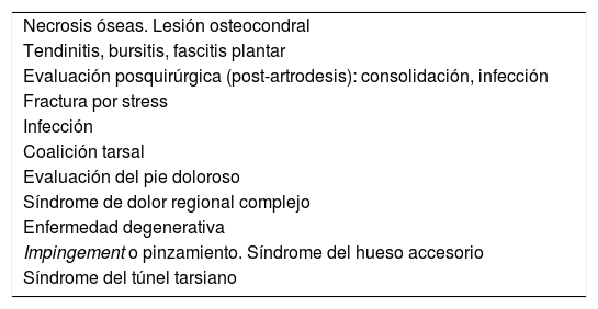
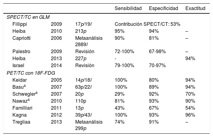
La patología del pie y tobillo es una de las más frecuentes del sistema musculoesquelético y de gran repercusión en la calidad de vida de los pacientes. El diagnóstico preciso supone un desafío clínico importante debido a que la compleja anatomía y la función del pie dificultan la localización del origen del dolor por un examen clínico de rutina. En el estudio de la patología del pie se han utilizado técnicas anatómicas (radiografía, resonancia magnética [RM], ultrasonido y tomografía computarizada [TC]) y funcionales (gammagrafía ósea [GO], tomografía de emisión de positrones [PET] y RM). La imagen híbrida combina las ventajas de los estudios morfológicos y funcionales de forma sinérgica, ayudando al clínico en la gestión de problemas complejos. En este artículo profundizamos en la anatomía y en la biomecánica del pie y tobillo y describimos las indicaciones potenciales de las técnicas hibridas actuales disponibles para el estudio de la patología del pie y tobillo.
Disorders of the foot and ankle are some of the most frequent ones affecting the musculoskeletal system and have a great impact on patients’ quality of life. Accurate diagnosis is an important clinical challenge because of the complex anatomy and function of the foot, that make it difficult to locate the source of the pain by routine clinical examination. In the study of foot pathology, anatomical imaging (radiography, magnetic resonance imaging [MRI], ultrasound and computed tomography [CT]) and functional imaging (bone scan, positron emission tomography [PET] and MRI) techniques have been used. Hybrid imaging combines the advantages of morphological and functional studies in a synergistic way, helping the clinician manage complex problems. In this article we delve into the anatomy and biomechanics of the foot and ankle and describe the potential indications for the current hybrid techniques available for the study of foot and ankle disease.
Artículo
Comprando el artículo el PDF del mismo podrá ser descargado
Precio 19,34 €
Comprar ahora


