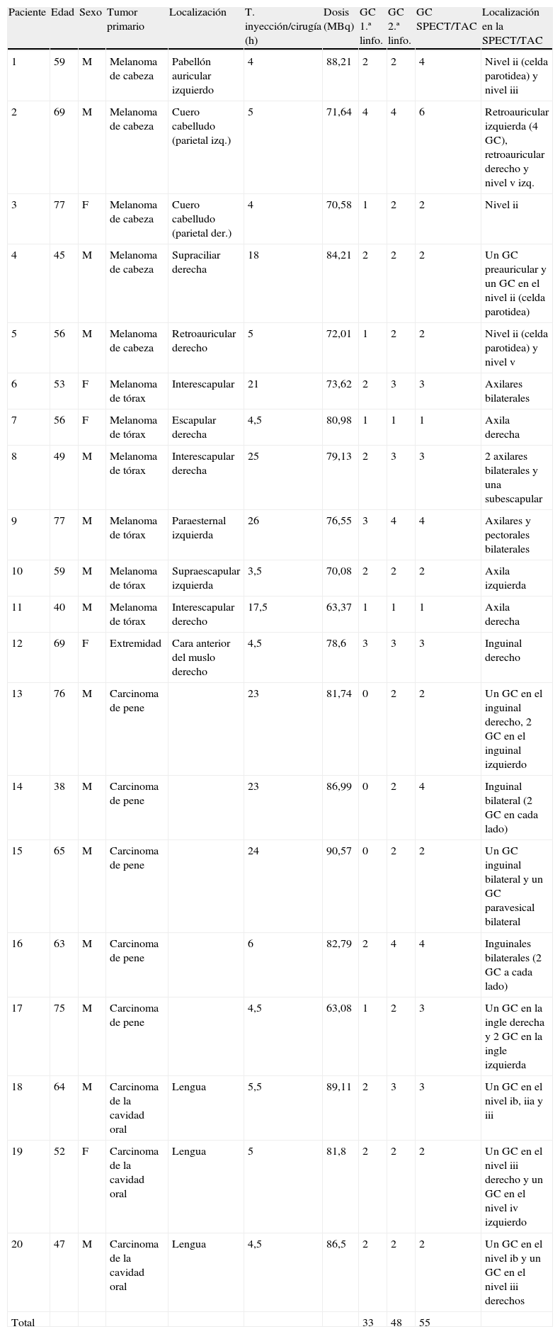
El verde de indocianina (ICG)-99mTc-nanocoloide es un novedoso trazador híbrido radioactivo y fluorescente para la biopsia selectiva del ganglio centinela (BSGC). Nuestro objetivo fue demostrar el valor añadido de este trazador en una serie de pacientes con diferentes neoplasias.
Material y métodosSe incluyeron consecutivamente 20 pacientes (con carcinoma de pene, cavidad oral y melanoma) entre marzo y mayo de 2012. A todos se les realizó una gammagrafía planar a los 15 y 120 min tras la inyección de ICG-99mTc-nanocoloide y posteriormente un SPECT/TAC. En quirófano se inyectó 1ml de colorante vital (blue dye) a 14 pacientes. Durante la cirugía, los ganglios centinelas (GC) fueron localizados utilizando la sonda gammadetectora y visualizados por fluorescencia y colorante vital. Finalmente, se confirmó la extirpación completa de los GC con la gammacámara portátil.
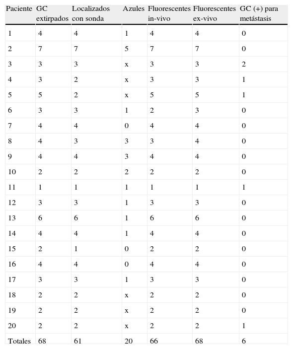
ResultadosMediante una SPECT/TAC se identificó al menos un GC por paciente. Todos los GC (total: 68, 100%) fueron extirpados utilizando la combinación de guía radiofluorescente: 89,7% se localizaron con la sonda gammadetectora. Los ganglios restantes, situados cerca del punto de inyección, fueron ubicados por fluorescencia. Durante la cirugía, un 97% del total de GC fueron fluorescentes y solo el 39,2% azules. ex-vivo, todos los GC fueron radioactivos y fluorescentes. En 5 pacientes el GC fue metastásico.
ConclusiónAplicar una guía radiofluorescente para la BSGC es posible utilizando el ICG-99mTc-nanocoloide. Este enfoque híbrido combina los beneficios de ambas modalidades. La imagen fluorescente mejora la detección visual del GC respecto al colorante vital y demuestra ser especialmente útil cuando el GC está cerca del punto de inyección.
Indocyanine green (ICG)-99mTc-nanocolloid is a novel hybrid fluorescent radioactive tracer for sentinel node (SN) biopsy. This study has aimed to evaluate the added value of this novel versatile tracer in a series of patients with different malignancies.
Material and methodsTwenty patients (with penile carcinoma, oral cavity tumors, melanoma) were consecutively included between March–May 2012. Planar lymphoscintigraphy was performed 15min and 2h after injection of ICG-99mTc-nanocolloid followed by SPECT/CT. Blue dye (1ml) was injected in 14 patients in surgery room. Intraoperatively, SNs were localized using a gamma probe and visualized by optical SN-detection using blue dye and fluorescence imaging. Finally, a portable gamma camera was used to confirm complete SN removal.
ResultsAt least one SN was identified by SPECT/CT in all patients. All SNs (total 68, 100%) were excised using a combination of radio- and fluorescence guidance: 89.7% were intraoperatively localized with the gamma probe. The remaining SNs, located near the injection site, were localized using fluorescence imaging. During the surgery, 97% of the SNs were fluorescent while only 39.2% were stained blue. Ex vivo, all SNs were both radioactive and fluorescent. The SN was positive in 5 patients.
ConclusionSynchronous radio- and fluorescence guided SN biopsy is feasible using ICG-99mTc-nanocolloid. This hybrid approach combines the beneficial properties of both modalities. Adding fluorescence imaging improves optical SN detection compared to blue dye. It has been shown to be especially useful in the localization of SNs near the injection site.
Artículo
Comprando el artículo el PDF del mismo podrá ser descargado
Precio 19,34 €
Comprar ahora


