


In this series, we analyze the diagnostic efficacy of serial voiding urosonography (VUS) with second-generation contrast, combined harmoniously and specifically with contrast technology, in the examination of the urinary tract in children. This examination includes the diagnosis and follow-up for vesicoureteral reflux (VUR) and urethral disorders, mainly those of the posterior urethral valve (PUV).
Patients and methodsAfter obtaining informed consent, a prospective study was conducted using urosonography with second-generation contrast (sulphur hexafluoride microbubbles, SonoVue®) from November 2014 to October 2015 (1 year) in pediatric patients with suspected VUR or PUV impairment. For patients with a high suspicion of VUR and in cases of PUV, we also conducted simultaneous voiding cystourethrography (VCUG).
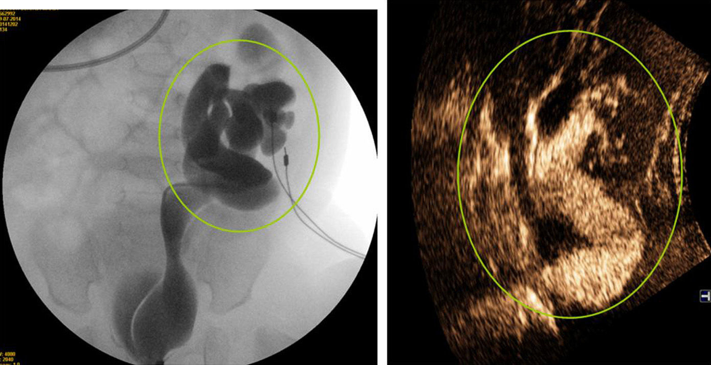
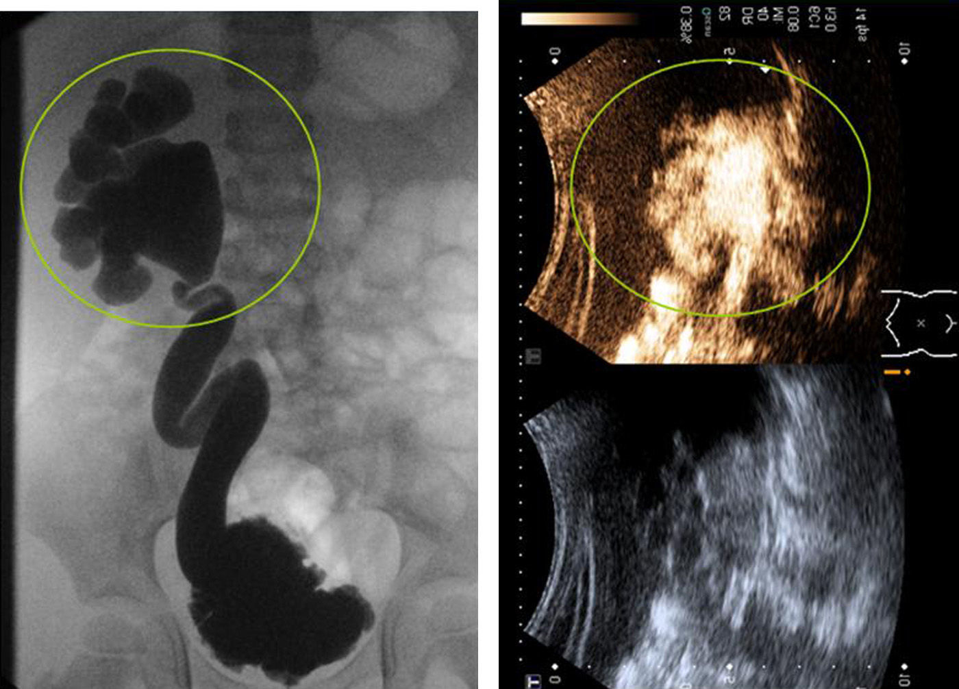
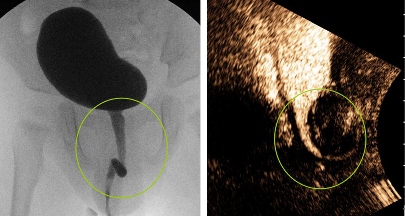
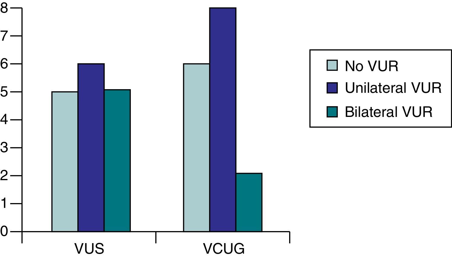
ResultsWe studied 40 patients (80 renal units) between the ages of 2 months and 13 years (median age, 14 months). The indication for the test was a suspected VUR (36 patients, group A) and PUV follow-up (4 patients, group B). The test was correlated with VCUG in 16 patients (12 cases with high suspicion of VUR in group A and with 4 cases of PUV in group B). The visualization of the urethra was appropriate in cases of dilation or urethral stricture. For 3 of these patients with bilateral VUR demonstrated in the serial VUS, the VCUG showed only unilateral VUR in 2 of the patients and no VUR in 1 of the patients (κ=.73).
DiscussionWe have shown that the visualization of the urethra is no longer a limitation and that serial VUS can be superior to conventional VCUG in diagnosing VUR.
En esta serie queremos analizar la eficacia diagnóstica de la urosonografía miccional seriada (UMS) con contraste de segunda generación, asociado a tecnología en modo armónico y específica para contraste, en el estudio de la vía urinaria en pediatría: diagnóstico y seguimiento de RVU, y también de anomalías uretrales, principalmente de las válvulas de la uretra posterior (VUP).
Pacientes y métodosSe realizó, previo consentimiento informado, estudio prospectivo con urosonografía con contraste de 2.ª generación (microburbujas de hexafluoruro de azufre, SonoVue®) en el periodo comprendido entre noviembre de 2014 a octubre de 2015 (un año) en pacientes pediátricos con sospecha de RVU, o alteración de la vía urinaria inferior (VUP). En pacientes con alta sospecha de RVU, y en los casos de VUP, se realizó además cistouretrografía miccional (CUMS) simultánea.
ResultadosFueron estudiados 40 pacientes (80 unidades renales) de entre 2 meses y 13 años (mediana 14 meses). La indicación de la prueba fue: sospecha de RVU (36 pacientes, grupo A) y seguimiento de VUP (4 pacientes, grupo B). Se correlacionó con CUMS en 16 pacientes (12 casos con alta sospecha de RVU en el grupo A y con los 4 casos de VUP del grupo B). La visualización de la uretra fue adecuada en los casos de dilatación o estenosis uretral. En 3 de estos pacientes con RVU bilateral en UMS en la CUMS solo se apreciaba de forma unilateral en 2 de los casos y sin RVU en uno; κ=0,73.
DiscusiónHemos comprobado que la visualización de la uretra ya no es una limitación, y que la UMS puede ser superior a la CUMS convencional en el diagnóstico del RVU.
Artículo

Comprando el artículo el PDF del mismo podrá ser descargado
Precio 19,34 €
Comprar ahora
