metricas
covid
Buscar en
Annals of Hepatology
Toda la web
Inicio Annals of Hepatology A giant hepatic hemangioma treated successfully with hepatic enucleation
Journal Information
Vol. 8. Issue 4.
Pages 377-378 (October - December 2009)
Share
Share
Download PDF
More article options
Vol. 8. Issue 4.
Pages 377-378 (October - December 2009)
Open Access
A giant hepatic hemangioma treated successfully with hepatic enucleation
Visits
1292
Gustavo Lopez-Arce Angeles
,****,
Corresponding author
glopezarce@gmail.com

Correspondence and reprint request:
, J. Barahona-Garrido*,****, F.I. Tellez-Avila*,****, L. Burgos-Zuleta**,****, T. Lopez-Placier**,****, F.J. Llamas-Gutiérrez***,****, A. Torre-Delgadillo*,****
* Departments of Gastroenterology. Nutrición Salvador Zubirán. Mexico City, Mexico
** Radiology. Nutrición Salvador Zubirán. Mexico City, Mexico
*** Pathology. Instituto Nacional de Ciencias Médicas y Nutrición Salvador Zubirán. Mexico City, Mexico
**** Universidad Nacional Autónoma de México, Mexico City, Mexico
This item has received

Under a Creative Commons license
Article information
Full Text
Bibliography
Download PDF
Statistics
Figures (3)
Show moreShow less
Full Text

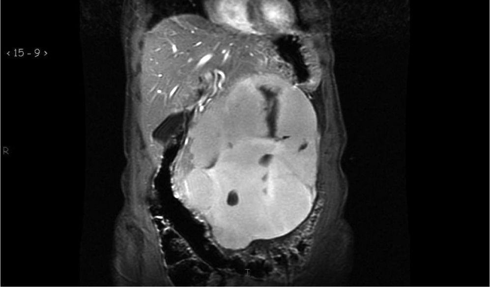
Hepatic hemangiomas (HH) are the first cause of benign hepatic tumors, their prevalence varies from 3% to 20% in general population.1 Giant HH are those greater than 4 cm; they account for only 10% of all HH. HH are usually asymptomatic. Computed tomography (CT) and magnetic resonance image (MRI) characteristic finding is the centripetal enhancement of contrast.2 Histological characteristic findings are spongy appearance with blood filled vascular channels lined by endothelium; thrombi are frequent.3 Surgical treatment (resection or enucleation) is recommended in symptomatic patients.1

A 56-year-old woman was evaluated because she suffered for systemic arterial hypertension, but she also complaints for heartburn, occasional abdominal discomfort, and abdominal distension. A distended abdomen and a large mass were noted by palpation. Abdominal ultrasound revealed a tumor of the liver. Contrast enhanced CT and MRI confirmed characteristic features of giant HH (Figures 1-2). Blood count cells and liver tests were normal. Enucleation was performed without complications. Histology confirmed diagnosis of HH (Figure 3). She is being asymptomatic at 12 months of follow up.

Figure 1.

MRI T2-weighted coronal reconstruction. A giant HH (20 cm) displaces adjacent structures.

(0.04MB).
Figure 2.

Gadolinium-contrasted MRI T1-weighted. In panel A peripheral enhancement of gadolinium is observed. Panel B and C reveals a centripetal pattern. Panel D shows total enhancement of HH. Note a typical centripetal slow filling pattern characteristic of HH.

(0.06MB).
Figure 3.

Microscopic characteristics of HH. Upper. Normal liver. Middle to bottom. Hemangioma; formed by blood filled vascular channels, lined by a single layer of flat endothelial cells supported by fibrous tissue (H & E stain. 10 X).

(0.24MB).
References
[1.]
Bahirwani R., Reddy KR..
The evaluation of solitary liver masses.
Aliment Pharmacol Ther, 28 (2008), pp. 953-965
[2.]
Choi B.Y., Nguyen MH..
The diagnosis and management of benign hepatic tumors.
J Clin Gastroenterol, 39 (2005), pp. 401-412
[3.]
Bioulac-Sage P., Laumonier H., Laurent C., et al.
Benign and malignant vascular tumors of the liver in adults.
Sem Liv Dis, 28 (2008), pp. 302-314
Copyright © 2009. Fundación Clínica Médica Sur, A.C.
Article options
es en pt

¿Es usted profesional sanitario apto para prescribir o dispensar medicamentos?

Are you a health professional able to prescribe or dispense drugs?

Você é um profissional de saúde habilitado a prescrever ou dispensar medicamentos

Quizás le interese:
10.1016/j.aohep.2023.100975
No mostrar más