metricas
covid
Buscar en
Archivos de la Sociedad Española de Oftalmología
Toda la web
Inicio Archivos de la Sociedad Española de Oftalmología Evaluación del músculo ciliar y del grosor escleral anterior en miopes altos m...
Journal Information
Vol. 99. Issue 1.
Pages 16-22 (January 2024)
Share
Share
Download PDF
More article options
Visits
31
Vol. 99. Issue 1.
Pages 16-22 (January 2024)
Artículo original
Evaluación del músculo ciliar y del grosor escleral anterior en miopes altos mediante tomografía de coherencia óptica
Assessment of the ciliary muscle and scleral anterior thickness in high myopia by optical coherence tomography
Visits
31
J.I. Fernández-Vigoa,b,
Corresponding author
jfvigo@hotmail.com

Autor para correspondencia.
, S. Fernández-Aragóna,b, Lucía de-Pablo-Gómez-de-Liañob,c, C. Fernández-Vigob, I. Almorín-Fernández-Vigod, J. García-Feijóoa, J.A. Fernández-Vigob,e
a Departamento de Oftalmología, Hospital Clínico San Carlos, Instituto de Investigación Sanitaria (IdISSC), Madrid, España
b Centro Internacional de Oftalmología Avanzada, Madrid, España
c Departamento de Oftalmología, Hospital 12 de Octubre, Madrid, España
d Centro Internacional de Oftalmología Avanzada, Badajoz, España
e Departamento de Oftalmología, Universidad de Extremadura, Badajoz, España
This item has received
Article information
Abstract
Full Text
Bibliography
Download PDF
Statistics
Figures (1)
Tables (2)
Tabla 1. Dimensiones del músculo ciliar según los diferentes grupos clasificados por la refracción. Media±DE (rango)
Tabla 2. Dimensiones del grosor escleral anterior según los diferentes grupos clasificados por la refracción. Media±DE (rango)
Show moreShow less
Resumen
Propósito

Evaluar las dimensiones del músculo ciliar (MC) y del grosor escleral anterior (AST) in vivo en miopes altos mediante tomografía de coherencia óptica de fuente de barrido (SS-OCT) y comparar con sujetos emétropes e hipermétropes.
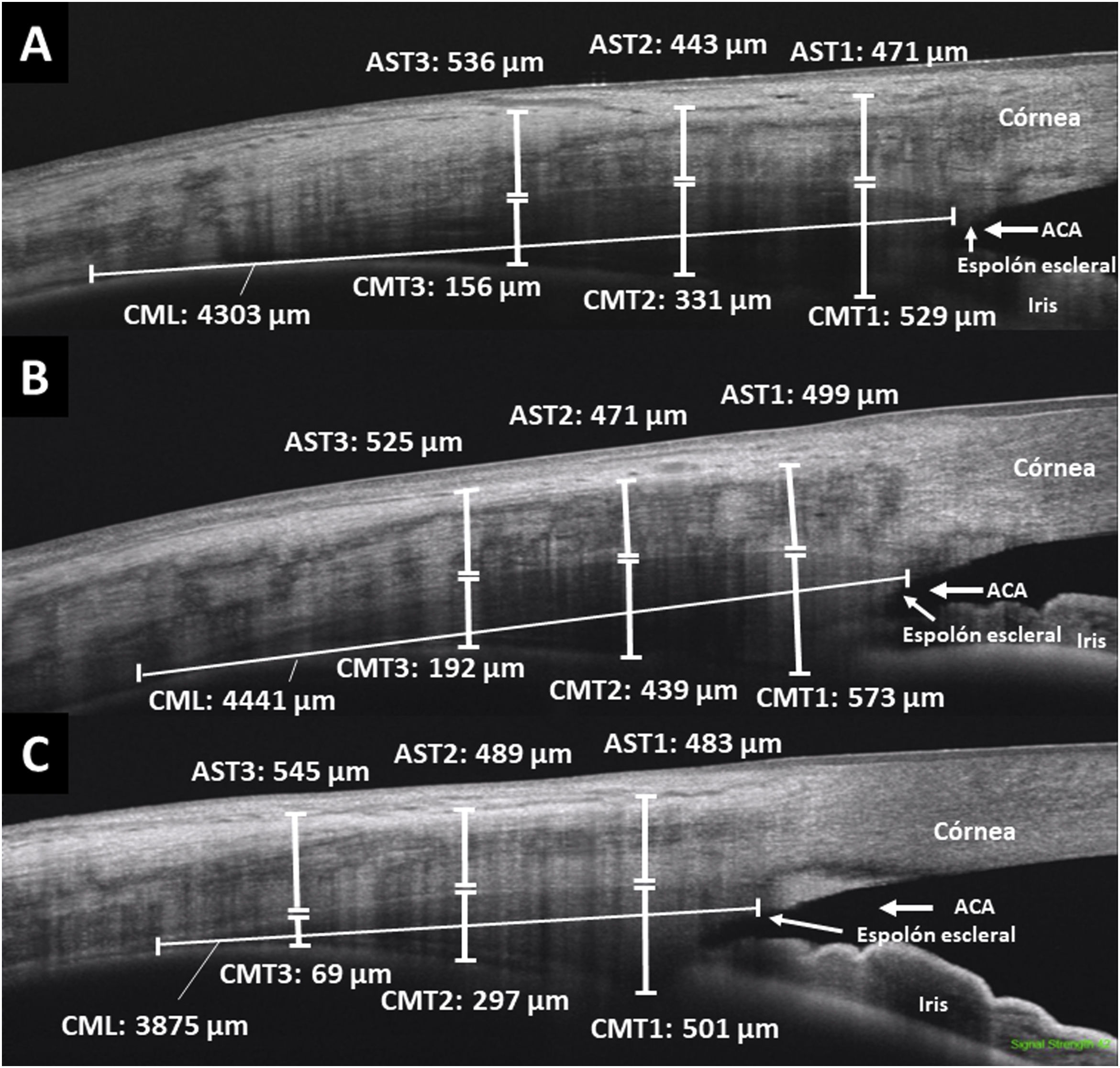
Métodos

Estudio transversal en el que se incluyeron 34 miopes altos (≥−6dioptrías [D]), 90 emétropes (−1 a +1D) y 38 hipermétropes (≥+3,5D). Se midieron el grosor del MC (CMT) y el AST en los cuadrantes temporal y nasal a 1, 2 y 3mm del espolón escleral utilizando la SS-OCT. Además, se evaluó la longitud del MC (CML).
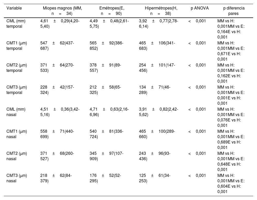
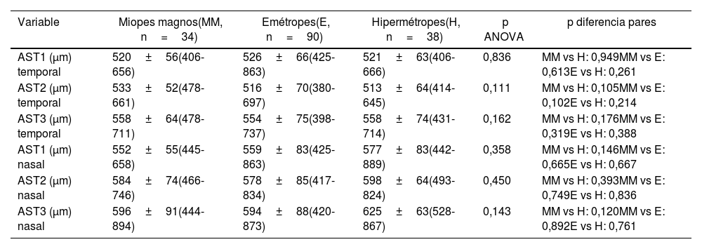
Resultados

Las dimensiones tanto del CML como del CMT en cualquiera de sus puntos de medida fueron mayores en miopes altos y en emétropes que en hipermétropes, tanto en el cuadrante nasal como en el temporal (p<0,001). Sin embargo, no existieron diferencias entre miopes magnos y emétropes para ninguno de los parámetros (p0,076), salvo para el CMT a 3mm en temporal (p<0,001). No existieron diferencias en el AST entre miopes altos, emétropes e hipermétropes, en ninguno de los puntos de medida ni cuadrantes estudiados (p>0,05).

Conclusiones

La SS-OCT permite medir el MC in vivo, no observándose diferencias en sus dimensiones entre miopes altos y emétropes, pero sí que fueron menores en hipermétropes. En la medida de la esclera anterior no se observaron diferencias entre los tres grupos analizados según la refracción.

Palabras clave:
Músculo ciliar
Cuerpo ciliar
Tomografía de coherencia óptica
Alta miopía
Esclera anterior
Abstract
Purpose

To assess ciliary muscle (CM) and anterior scleral thickness (AST) dimensions in vivo in high myopia using swept-source optical coherence tomography (SS-OCT) and to compare with emmetropic and hyperopic subjects.

Methods

Cross-sectional study that included 34 high myopic patients (≥−6 diopters [D]), 90 emmetropes (−1 to +1D) and 38 hyperopic patients (≥+3.5D). CM thickness (CMT) and AST were measured in the temporal and nasal quadrants at 1, 2, and 3mm from the scleral spur using SS-OCT. In addition, the length of the CM (CML) was evaluated.

Results

The dimensions of the CML and the CMT at any of their measurement points were greater in high myopes and emmetropes than in hyperopes, both in the nasal and temporal quadrants (P<.001). However, there were no differences between high myopes and emmetropes for any of the parameters (P.076) except for the CMT at 3mm in the temporal quadrant (P<.001). There were no differences in the AST between high myopes, emmetropes and hyperopes, in any of the measurement points or quadrants studied (P>.05).

Conclusions

The SS-OCT allows to measure the CM in vivo, not observing differences in its dimensions between high myopes and emmetropes, but they were smaller in hyperopes. In the measurement of the anterior sclera, no differences were observed between the three groups analyzed according to refraction.

Keywords:
Ciliary muscle
Ciliary body
Optical coherence tomography
High myopia
Anterior sclera

Article

These are the options to access the full texts of the publication Archivos de la Sociedad Española de Oftalmología
Subscriber
Subscriber

If you already have your login data, please click here .

If you have forgotten your password you can you can recover it by clicking here and selecting the option “I have forgotten my password”
Subscribe
Subscribe to

Archivos de la Sociedad Española de Oftalmología

Purchase
Purchase article

Purchasing article the PDF version will be downloaded

Price 19.34 €

Purchase now
Contact
Phone for subscriptions and reporting of errors
From Monday to Friday from 9 a.m. to 6 p.m. (GMT + 1) except for the months of July and August which will be from 9 a.m. to 3 p.m.
Calls from Spain
932 415 960
Calls from outside Spain
+34 932 415 960
E-mail
Article options
es en pt

¿Es usted profesional sanitario apto para prescribir o dispensar medicamentos?

Are you a health professional able to prescribe or dispense drugs?

Você é um profissional de saúde habilitado a prescrever ou dispensar medicamentos