metricas
covid
Buscar en
Archivos de la Sociedad Española de Oftalmología
Toda la web
Inicio Archivos de la Sociedad Española de Oftalmología Actualización de la utilidad de la tomografía de coherencia óptica para el es...
Journal Information
Vol. 94. Issue 10.
Pages 478-490 (October 2019)
Share
Share
Download PDF
More article options
Visits
237
Vol. 94. Issue 10.
Pages 478-490 (October 2019)
Revisión
Actualización de la utilidad de la tomografía de coherencia óptica para el estudio del ángulo iridocorneal
Update on the usefulness of optical coherence tomography in assessing the iridocorneal angle
Visits
237
B. Kudsieha,b,
Corresponding author
bacharkudsieh@gmail.com

Autor para correspondencia.
, J.I. Fernández-Vigob,c, J. Vila-Arteagad,e, J. Aritz Urcolaf,g, J.M. Martínez-de-la-Casac, J. García-Feijóoc, J.M. Ruiz-Morenoa, J.Á. Fernández-Vigob,h
a Departamento de Oftalmología, Hospital Universitario Puerta de Hierro Majadahonda, Madrid, España
b Centro Internacional de Oftalmología Avanzada, Madrid, España
c Departamento de Oftalmología, Hospital Clínico San Carlos, Instituto de Investigación Sanitaria (IdISSC), Madrid, España
d Hospital La Fe, Valencia, España
e Clínica Vila-Innova Ocular, Valencia, España
f Departamento de Oftalmología, Hospital Universitario de Álava, Vitoria, España
g Begitek Clínica Oftalmológica, Innova Ocular, Donostia-San Sebastián, España
h Departamento de Oftalmología, Universidad de Extremadura, Badajoz, España
This item has received
Article information
Abstract
Full Text
Bibliography
Download PDF
Statistics
Figures (10)
Show moreShow less
Tables (4)
Tabla 1. Características de los principales dispositivos de tomografía de coherencia óptica
Tabla 2. Porcentaje de identificación de las estructuras angulares mediante tomografía de coherencia óptica de segmento anterior
Tabla 3. Parámetros empleados en las mediciones de la región angular mediante tomografía de coherencia óptica (OCT)
Tabla 4. Utilidad de la exploración mediante tomografía de coherencia óptica (OCT) del ángulo iridocorneal
Show moreShow less
Resumen

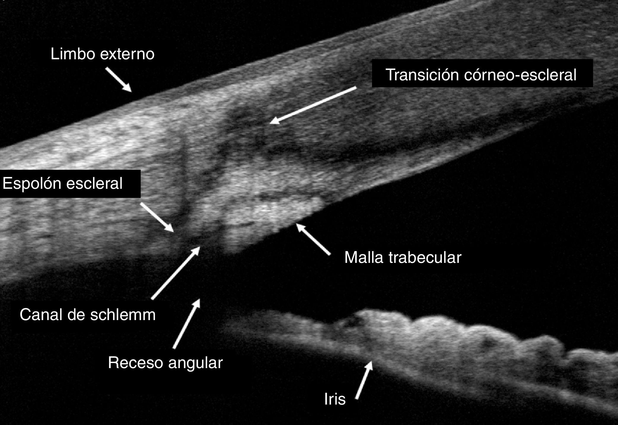
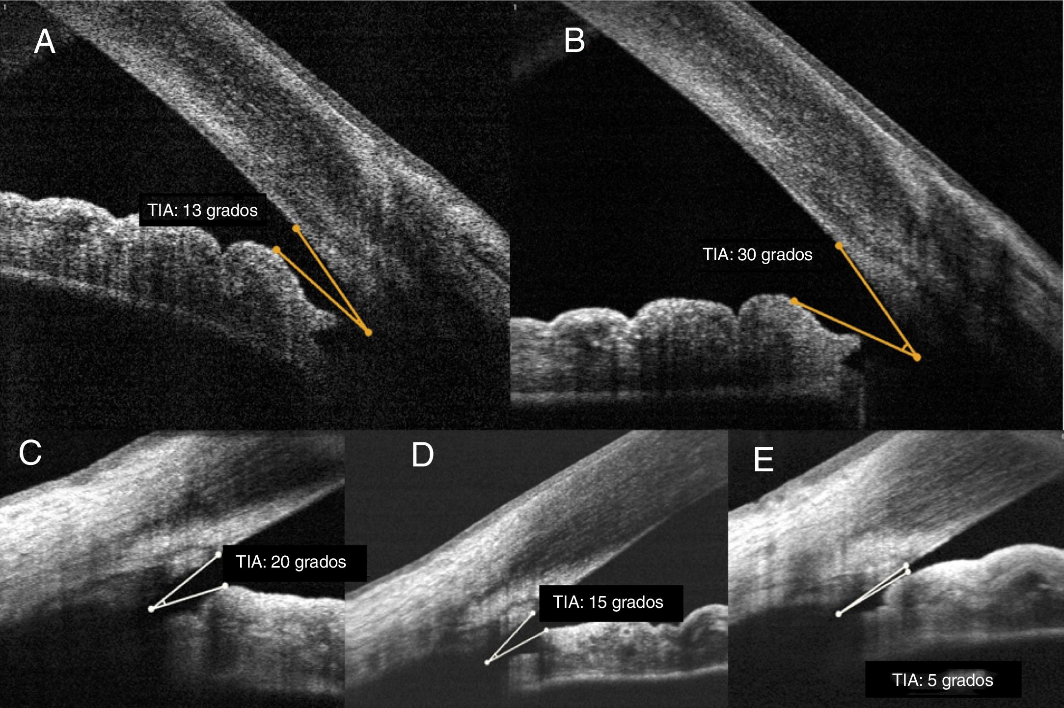
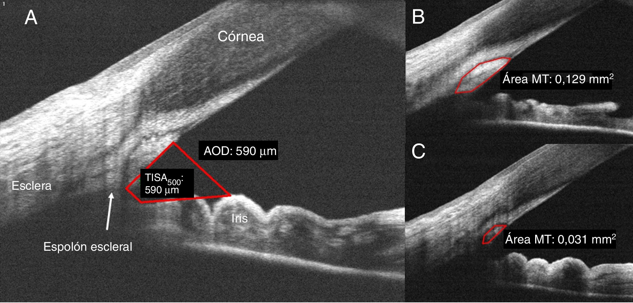
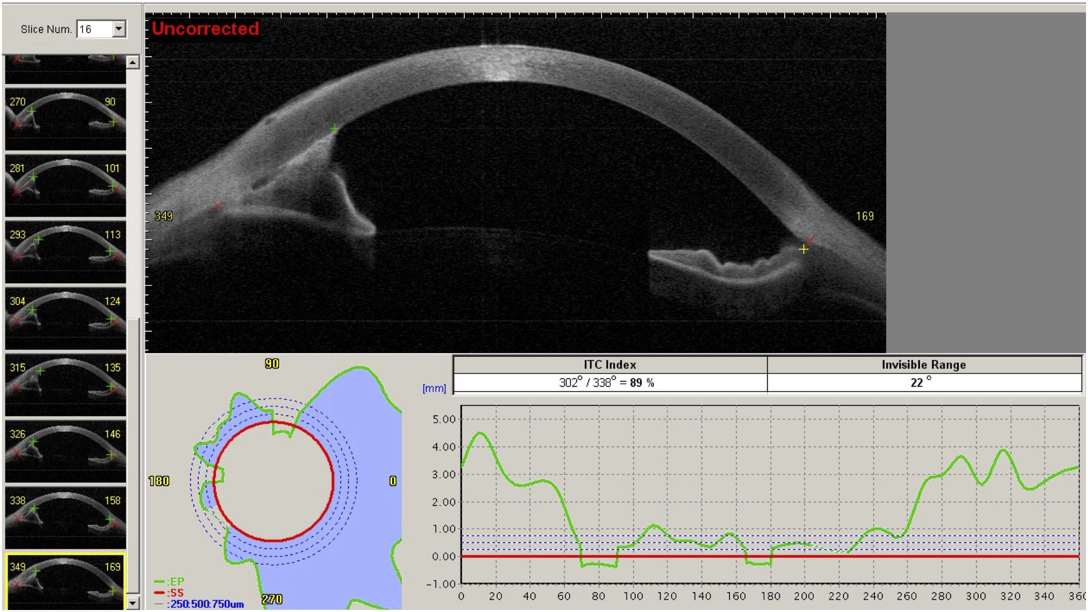
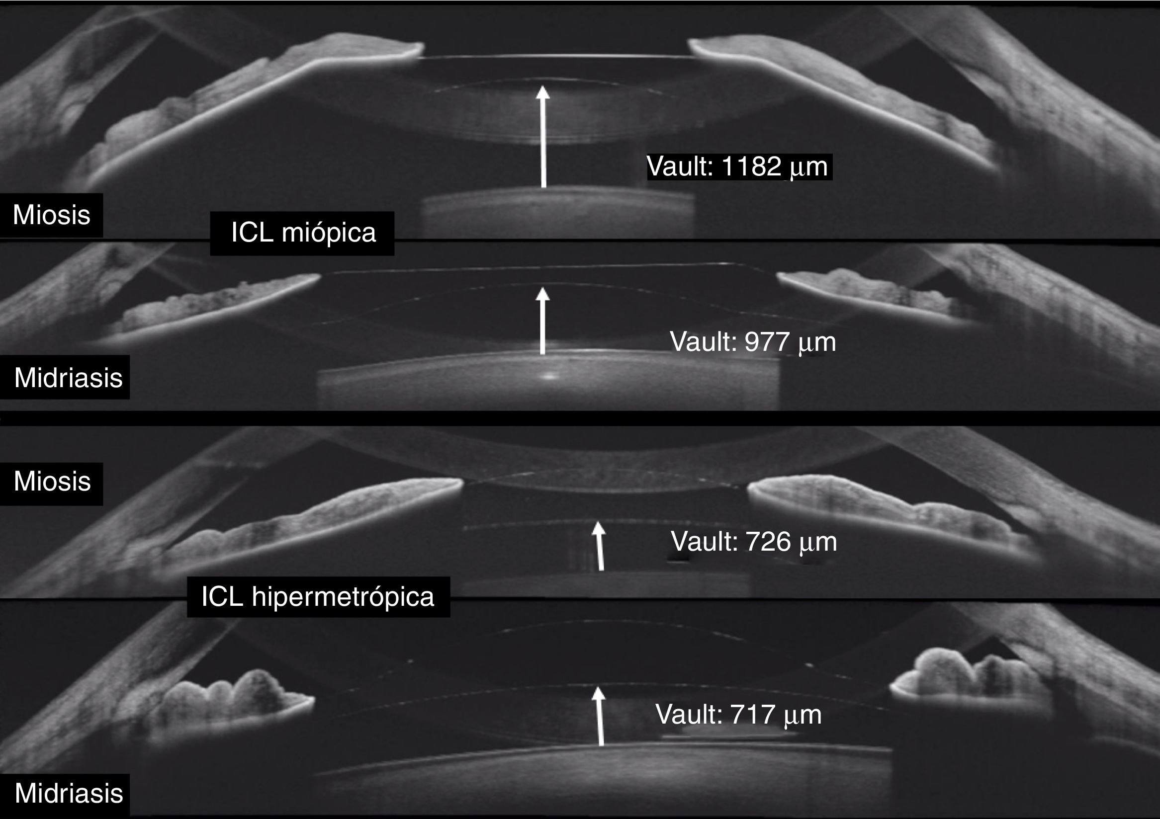
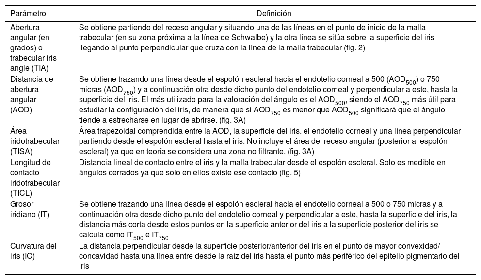
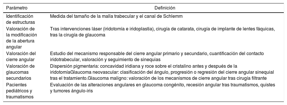
El ángulo iridocorneal por sus implicaciones en la fisiopatología del drenaje del humor acuoso es una estructura fundamental de la cámara anterior. La tomografía de coherencia óptica de segmento anterior es una técnica rápida y no invasiva que obtiene imágenes de los tejidos vivos con una alta resolución permitiendo conocer la anatomía normal del ángulo, sus alteraciones y los cambios que se producen en el mismo tras diferentes intervenciones terapéuticas. La tecnología de la tomografía de coherencia óptica de segmento anterior ha ido evolucionando hasta ofrecer imágenes que permiten identificar y cuantificar estructuras angulares claves en sujetos sanos y en pacientes con glaucoma, especialmente la malla trabecular y el canal de Schlemm, lo que puede contribuir a ampliar el conocimiento de la fisiopatología del glaucoma. Además, permite cuantificar la abertura angular con unos parámetros objetivos descritos en los últimos años, entre los que destacan el ángulo iridotrabecular, la distancia de abertura angular y el área iridotrabecular. La tomografía de coherencia óptica de segmento anterior presenta múltiples utilidades en el estudio de los distintos mecanismos del cierre angular, la evaluación de los cambios angulares tras la realización de una iridotomía láser o iridoplastia, cirugía de la catarata o el implante de lentes fáquicas.

Palabras clave:
Ángulo iridocorneal
Tomografía de coherencia óptica
Glaucoma
Abertura angular
Malla trabecular
Canal de Schlemm
Abstract

The iridocorneal angle, due to its implications in the physiopathology of aqueous humour drainage, is a fundamental structure of the anterior chamber. Anterior segment optical coherence tomography is a rapid and non-invasive technique that obtains images in vivo. The high resolution allows it to analyse the normal anatomy of the angle, any alterations, and the changes that occur after different therapeutic interventions. Anterior segment optical coherence tomography technology has evolved to provide images that allow the identification and quantification of the angular structures in healthy subjects and in glaucoma patients, and especially the trabecular meshwork and the Schlemm's canal. It also enables the angle width to be quantified, with some objective parameters that have been standardised in recent years, such as the trabecular-iris angle, the angle opening distance, and the trabecular-iris area. This technique has multiple uses in the study of the different mechanisms of angle closure, the evaluation of changes after a laser peripheral iridotomy or iridoplasty after cataract surgery, as well as after the implantation of phakic lenses.

Keywords:
Iridocorneal angle
Optical coherence tomography
Glaucoma
Angle width
Trabecular meshwork
Schlemm's canal

Article

These are the options to access the full texts of the publication Archivos de la Sociedad Española de Oftalmología
Subscriber
Subscriber

If you already have your login data, please click here .

If you have forgotten your password you can you can recover it by clicking here and selecting the option “I have forgotten my password”
Subscribe
Subscribe to

Archivos de la Sociedad Española de Oftalmología

Purchase
Purchase article

Purchasing article the PDF version will be downloaded

Price 19.34 €

Purchase now
Contact
Phone for subscriptions and reporting of errors
From Monday to Friday from 9 a.m. to 6 p.m. (GMT + 1) except for the months of July and August which will be from 9 a.m. to 3 p.m.
Calls from Spain
932 415 960
Calls from outside Spain
+34 932 415 960
E-mail
Article options
es en pt

¿Es usted profesional sanitario apto para prescribir o dispensar medicamentos?

Are you a health professional able to prescribe or dispense drugs?

Você é um profissional de saúde habilitado a prescrever ou dispensar medicamentos