


Alopecia areata incognita, also known as diffuse alopecia areata, is a rare form of alopecia areata described predominantly in young women. In cases of alopecia areata incognita, the typical patchy distribution of hair loss in classical alopecia areata is absent, but abrupt and intense hair loss is characteristic. While the clinical picture presented by this disease closely resembles that of telogen effluvium, specific clinical and dermoscopic findings of alopecia areata are invariably present along the disease course.1 Prognosis is generally favorable, especially as compared to certain variants of alopecia areata, namely, alopecia areata totalis, universalis and ophiasic areata.
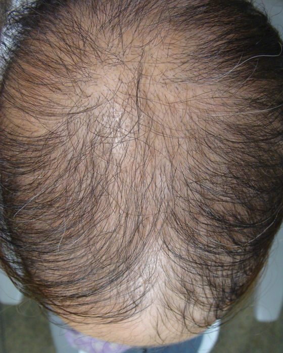
CASE REPORTA 23-year-old Brazilian woman was referred with rapidly progressing hair loss that became apparent over a period of two weeks (Figure 1). The patient was otherwise healthy, but she noticed that her hair loss began after a stressful business trip. Her personal history included trichotillomania, which was diagnosed before age 20 and completely regressed after one year on antidepressive agents. The patient was not taking any other medications.

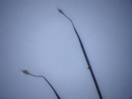
Dermatological examination revealed diffuse hair loss affecting the entire scalp with no areas of patchy hair loss. With only a gentle pull, most of her hair could be easily removed. So-called ‘exclamation point’ hairs and vellus hairs could be observed under the microscope, and the scalp did not present erythema or scaling (Figure 2). The patient's eyebrows, eyelashes and body hair were completely normal. Moreover, no nail or other skin abnormalities were observed.

Dermoscopy of the scalp revealed yellow dots. A scalp biopsy specimen showed few mononuclear cells around the hair follicles in the papillary dermis and an increased proportion of telogens and vellus/miniaturized hairs. Complete blood count, serum biochemistry tests, and thyroid hormones were within normal limits.
The patient was treated with a class 1 topical steroid (clobetasol) every other night under occlusion; the cream was washed off in the morning. Biotin (10 mg per os) was taken daily. After 12 weeks, hair regrowth was evident over the entire scalp, but the fronto-parietal regions showed a lower hair density, similar to an androgenetic pattern (Figure 3). No recurrences have occurred during the 24-month follow-up period.
DISCUSSION
Alopecia areata incognita (AAI) was first described by Rebora2 in 1987. The disorder has an extremely acute onset with subsequent diffuse hair loss that occurs within a few weeks. Similar cases have been described under different names, including “acute alopecia totalis”3 and “acute diffuse and total alopecia of the female scalp”,1,4 which are identical to AAI. AAI is more common in patients under the age of 40, especially in those from 20 to 40 years of age. A strong female predominance (86.6%) is evident in the 112 cases that have been reported.
Although clinically different from other forms of alopecia areata, the histopathologic findings of AAI are similar to the classical forms of the disease and include its variation with disease stage.5 The most consistent finding in acute AAI scalp biopsies is an inflammatory infiltrate around the terminal hair bulb. This infiltrate gradually decreases with chronicity and concentrates around either only the miniaturized follicles or the follicular stelae (streamers).6 Additionally, a reversal in the anagen-telogen and terminal-vellus ratios is always observed and may be the only evidence suggesting the diagnosis in long-standing cases.7 Follicular density is preserved in the acute and subacute stages, but it may decrease over time.
Tosti et al.7 reported 70 patients with AAI, of whom 50 were histologically confirmed. In all such cases, the dermoscopic findings were suggestive of AAI, showing diffuse, round or polycyclic yellow dots, which is a specific feature of alopecia areata, and regrowing, tapered, terminal hairs. Choi and Ihm3 classified a group of 13 patients (3 males and 10 females) who experienced acute diffuse hair loss over the entire scalp as having acute alopecia totalis. The time from the initial onset of the excessive hair loss to total hair loss was two months on average, which is similar to our patient. Sato-Kawamura4 et al. described 9 female cases of an acute and diffuse hair loss, which they termed “acute diffuse and total alopecia of the female scalp”. The histology of the lesions was indistinguishable from that of alopecia areata, except for a remarkable eosinophilic infiltrate. They treated eight cases mainly with systemic steroids, while a single case was treated with topical steroids. A complete regrowth of hair was observed within 6 months in 8 out of 9 cases. Even the case treated with only topical steroids showed a favorable prognosis. Inui1 et al. dermoscopically examined 20 female cases of AAI diagnosed based on clinical and histopathological findings. They classified cadaverized hairs, exclamation mark hairs and broken hairs as specific diagnostic markers for diffuse and total alopecia of the female scalp and found a sensitivity of almost 96% when a combination of yellow dots and/or short vellus hairs were present in the dermoscopy.
In our case, vertical sections showed a very subtle perifollicular lymphocytic infiltrate around the sebaceous glands and an increased number of catagens and miniaturized follicles. Horizontal sections confirmed the increase in miniaturized follicles and also showed an inverted anagen-telogen ratio. Additionally, the total hair follicle density was normal.
As with the histopathology, dermoscopy of AAI cases has resulted in findings similar to classic AA that also depended on disease activity.1 Typical findings include multiple yellow dots, short regrowing hair, dystrophic hairs, exclamation point hairs and cadaverized hairs.7 When the methods are considered separately, their sensitivity and specificity are still controversial in the literature, but the combination of two or more of them greatly improves the diagnostic specificity.
In this case, we observed a great number of typical signs (i.e., yellow dots, many exclamation point hairs, dystrophic hairs and some cadaverized hairs) over the entire scalp.
Etiopathogenic mechanisms of the disease remain controversial. Rebora et al. have suggested that a high percentage of hairs in the late phases of the hair cycle at the moment when the disease is triggered could possibly explain the particular distribution of this disease.2 This situation is found in androgenetic alopecia (AAG) and results from the shortening and synchronization of hair cycles diffusely across all scalp follicles. In fact, our patient did show a typical AAG pattern of hair regrowth after treatment.
Therapeutic approaches for AAI have mainly included steroids, such as intravenous methylprednisolone pulse therapy, oral prednisone, intramuscular steroid injections and topical steroids; these approaches have a good prognosis. Our patient responded well to topical use of clobetasol cream under occlusion with oral biotin. No recurrence was observed.
Diffuse hair loss of variable intensity is a common complaint, especially among women. In such cases, the differential diagnosis generally includes telogen effluvium and androgenic alopecia. Although rare, AAI should be included in the differential diagnosis of acute and diffuse hair loss in order to avoid unnecessary exams and to allow adequate treatment of this distressing hair disorder.