


Epithelioid hemangioendothelioma is a rare vascular tumor with an estimated prevalence of less than 1 in 1 million.1 It can have several locations, with the pulmonary being one of the most common.2 The typical radiological image shows multiple pulmonary nodules, followed by parenchymal tumors with pleural invasion, reticulonodular opacities, and diffuse pleural thickening.3,4 The prognosis is unpredictable, and life expectancy can vary from 1 to 15 years.2
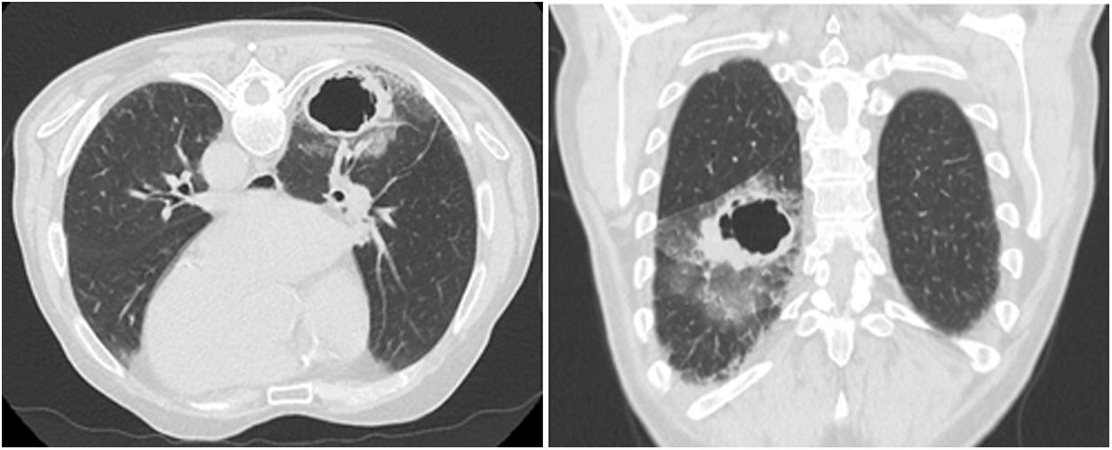
We present a 75-year-old man who was an active smoker with a smoking load of 50 pack-year units who was referred to the pulmonology outpatient department due to coughing and hemoptoic expectoration with several months of evolution. The patient had a personal history of chronic bronchitis and permanent atrial fibrillation. Chest radiography revealed air-fluid cavitation over the right hilum. A computed tomography (CT) scan of the chest detailed a 5cm excavated lesion on the right inferior lobe with thick walls and paraseptal emphysema in the upper lobes (Fig. 1).

Bronchoscopic examination revealed signs of past bleeding in the right B6 bronchus without direct signs of neoplasia. The bronchial aspirate microbiology revealed Rothia and Streptococcus viridans. There was no improvement with the antibiotic. At this point, the principal diagnostic hypothesis was lung cancer, and a transthoracic lung aspiration puncture guided by CT was performed in an attempt to obtain a histopathological diagnosis. However, the cytology showed an area of fibrosis with a chronic inflammatory process. A CT of the brain and a PET scan hadn’t identified extra pulmonary metastases. Yet, in the PET scan, a large cavitated and intensely hypermetabolic nodular formation in the right lower lobe (52mm×36mm/SUV 15.5) with an apparent satellite lesion was described. Besides, right broncho hilar lymph nodes with slightly increased FDG F18 uptake (2.5) and a 5mm pulmonary nodule on the left were also found. The patient underwent endobronchial ultrasonography (EBUS) and another bronchoscopy with transbronchial lung biopsies. The lymph nodes punctured in the EBUS were negative for malignancy. The cytology of transbronchial lung biopsies mentioned a suspected malignancy with atypia. For diagnostic and therapeutic purposes, the patient underwent a right lower lobectomy. Finally, the surgical specimen histopathology revealed a high-grade epithelioid hemangioendothelioma with expression of TFE-3. The patient was followed by the Oncology outpatient department with the indication for surveillance. Unfortunately, the patient died a year later from aspiration pneumonia in the context of a stroke.
High-grade epithelioid hemangioendothelioma is a rare pathology with a wide differential diagnosis, ranging from granulomatous pathology to primary lung cancer. In this clinical case, the atypical imaging presentation is highlighted. In fact, to the best of our knowledge, it represents the first case with a cavitied lung lesion.
Patient consentInformed consent was obtained from the patient's relative for publication of the clinical data and images present in this manuscript.
Authors’ contributionAll authors have contributed to the elaboration and writing of the manuscript and they all approved its final version to be submitted.
FundingThe authors declare that no funding was received for this article.
Conflict of interestThe authors have no conflicts of interest to declare.
