metricas
covid
Buscar en
Radiología (English Edition)
Toda la web
Inicio Radiología (English Edition) Study of the right ventricle using magnetic resonance imaging
Información de la revista
Vol. 54. Núm. 3.
Páginas 231-245 (mayo - junio 2012)
Compartir
Compartir
Descargar PDF
Más opciones de artículo
Visitas
8555
Vol. 54. Núm. 3.
Páginas 231-245 (mayo - junio 2012)
Update in radiology
Study of the right ventricle using magnetic resonance imaging
Estudio del ventrículo derecho mediante resonancia magnética
Visitas
8555
A. Capelastegui Alber
Autor para correspondencia
acapelastegui@osatek.es

Corresponding author.
, E. Astigarraga Aguirre, M.A. de Paz, J.A. Larena Iturbe, T. Salinas Yeregui
Osatek, Hospital de Galdakao, Vizcaya, Spain
Este artículo ha recibido
Información del artículo
Resumen
Texto completo
Bibliografía
Descargar PDF
Estadísticas
Figuras (12)
Mostrar másMostrar menos
Tablas (2)
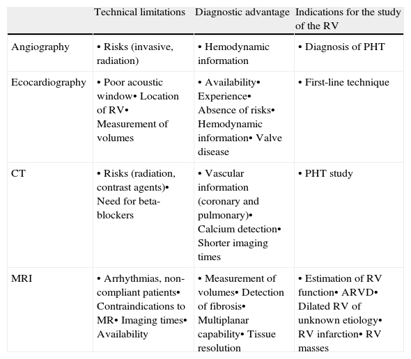
Table 1. Imaging techniques for the study of the RV.
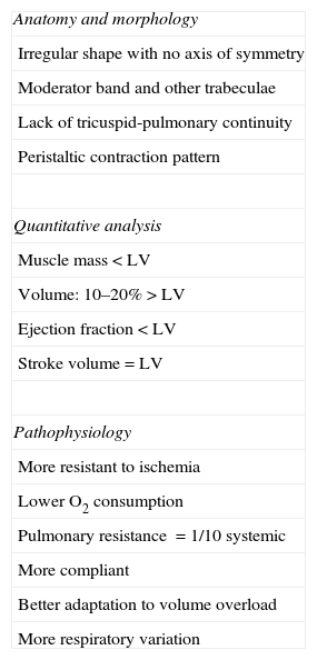
Table 2. Specific differences of the RV compared with LV.
Mostrar másMostrar menos
Material adicional (12)
Abstract

Magnetic resonance (MR) imaging has proven efficacy in the study of the heart. Its clinical applications are directed primarily at the study of the left ventricle, and the right ventricle is relegated to the background. This article reviews the anatomy and physiology of the right ventricle, as well as the manifestations of most common diseases affecting this chamber of the heart: infarction, cardiomyopathy, masses, and right heart failure. Knowing the distinctive features of the right ventricle with respect to the left and the particularities of the MR imaging protocol results in better technical performance in cases in which the reason for the examination or imaging findings point to the right ventricle. The importance of the right ventricle in the management of cardiopulmonary disease is growing and MR imaging can provide clinicians with the support they need.

Keywords:
Magnetic resonance imaging
Heart
Right ventricle
Right heart
Cardiac imaging
Resumen

La resonancia magnética (RM) es una técnica de probada eficacia en el estudio del corazón. Sus aplicaciones clínicas se dirigen preferentemente al estudio del ventrículo izquierdo, quedando el ventrículo derecho relegado a un segundo plano. Este artículo ofrece una revisión de la anatomía y fisiología del ventrículo derecho, así como de las manifestaciones de la afección más frecuente en esta cámara cardíaca: infarto, miocardiopatías, masas y fallo cardíaco derecho. El conocimiento de los rasgos diferenciales del ventrículo derecho con respecto al izquierdo y de las particularidades del protocolo de estudio mediante RM, consigue un mayor rendimiento de la técnica en aquellos casos en que el motivo de petición o los hallazgos de imagen apuntan al ventrículo derecho. La RM reúne características para apoyar desde la imagen el protagonismo creciente que los clínicos están otorgando al ventrículo derecho en el manejo de las enfermedades cardiopulmonares.

Palabras clave:
Resonancia magnética
Corazón
Ventrículo derecho
Corazón derecho
Imagen cardíaca

Artículo

Opciones para acceder a los textos completos de la publicación Radiología (English Edition)
Suscriptor
Suscriptor de la revista

Si ya tiene sus datos de acceso, clique aquí.

Si olvidó su clave de acceso puede recuperarla clicando aquí y seleccionando la opción "He olvidado mi contraseña".
Comprar
Comprar acceso al artículo

Comprando el artículo el PDF del mismo podrá ser descargado

Precio 19,34 €

Comprar ahora
Contactar
Teléfono para suscripciones e incidencias
De lunes a viernes de 9h a 18h (GMT+1) excepto los meses de julio y agosto que será de 9 a 15h
Llamadas desde España
932 415 960
Llamadas desde fuera de España
+34 932 415 960
E-mail
Opciones de artículo
es en pt

¿Es usted profesional sanitario apto para prescribir o dispensar medicamentos?

Are you a health professional able to prescribe or dispense drugs?

Você é um profissional de saúde habilitado a prescrever ou dispensar medicamentos