metricas
covid
Buscar en
Radiología
Toda la web
Inicio Radiología Diastematomielia en imágenes: consideraciones clínicas y radiológicas de esta...
Journal Information
Share
Share
Download PDF
More article options
Visits
9
RADIOLOGÍA EN IMÁGENES
Available online 10 September 2024
Diastematomielia en imágenes: consideraciones clínicas y radiológicas de esta anomalía espinal
Diastematomyelia on imaging: Clinical and radiological considerations for this spinal abnormality
Visits
9
P. Briceño Torralba
Corresponding author
palomabtvo@gmail.com

Autor para correspondencia.
, E. Sierra Beltrán, A. Aranaz Murillo, A.C. Vela Marín, C. Bernal Lafuente, P. Seral Moral
Servicio de Radiología, Hospital Universitario Miguel Servet, Zaragoza, España
This item has received
Article information
Abstract
Full Text
Bibliography
Download PDF
Statistics
Figures (11)
Show moreShow less
Tables (1)
Tabla 1. Clasificación funcional neurológica según Huang et al.13
Resumen

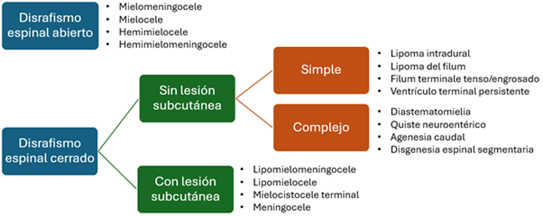
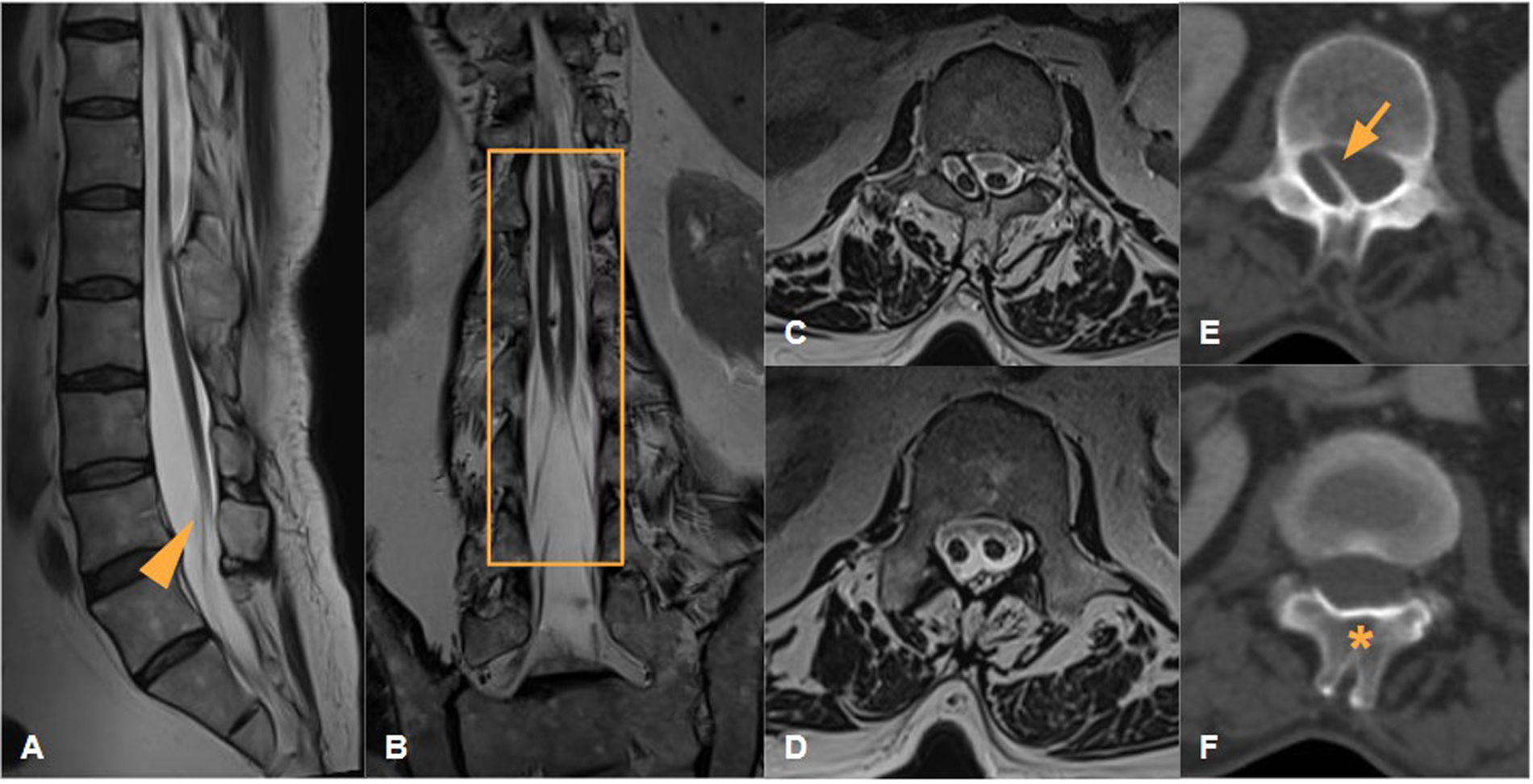
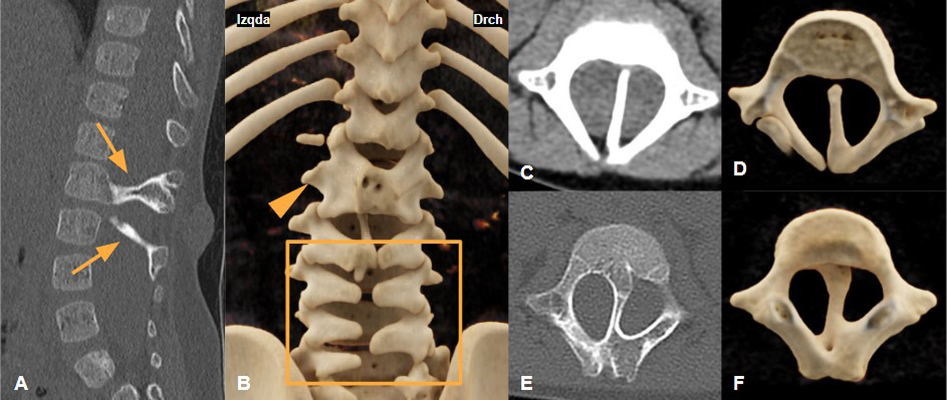
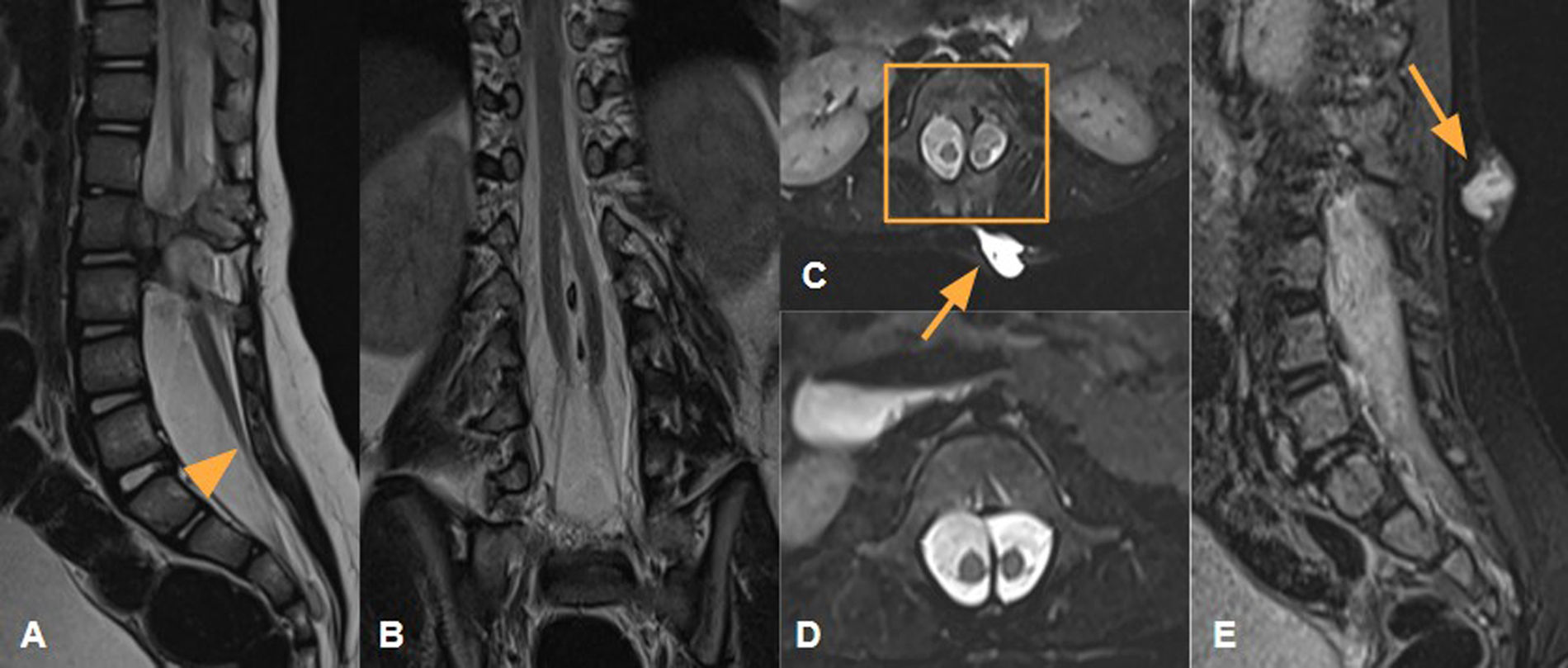
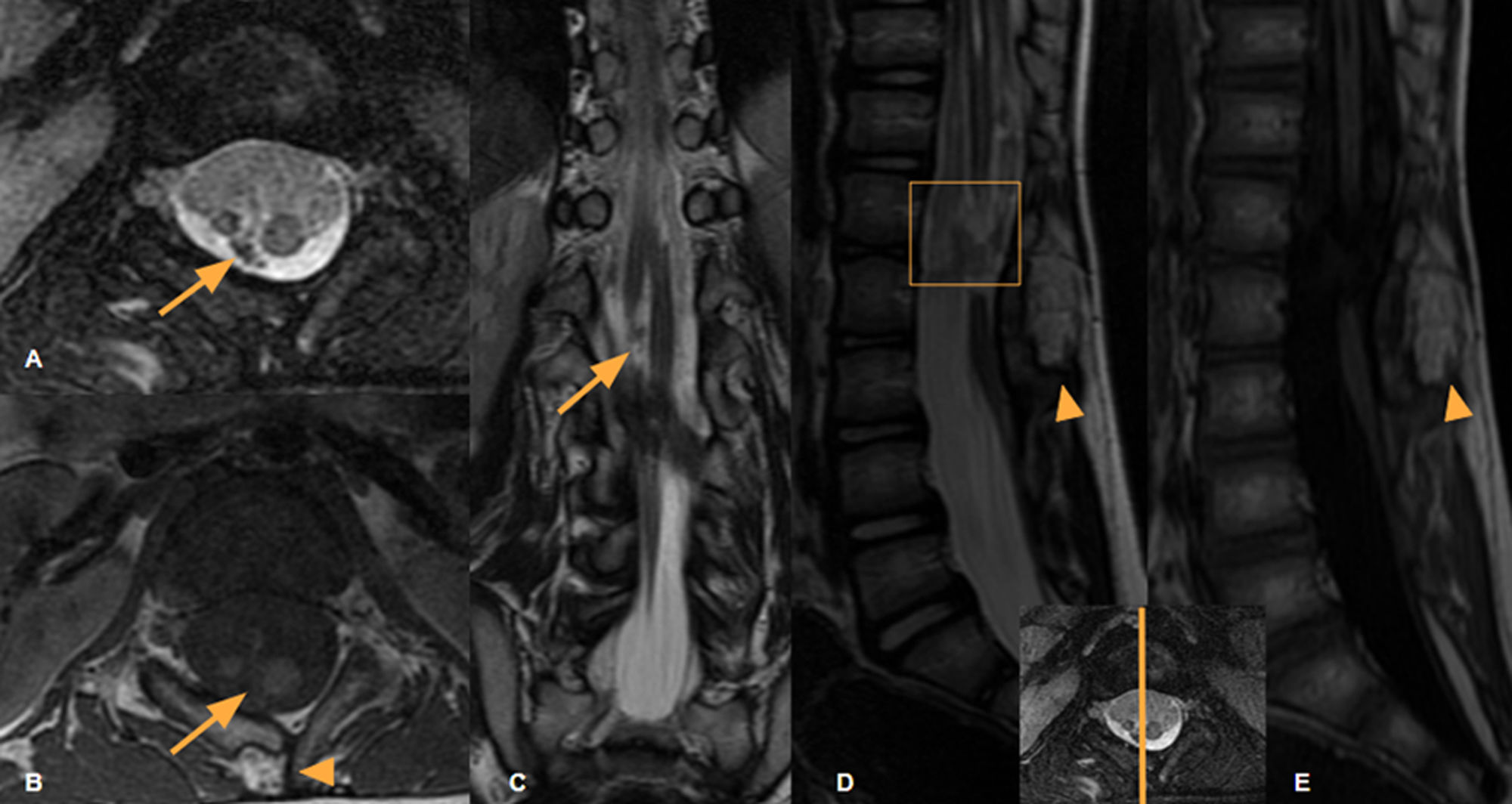
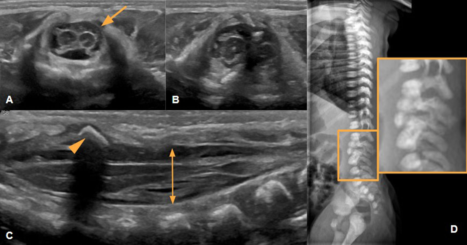
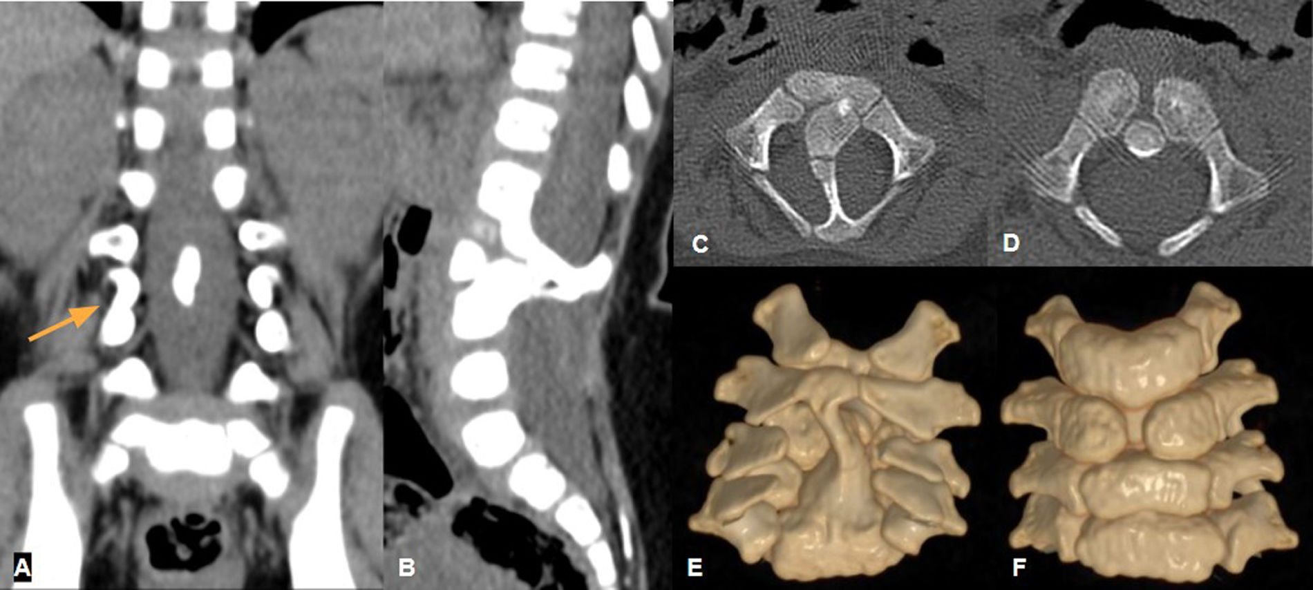
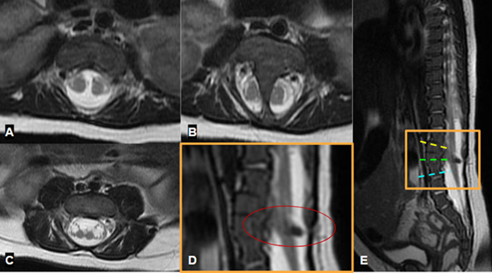
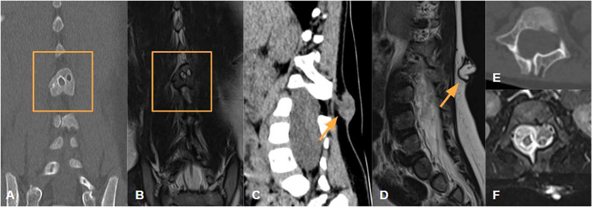
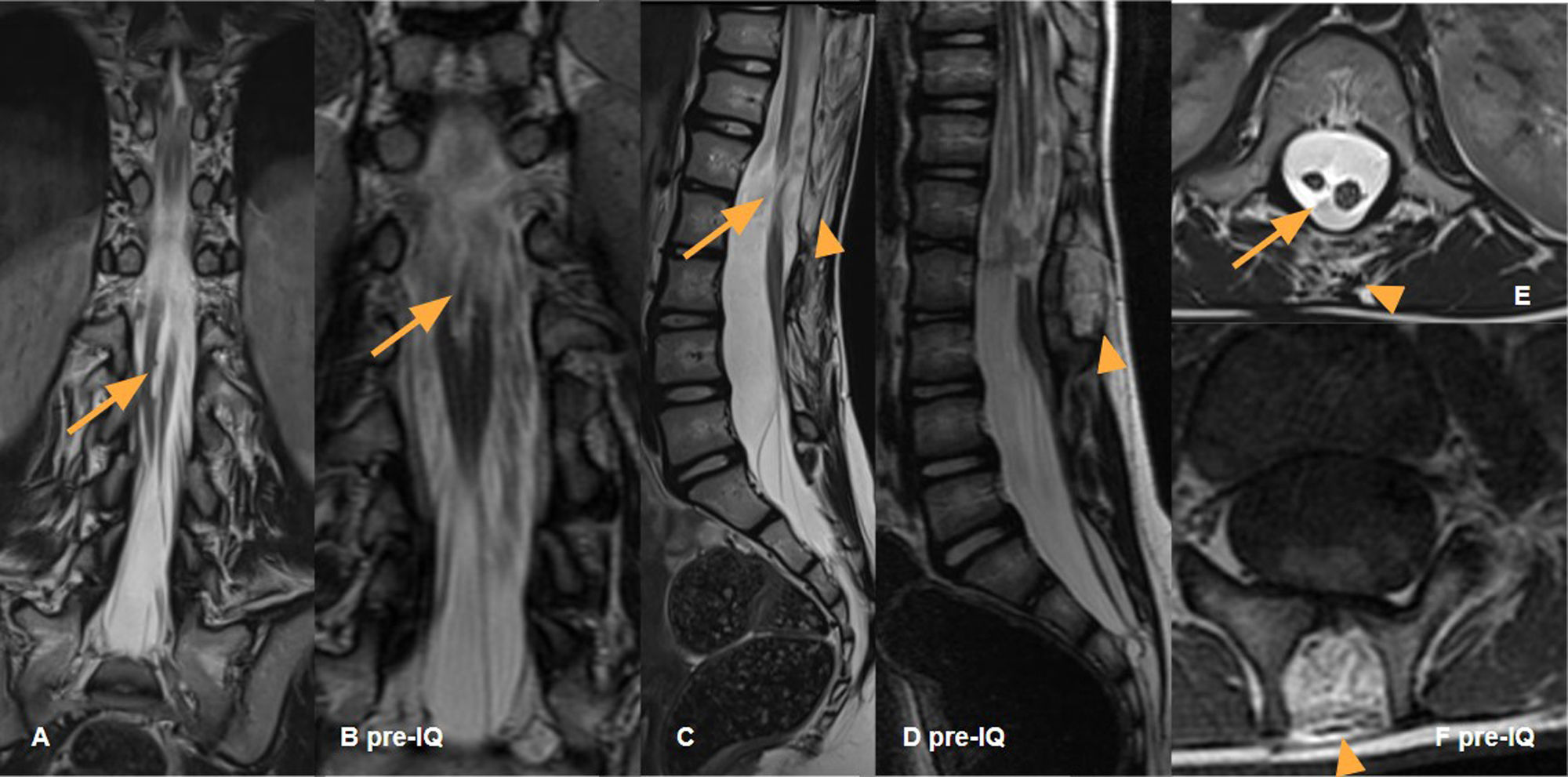
La diastematomielia, una rara anomalía congénita de la columna vertebral, se caracteriza por la división de la médula espinal y a menudo se asocia con otras malformaciones espinales y cutáneas. Su diagnóstico se realiza principalmente mediante ecografía prenatal y resonancia magnética (RM) fetal. La evaluación posnatal se lleva a cabo con tomografía computarizada (TC) y RM, que son esenciales para una evaluación integral y la planificación terapéutica. El pronóstico depende de la presencia de síntomas neurológicos, cuya gravedad puede correlacionarse con hallazgos radiológicos específicos. El tratamiento varía desde la cirugía en casos sintomáticos hasta la observación en pacientes asintomáticos. Este artículo revisa la diastematomielia aportando imágenes ilustrativas de TC y RM posnatal para recalcar la importancia de su evaluación integral para garantizar un diagnóstico preciso que guíe el manejo clínico adecuado.

Palabras clave:
Diastematomielia
Defectos del tubo neural
Disrafismos espinales
Diagnóstico
Radiología
Resonancia magnética
Tomografía computarizada
Abstract

Diastematomyelia, a rare congenital anomaly of the spine, is characterised by the separation of the spinal cord and is often associated with other spinal and cutaneous malformations. Diagnosis is made primarily by prenatal ultrasound and fetal Magnetic Resonance Imaging (MRI). Postnatal evaluation is carried out using Computed Tomography (CT) and MRI, and this is essential for a comprehensive assessment and appropriate treatment planning. Prognosis depends on whether or not neurological symptoms are present, and its severity may correlate with specific radiological findings. Treatment ranges from surgery in symptomatic cases to observation in asymptomatic patients. This article reviews diastematomyelia, providing illustrative images from postnatal CT and MRI to emphasize the importance of carrying out a comprehensive assessment to ensure that the diagnosis is accurate and clinical management is thus appropriately guided.

Keywords:
Diastematomyelia
Neural Tube Defects
Spinal Dysraphism
Diagnosis
Radiology
Magnetic Resonance Imaging
Computed Tomography

Article

These are the options to access the full texts of the publication Radiología
Subscriber
Subscriber

If you already have your login data, please click here .

If you have forgotten your password you can you can recover it by clicking here and selecting the option “I have forgotten my password”
Subscribe
Subscribe to

Radiología

Purchase
Purchase article

Purchasing article the PDF version will be downloaded

Price 19.34 €

Purchase now
Contact
Phone for subscriptions and reporting of errors
From Monday to Friday from 9 a.m. to 6 p.m. (GMT + 1) except for the months of July and August which will be from 9 a.m. to 3 p.m.
Calls from Spain
932 415 960
Calls from outside Spain
+34 932 415 960
E-mail
Article options
es en pt

¿Es usted profesional sanitario apto para prescribir o dispensar medicamentos?

Are you a health professional able to prescribe or dispense drugs?

Você é um profissional de saúde habilitado a prescrever ou dispensar medicamentos

Quizás le interese:
10.1016/j.rx.2024.05.012
No mostrar más