Valorar la calidad de imagen y la dosis de radiación en tomografía computarizada (TC) de peñascos adquiridos con una TC multidetector (TCMD) con filtro de estaño, detectores de alta resolución y reconstrucción iterativa, comparándola con otro equipo sin filtro de estaño y con reconstrucción por retroproyección filtrada.
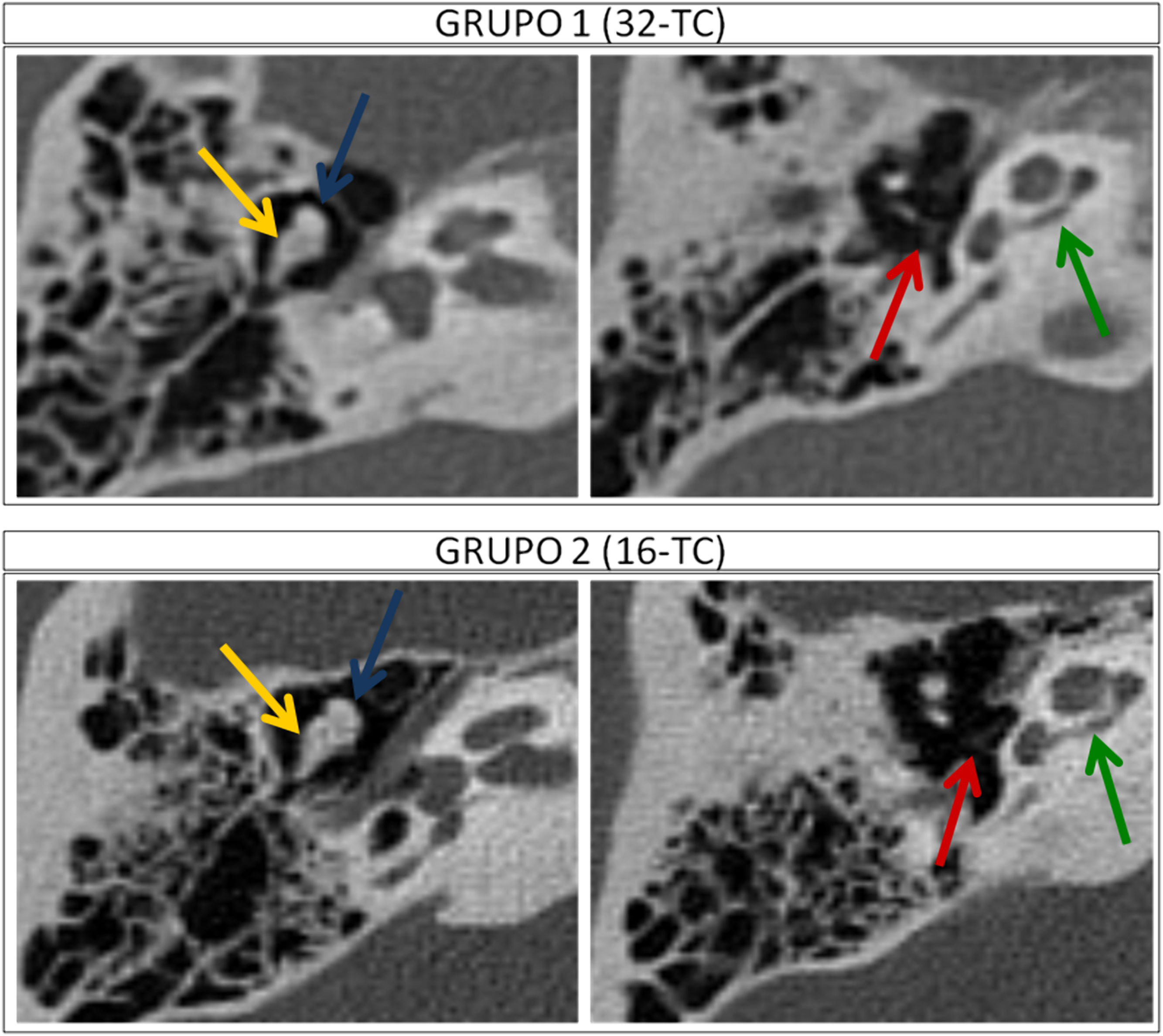
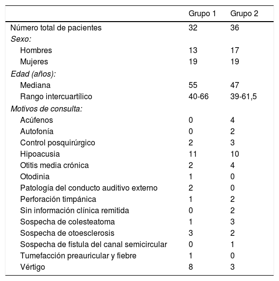
Material y métodosSe incluyeron retrospectivamente 32 pacientes con TC de peñascos, realizadas con dosis ultrabaja en una 32-TCMD (130 kV con filtro de estaño y reconstrucción iterativa). Se compararon con 36 estudios realizados en una 16-TCMD (120 kV y retroproyección filtrada). Se cuantificó la densidad muscular, ósea y el ruido de fondo, y se calculó la relación señal/ruido. Para valorar la calidad de imagen, dos radiólogos evaluaron de forma subjetiva e independiente la visualización de las diferentes estructuras del oído (0=no se visualiza; 3=se identifica y delimita perfectamente). Se calculó el coeficiente de concordancia interobservador kappa. Utilizando un software comercial, se cuantificó a diferentes niveles anatómicos la dosis efectiva con el producto dosis-longitud.
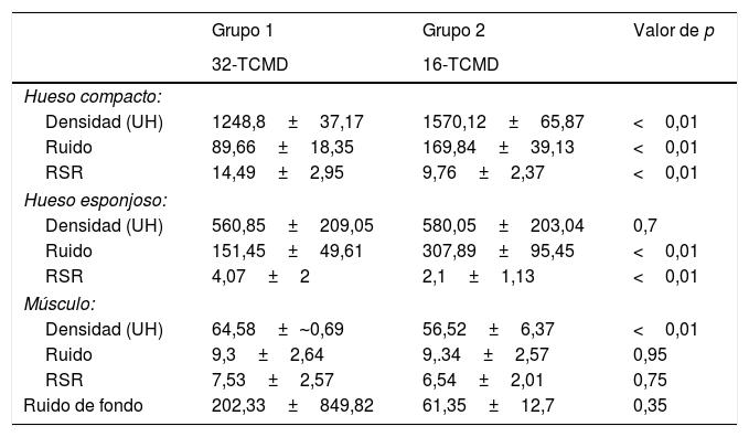
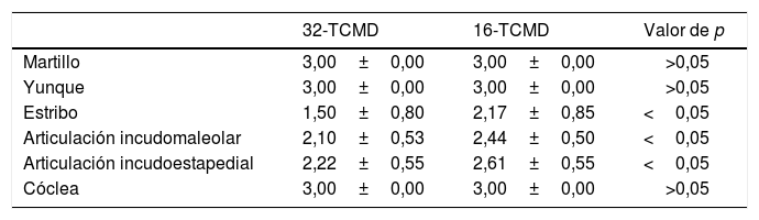
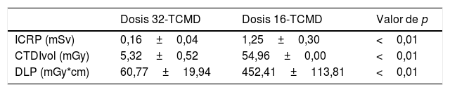
ResultadosEn el análisis cuantitativo de las imágenes no se observaron diferencias significativas en el ruido de fondo. En el análisis cualitativo se obtuvo una puntuación subjetiva similar o ligeramente menor en la delimitación de las diferentes estructuras de la cadena osicular y cóclea en la 32-TCMD, con diferencias estadísticamente significativas. La dosis media efectiva fue de 0,16±0,04 mSv para la 32-TCMD frente a 1,25±0,30 mSv para la 16-TCMD. Conclusiones: La utilización de equipos con filtro de estaño, detectores de alta resolución y reconstrucción iterativa permiten obtener TC con dosis de radiación ultrabaja (0,16±0,04 mSv) con una calidad de imagen adecuada para valorar las estructuras de los peñascos.
To compare image quality and radiation dose in computed tomography (CT) studies of the petrous part of the temporal bone done with a scanner using a tin filter, high-resolution detectors, and iterative reconstruction versus in studies done with another scanner without a tin filter using filtered back projection.
Material and methodsThis retrospective study compared CT studies in 32 patients who underwent ultralow-dose CT of the petrous part of the temporal bone in a 32-detector CT scanner (130 kV with a tin filter and iterative reconstruction) and in 36 patients who underwent the studies in a 16-detector CT scanner (120 kV and filtered back projection). We quantified the densities of muscle and bone tissues and background noise, and we calculated the signal-to-noise ratio. To evaluate image quality, two radiologists working independently subjectively evaluated the visualization of the different structures of the ear on a four-point scale (0=not visible; 3=perfectly identifiable and delimited), and we calculated the coefficient of interobserver concordance (k). Using commercial software, we quantified the effective dose of radiation at different anatomic levels with the dose-length product.
ResultsIn the quantitative analysis, no significant differences were observed in background noise. In the qualitative analysis, the score on the subjective evaluation was similar or slightly lower for the delimitation of the different structures in the ossicular chain and cochlea in the studies done with the 32-detector scanner, with statistically significant differences. The mean effective dose of radiation was 0.16±0.04 mSv for the 32-detector scanner versus 1.25±0.30 mSv for the 16-detector scanner.
ConclusionsUsing scanners with tin filters, high-resolution detectors, and iterative reconstruction makes it possible to obtain images with adequate quality for the evaluation of the structures of the petrous part of the temporal bone with ultralow doses of radiation (0.16±0.04 mSv).