


Madelung’s deformity is a type of wrist abnormality due to premature closure of the distal radius physis. It can be associated with a wide spectrum of pathologies, and 2 main groups have been differentiated, true-Madelung and pseudo-Madelung deformity, the presence of Vickers ligament and/or an anomalous radiotriquetral ligament being features of the former. We present the case of a child with a bilateral Madelung deformity, in the context of a Léri-Weill syndrome.
La deformidad de Madelung es un tipo de anomalía de la muñeca, secundaria a un cierre prematuro de la fisis distal del radio. Puede estar asociada a un amplio espectro de enfermedades, diferenciándose 2 grandes grupos: la verdadera y la pseudodeformidad de Madelung. Lo que caracteriza al primer grupo es la presencia del ligamento de Vickers o de un ligamento radiopiramidal anómalo. Se presenta el caso de un niño con una deformidad de Madelung bilateral, en el contexto de un síndrome de Léri-Weill.
Madelung deformity is a type of wrist anomaly defined as a growth disorder of the anteromedial region of the distal epiphysis of the radius.1 It can be congenital, post-traumatic or associated with various bone dysplasias, such as Ollier’s disease or multiple hereditary exostoses,2–4 and represents less than 2% of hand deformities in pediatrics.3
Women are affected more frequently than men,1 with a 4:1 ratio,2,3 and it does not usually occur until adolescence5 (10–14 years); it can be bilateral in 50%–66% of patients.1
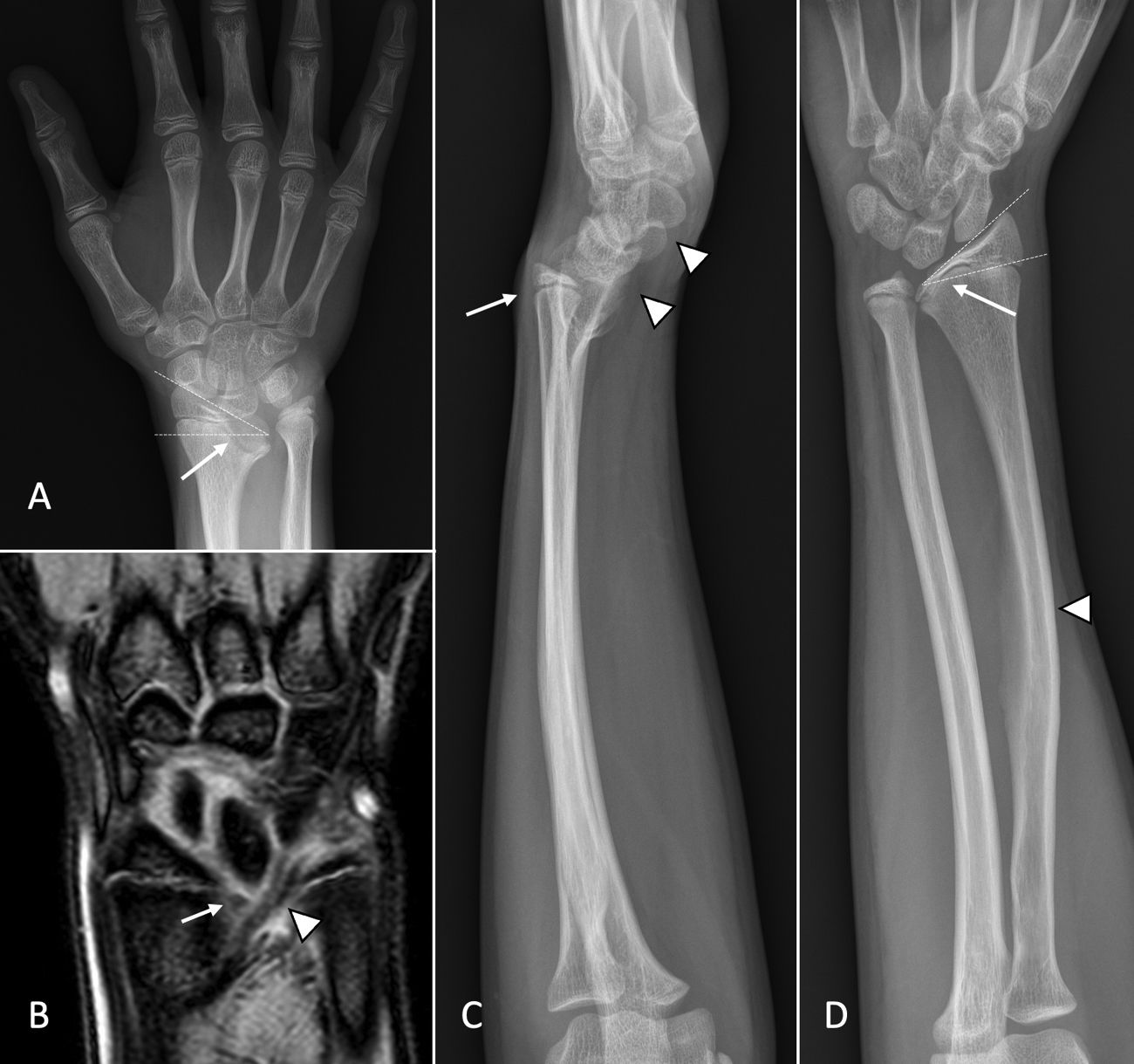
We present the case of a 13-year-old boy with a personal history of mild/moderate mitral insufficiency, under study by endocrinology due to short stature, who attended the emergency room due to pain in the left wrist during flexion and extension, of 2 months of evolution, without previous trauma; for this reason an X-ray was performed (Fig. 1A), in which a morphological alteration of the distal epiphysis of the radius was observed, and given the doubt that a bone lesion existed, it was recommended to perform a scheduled magnetic resonance imaging scan (Fig. 1B).
 and of the right wrist and forearm (D). An increase in the angle of radial tilt of about 36° in the left side and 28° in the right side is observed (dotted lines in A and D), with a proximal migration of the lunate bone that confers a triangular morphology of the carpus (arrows in A and D), in addition to shortening and bowing of the diaphysis of the right radius (arrowhead in D). The lateral radiograph of the left wrist and forearm (C) shows a volar tilt of the radius and a volar displacement of the carpus (arrowheads), as well as a dorsal subluxation of the ulnar head (arrow). Coronal plane magnetic resonance image in gradient echo sequence (B), in which the Vickers ligament (arrow) and the radiopyramidal ligament (arrowhead) are observed.")
Anteroposterior radiograph of the left wrist (A) and of the right wrist and forearm (D). An increase in the angle of radial tilt of about 36° in the left side and 28° in the right side is observed (dotted lines in A and D), with a proximal migration of the lunate bone that confers a triangular morphology of the carpus (arrows in A and D), in addition to shortening and bowing of the diaphysis of the right radius (arrowhead in D). The lateral radiograph of the left wrist and forearm (C) shows a volar tilt of the radius and a volar displacement of the carpus (arrowheads), as well as a dorsal subluxation of the ulnar head (arrow). Coronal plane magnetic resonance image in gradient echo sequence (B), in which the Vickers ligament (arrow) and the radiopyramidal ligament (arrowhead) are observed.
The exploration revealed a “bayonet” deformity of both wrists, more marked on the left, for which the study was completed with anteroposterior and lateral X-rays of both forearms and wrists (Fig. 1C and D), in which a curved radius was visualized, with a shortening of the ulnar aspect of its distal end, an increase in the radial inclination angle, dorsal subluxation of the ulnar head and proximal and volar migration of the lunate bone.
A genetic study of the patient was requested, in which a heterozygous deletion was identified and considered pathogenic, since it included the SHOX gene, as well as its expression regulatory elements, which cause Léri-Weill dyschondrosteosis and idiopathic short stature. Due to the clinical condition of the patient, it was decided to follow conservative treatment with follow-up.
Madelung deformity is especially common in Léri-Weill dyschondrosteosis,2 a type of mesomelic dysplasia secondary to mutations in the SHOX3 gene, which should be suspected when the wrist deformity is bilateral and is accompanied by short stature.
Both in the Madelung deformity and in the pseudo-Madelung type deformities, premature closure of the volar and medial aspects of the distal physis of the radius occurs.2 The key to differentiating them is the presence, in the true forms, of an anomalous hypertrophic volar radioulnar ligament or Vickers ligament,2,4,6 as well as an anomalous and hypertrophic volar radiopyramidal ligament, recently described.2,4
Radiographic criteria for classic Madelung deformity include dorsal subluxation of the ulnar head, volar tilt of the distal radius, increased radial tilt angle of more than 25°, triangular morphology of the carpus, and the presence of the Vickers ligament.6
In the anteroposterior projection, a flame-shaped radiolucent notch can also be visualized in the medial radial metaphysis, where the Vickers ligament originates,2 although it is better assessed in magnetic resonance imaging.
Usually this abnormality occurs asymptomatically, however, in addition to the cosmetic consequences, it can present clinically with pain, loss of grip strength and reduced range of motion.1,2
When it comes to therapeutic management, it is necessary to take into account both age and functionality. When deciding to opt for surgical treatment, it usually includes a corrective osteotomy of the radial deformity combined with a release of the anomalous ligaments.2,3 In this case, a computed tomography with 3D reconstructions can be very useful for surgical planning.3
Early diagnosis of this abnormality is very important to adopt a good therapeutic approach and prevent progression as well as to rule out a possible hereditary origin and provide adequate genetic counseling for future generations.
Although it has been described as defining sign of true Madelung deformity, the presence of the Vickers ligament and the volar radiopyramidal ligament, being the most extensive series the one provided by Hanson et al. with 8 patients,6 only one case like ours has been reported in the literature, by De Leucio et al.,4 in which a Léri-Weill dyschondrosteosis has been genetically confirmed.
Ethical considerationsThe authors have the informed consent of the patients.
Conflict of interestThe authors declare that they have no conflict of interest for the preparation of this article.
