metricas
covid
Buscar en
Revista Colombiana de Reumatología (English Edition)
Toda la web
Inicio Revista Colombiana de Reumatología (English Edition) Primary Sjögren’s syndrome: Autoantibodies and their relationship to clinical...
Journal Information
Visits
7
Original Investigation
Full text access
Available online 1 July 2024
Primary Sjögren’s syndrome: Autoantibodies and their relationship to clinical manifestations and histology of minor salivary glands
Síndrome de Sjögren primario: autoanticuerpos y su relación con las manifestaciones clínicas y la histología de las glándulas salivales menores
Visits
7
Carlos Agudelo-Cardonaa, Julián Naranjo-Millánb,
Corresponding author
juliann.06@hotmail.com

Corresponding author.
, Julio Martínez-Echeverric, Natalia Prieto-Rayod, Nancy Barrerae, Carlos Arteaga-Unigarrof
a Departamento de Reumatología, Facultad de Ciencias de la Salud, Universidad de Antioquia, Medellín, Colombia
b Departamento de Reumatología, Facultad de Ciencias de la Salud, Universidad Icesi, Cali, Colombia
c Departamento de Medicina interna, Escuela de Medicina, Universidad del Rosario – Fundación Cardioinfantil, Bogotá, Colombia
d Departamento de Reumatología, Facultad de Medicina, Universidad Nacional de Colombia, Bogotá, Colombia
e Departamento de Reumatología, Fundación Instituto de Reumatología Fernando Chalem, Bogotá, Colombia
f Departamento de Reumatología, Hospital Universitario Mayor - Mederi, Bogotá, Colombia
This item has received
Article information
Abstract
Full Text
Bibliography
Download PDF
Statistics
Tables (5)
Table 1. Sociodemographic features.
Table 2. Presence and levels of autoantibodies.
Table 3. Comparison of antibody profile between men and women.
Table 4. Antibody profile. Diagnosis before and after 50 years of age.
Table 5. Association between autoantibodies with glandular and extraglandular manifestations.
Show moreShow less
Abstract
Introduction

To evaluate the association between autoantibodies with clinical manifestations (extraglandular and glandular) and histopathological findings of minor salivary gland biopsy in primary Sjögren’s syndrome.
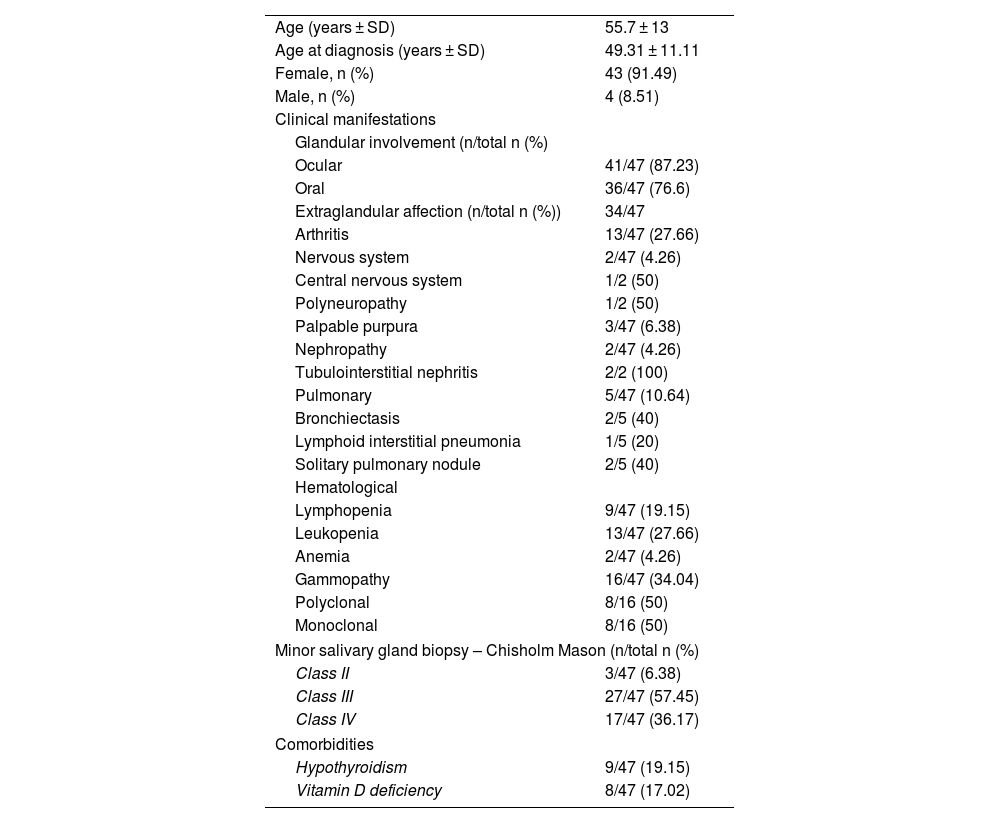
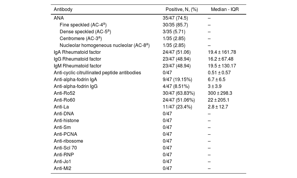
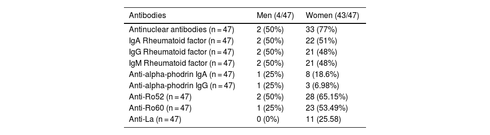
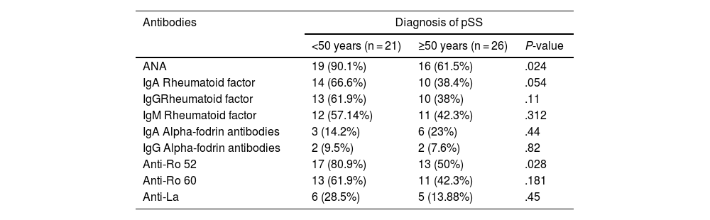
Materials and methods

Observational, descriptive, and cross-sectional study. Forty-seven patients with pSS according to the ACR/EULAR 2016 criteria were included. A face-to-face survey, a review of medical records, and the measurement of autoantibodies (Ab) anti-Ro 52, anti-Ro 60, anti-La, antinuclear antibodies (ANA), rheumatoid factor (RF) IgA, IgG and IgM, and anti-alpha fodrin IgA and IgG were done. Characterization of the population and analysis of the association between clinical characteristics, autoantibodies, and histopathology were performed.
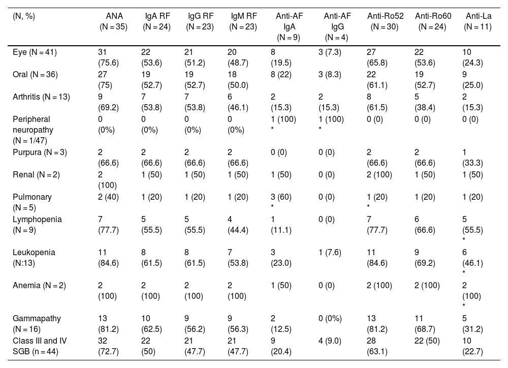
Results

Association of anti-alpha fodrin IgA and anti-Ro 52 Ab was found with pulmonary involvement (P = .014 and P = .031 respectively) and anti-La antibodies with haematological manifestations, specifically leukopenia (P = .011), lymphopenia (P = .023), and anaemia (P = .09). We found no association between the histopathological findings of the minor salivary gland biopsy and extraglandular manifestations.

Conclusions

The activation of B cells, reflected in the increased production of autoantibodies, is related to extraglandular manifestations in pSS, which is observed more frequently in patients with earlier diagnosis.

Keywords:
Sjogren's syndrome
Autoantibodies
Salivary glands
Autoimmunity
Histology
Glandular
Extraglandular
Resumen
Introducción

Evaluar la asociación entre los autoanticuerpos, las manifestaciones clínicas (extraglandulares y glandulares) y los hallazgos histopatológicos de la biopsia de glándula salival menor en el síndrome de Sjögren primario (SSp).

Materiales y métodos

Estudio observacional, descriptivo, de corte transversal. Se incluyeron 47 pacientes con diagnóstico de SSp por criterios ACR/Eular 2016. Se hizo una encuesta presencial, así como una revisión de la historia clínica y la medición de los autoanticuerpos (Ac) anti-Ro 52, anti-Ro 60, anti-La, anticuerpos antinucleares (AAN), factor reumatoide (FR) IgA, IgG e IgM y anti-alfa fodrina IgA e IgG. Se hizo la caracterización de la población y el análisis de la asociación entre las características clínicas, los autoanticuerpos y la histopatología.

Resultados

Se encontró asociación de Ac anti-alfa fodrina IgA y anti-Ro 52 con el compromiso pulmonar (P = ,014 y P = ,031, respectivamente) y de los anticuerpos Anti-La con manifestaciones hematológicas, específicamente leucopenia (P = ,011), linfopenia (P = ,023) y anemia (P = ,09). No se encontró asociación entre los hallazgos histopatológicos de la biopsia de glándula salival menor y las manifestaciones extraglandulares.

Conclusiones

La activación de las células B, que se refleja en una mayor producción de autoanticuerpos, se relaciona con manifestaciones extraglandulares en el SSp, las cuales se observan con mayor frecuencia en pacientes con diagnóstico más temprano.

Palabras clave:
Síndrome de Sjogren
Autoanticuerpos
Glándulas salivales
Autoinmunidad
Histología
Glandular
Extraglandular
Full Text
Introduction

Sjögren syndrome (SS) is a systemic autoimmune disease characterized by lymphocytic infiltration of the exocrine glands. It is associated with circulating antibodies due to B-cell hyperreactivity and is termed primary SS (pSS) when it does not occur alongside other autoimmune diseases.1,2

Prevalence studies of rheumatic diseases in Colombia have estimated a range between 0.08% (95% CI: 0.02%−0.27%)3 and 0.12%,4 like the prevalence described in other Latin American countries such as Brazil (0.17%; 95%CI: 0.02−0.59).5 Primary SS predominantly affects females, with an estimated female-to-male ratio of 4.63:1 in Colombia. Various studies have shown an increased incidence beyond age 50.4,6

Primary SS presents a wide clinical spectrum, ranging from organ-specific involvement, primarily affecting the salivary glands, to multisystem involvement in 30%–40% of patients. Extraglandular manifestations, reflecting systemic disease activity, can occur in various organs and systems, including musculoskeletal, hematological, vascular, pulmonary, renal, cutaneous, and both central and peripheral nervous systems.7,8

Identifying markers to predict extraglandular manifestations in pSS has recently gained interest, as these reflect phenotypes with greater disease severity and higher risk of morbidity and mortality. Different studies have addressed this topic, but results have been controversial due to differences in population, sample size, and classification criteria.1,3,4

This study aimed to assess the association between autoantibodies (Ab), clinical manifestations (both extraglandular and glandular), and histopathological findings of minor salivary gland biopsies in pSS. To our knowledge, this is the first study on this topic in a Latin American population.

Materials and methods

An observational, analytical, cross-sectional study was conducted. Patients over 18 years old with a diagnosis of pSS who attended the Fernando Chalem Institute of Rheumatology and Immunology Foundation in Bogotá Colombia, were included according to the 2016 American College of Rheumatology (ACR)/European League Against Rheumatism (EULAR) Classification Criteria, between June 2018 and June 2019. Minor salivary gland biopsies were performed using the open dissection technique described by Caporali et al.9 According to the standardized criteria of Fisher et al., at least four minor salivary glands, each greater than 2 mm, were obtained.10 The samples were fixed and processed in paraffin, cut into 4um sections, and stained with hematoxylin and eosin. The lymphocytic infiltrates in each section were scored by the pathologists according to the Chisholm-Mason classification; grades 3 and 411 were considered positive for focal lymphocytic sialadenitis.

Subjects with other autoimmune diseases, those participating in research protocols, or those who did not attend for sample collection and completion of the sociodemographic survey were excluded.

Serological samples were taken for antibody processing using indirect immunofluorescence for antinuclear antibodies (ANA) and enzyme-linked immunosorbent assay (ELISA) for IgA, IgG, and IgM rheumatoid factor (RF), anti-citrullinated peptide antibodies, anti-alpha-fodrin IgA and IgG, anti-Ro60, anti-Ro52, anti-La, anti-DNA, anti-histone, anti-Sm, anti-ribosomes, anti-ScL 70, anti-RNP, anti-M2, and anti-Ku. Antibody measurement and minor salivary gland biopsy were performed at different times.

Patient information was obtained through face-to-face surveys and electronic medical records. Sociodemographic data, clinical manifestations, and histopathological results of minor salivary gland biopsies were collected.

Statistical analysis

Univariate analyses to describe the study population were conducted. Categorical variables were assessed using frequencies, while normality tests, primarily the Kolmogorov-Smirnov test, were applied to continuous quantitative variables. Parametric data are presented as means t-standard deviation (SD), whereas non-parametric data are described using medians and interquartile ranges.

Bivariate analyses were carried out to assess the association between the degree of glandular and extraglandular involvement and autoantibodies. Subgroups were analyzed based on sex, mean age at diagnosis, and their association with autoantibodies. Given the higher incidence of pSS in patients over 50 years, a subgroup analysis was performed, comparing the antibody profile in patients diagnosed early (<50 years) with those diagnosed later (≥50 years).

Chi-square or Fisher's exact tests were used for dichotomous independent variables. A P-value <.05 was considered statistically significant. All analyses were performed using Stata/SE 14.0.

Ethical considerations

Written informed consent was obtained from all participants, and the consent forms are in the custody of the corresponding author.

The research complies with current bioethical regulations and was authorized by the ethics committee of the Fernando Chalem Institute of Rheumatology.

ResultsDemographic and clinical features

Of the 88 subjects enrolled in the study, 41 were excluded for the following reasons: not signing the informed consent, not attending the sample collection, completing the sociodemographic survey, or not responding to telephone calls or emails.

The average age was 55.7 years (SD ± 13), and the mean age at diagnosis of pSS was 49.3 years (SD ± 11.11). A total of 43 individuals (91.49%) were female (Tables 1 and 2).

Table 1.

Sociodemographic features.

Age (years ± SD)  55.7 ± 13 
Age at diagnosis (years ± SD)  49.31 ± 11.11 
Female, n (%)  43 (91.49) 
Male, n (%)  4 (8.51) 
Clinical manifestations
Glandular involvement (n/total n (%)
Ocular  41/47 (87.23) 
Oral  36/47 (76.6) 
Extraglandular affection (n/total n (%))  34/47 
Arthritis  13/47 (27.66) 
Nervous system  2/47 (4.26) 
Central nervous system  1/2 (50) 
Polyneuropathy  1/2 (50) 
Palpable purpura  3/47 (6.38) 
Nephropathy  2/47 (4.26) 
Tubulointerstitial nephritis  2/2 (100) 
Pulmonary  5/47 (10.64) 
Bronchiectasis  2/5 (40) 
Lymphoid interstitial pneumonia  1/5 (20) 
Solitary pulmonary nodule  2/5 (40) 
Hematological
Lymphopenia  9/47 (19.15) 
Leukopenia  13/47 (27.66) 
Anemia  2/47 (4.26) 
Gammopathy  16/47 (34.04) 
Polyclonal  8/16 (50) 
Monoclonal  8/16 (50) 
Minor salivary gland biopsy – Chisholm Mason (n/total n (%)
Class II  3/47 (6.38) 
Class III  27/47 (57.45) 
Class IV  17/47 (36.17) 
Comorbidities
Hypothyroidism  9/47 (19.15) 
Vitamin D deficiency  8/47 (17.02) 

SD: standard deviation.

Table 2.

Presence and levels of autoantibodies.

Antibody  Positive, N, (%)  Median - IQR 
ANA  35/47 (74.5)  – 
Fine speckled (AC-4a30/35 (85.7)  – 
Dense speckled (AC-5a3/35 (5.71)  – 
Centromere (AC-3a1/35 (2.85)  – 
Nucleolar homogeneous nucleolar (AC-8a1/35 (2.85)  – 
IgA Rheumatoid factor  24/47 (51.06)  19.4 ± 161.78 
IgG Rheumatoid factor  23/47 (48.94)  16.2 ± 67.48 
IgM Rheumatoid factor  23/47 (48.94)  19.5 ± 130.17 
Anti-cyclic citrullinated peptide antibodies  0/47  0.51 ± 0.57 
Anti-alpha-fodrin IgA  9/47 (19.15%)  6.7 ± 6.5 
Anti-alpha-fodrin IgG  4/47 (8.51%)  3 ± 3.9 
Anti-Ro52  30/47 (63.83%)  300 ± 298.3 
Anti-Ro60  24/47 (51.06%)  22 ± 205.1 
Anti-La  11/47 (23.4%)  2.8 ± 12.7 
Anti-DNA  0/47  – 
Anti-histone  0/47  – 
Anti-Sm  0/47  – 
Anti-PCNA  0/47  – 
Anti-ribosome  0/47  – 
Anti-Scl 70  0/47  – 
Anti-RNP  0/47  – 
Anti-Jo1  0/47  – 
Anti-Mi2  0/47  – 

ANA: Antinuclear antibodies; IQR: Interquartile range.

a

Nomenclature and classification of the International Consensus on ANA Standards (ICAP) in 2021.

The most frequent glandular involvement was ocular (n = 41; 87.23%), followed by oral (n = 36; 76.6%). Extraglandular manifestations occurred in 72% of the individuals, with the most frequent being hematological, mainly polyclonal gammopathy (n = 16; 34%) and leukopenia (n = 13; 27.6%), followed by joint involvement (n = 13; 27.66%). Other systems affected were pulmonary (n = 5; 10.64%), nervous (n = 2; 4.26%), cutaneous (n = 3; 6.38%), and renal (n = 2; 4.26%). In minor salivary gland biopsies, the most common histological subtype was grade 3 in 57.45% of cases.

Regarding comorbidities, 19.15% of the patients had hypothyroidism and 17.02% had vitamin D deficiency (Table 1).

Antibody presence and levels

Among the antibodies tested, the most detected in the population were ANA, with a prevalence of 74%. The predominant ANA pattern observed was fine speckled, accounting for 85.7% of cases (n = 35), followed by dense speckled at 5.71% (n = 3).

The most prevalent subtype of RF was IgA, comprising 51.6% (n = 24) of cases. Similarly, IgA was the most frequent subtype of anti-alpha-fodrin antibodies, detected in 19.15% (n = 9) of individuals. Notably, anti-Ro52 antibodies were found more frequently than anti-Ro60 antibodies, with prevalence rates of 63.83% (n = 30) and 51.06% (n = 24), respectively. Additionally, 11 individuals (23.4%) tested positive for anti-La antibodies. Conversely, tests for anti-cyclic citrullinated peptide, anti-DNA, anti-histone, anti-Sm, anti-PCNA, anti-ribosome, anti-Scl70, anti-RNP, anti-Jo1, and anti-Mi2 antibodies returned negative results (Table 2).

Female patients exhibited a higher seropositivity rate compared to male patients for ANA (77% vs. 50%), IgA RF (51% vs. 50%), anti-Ro52 (65.15% vs. 50%), and anti-La (25.58%) antibodies (refer to Table 3).

Table 3.

Comparison of antibody profile between men and women.

Antibodies  Men (4/47)  Women (43/47) 
Antinuclear antibodies (n = 47)  2 (50%)  33 (77%) 
IgA Rheumatoid factor (n = 47)  2 (50%)  22 (51%) 
IgG Rheumatoid factor (n = 47)  2 (50%)  21 (48%) 
IgM Rheumatoid factor (n = 47)  2 (50%)  21 (48%) 
Anti-alpha-phodrin IgA (n = 47)  1 (25%)  8 (18.6%) 
Anti-alpha-phodrin IgG (n = 47)  1 (25%)  3 (6.98%) 
Anti-Ro52 (n = 47)  2 (50%)  28 (65.15%) 
Anti-Ro60 (n = 47)  1 (25%)  23 (53.49%) 
Anti-La (n = 47)  0 (0%)  11 (25.58) 

A subgroup analysis was conducted among subjects diagnosed with the disease at an early age (under 50 years), and the antibody profiles were compared between both groups (early vs. late). Individuals diagnosed earlier demonstrated a higher positivity rate for all antibodies, except for anti-alpha-fodrin antibodies IgA. Notably, ANA (90.1% vs. 61.5%) and anti-Ro52 antibodies (80.9% vs. 50%) exhibited statistically significant differences when comparing both subgroups (refer to Table 4).

Table 4.

Antibody profile. Diagnosis before and after 50 years of age.

Antibodies  Diagnosis of pSS
  <50 years (n = 21)  ≥50 years (n = 26)  P-value 
ANA  19 (90.1%)  16 (61.5%)  .024 
IgA Rheumatoid factor  14 (66.6%)  10 (38.4%)  .054 
IgGRheumatoid factor  13 (61.9%)  10 (38%)  .11 
IgM Rheumatoid factor  12 (57.14%)  11 (42.3%)  .312 
IgA Alpha-fodrin antibodies  3 (14.2%)  6 (23%)  .44 
IgG Alpha-fodrin antibodies  2 (9.5%)  2 (7.6%)  .82 
Anti-Ro 52  17 (80.9%)  13 (50%)  .028 
Anti-Ro 60  13 (61.9%)  11 (42.3%)  .181 
Anti-La  6 (28.5%)  5 (13.88%)  .45 
Association of autoantibodies with glandular and extraglandular manifestations

Among the various autoantibodies examined, no statistically significant association was observed with glandular manifestations. The antibodies displaying the highest frequency of positivity were ANA, anti-Ro52, and anti-Ro60.

Regarding extraglandular involvement, polyneuropathy showed a significant association with anti-alpha-fodrin antibodies IgA (P = .0378) and IgG (P = .01), but it was identified in only one subject. Lung involvement exhibited a significant association with anti-alpha-fodrin IgA (P = .014) and anti-Ro52 (P = .031) antibodies. Hematological manifestations such as lymphopenia, leukopenia, and anemia were significantly associated with anti-La antibodies (P < .05). Grade III or IV biopsy results (44 out of 47 individuals) were considered positive according to the classification criteria, but no substantial association was found with the presence of autoantibodies when compared with class II results. Other extraglandular manifestations did not show any significant association with the antibodies studied (Table 5).

Table 5.

Association between autoantibodies with glandular and extraglandular manifestations.

(N, %)  ANA (N = 35)  IgA RF (N = 24)  IgG RF (N = 23)  IgM RF (N = 23)  Anti-AF IgA (N = 9)  Anti-AF IgG (N = 4)  Anti-Ro52 (N = 30)  Anti-Ro60 (N = 24)  Anti-La (N = 11) 
Eye (N = 41)  31 (75.6)  22 (53.6)  21 (51.2)  20 (48.7)  8 (19.5)  3 (7.3)  27 (65.8)  22 (53.6)  10 (24.3) 
Oral (N = 36)  27 (75)  19 (52.7)  19 (52.7)  18 (50.0)  8 (22)  3 (8.3)  22 (61.1)  19 (52.7)  9 (25.0) 
Arthritis (N = 13)  9 (69.2)  7 (53.8)  7 (53.8)  6 (46.1)  2 (15.3)  2 (15.3)  8 (61.5)  5 (38.4)  2 (15.3) 
Peripheral neuropathy (N = 1/47)  0 (0%)  0 (0%)  0 (0%)  0 (0%)  1 (100) *  1 (100) *  0 (0)  0 (0)  0 (0) 
Purpura (N = 3)  2 (66.6)  2 (66.6)  2 (66.6)  2 (66.6)  0 (0)  0 (0)  2 (66.6)  2 (66.6)  1 (33.3) 
Renal (N = 2)  2 (100)  1 (50)  1 (50)  1 (50)  1 (50)  0 (0)  2 (100)  1 (50)  1 (50) 
Pulmonary (N = 5)  2 (40)  1 (20)  1 (20)  1 (20)  3 (60) *  0 (0)  1 (20) *  1 (20)  1 (20) 
Lymphopenia (N = 9)  7 (77.7)  5 (55.5)  5 (55.5)  4 (44.4)  1 (11.1)  0 (0)  7 (77.7)  6 (66.6)  5 (55.5) * 
Leukopenia (N:13)  11 (84.6)  8 (61.5)  8 (61.5)  7 (53.8)  3 (23.0)  1 (7.6)  11 (84.6)  9 (69.2)  6 (46.1) * 
Anemia (N = 2)  2 (100)  2 (100)  2 (100)  2 (100)  1 (50)  0 (0)  2 (100)  2 (100)  2 (100) * 
Gammapathy (N = 16)  13 (81.2)  10 (62.5)  9 (56.2)  9 (56.3)  2 (12.5)  0 (0%)  13 (81.2)  11 (68.7)  5 (31.2) 
Class III and IV SGB (n = 44)  32 (72.7)  22 (50)  21 (47.7)  21 (47.7)  9 (20.4)  4 (9.0)  28 (63.1)  22 (50)  10 (22.7) 

ANA: antinuclear antibodies, RF: rheumatoid factor, anti-AF: alpha-fodrin antibodies SGB: salivary gland biopsy.

*

P-value <.05.

Discussion

pSS is a complex systemic autoimmune disease, with a prevalence between 0.01%−0.6% and an incidence between 1.1–5.3 cases per 100,000 inhabitants.12 Disease features vary according to sex (more common in women) and ethnicity (greater involvement in the Caucasian population, but earlier diagnosis in Blacks).13 Mortality is higher in men, related to extraglandular involvement.14–16

The present study characterized the population and evaluated the association between clinical manifestations and the presence of autoantibodies in pSS. The average age at the time of diagnosis was 49.3 years and the female: male ratio was 11:1. In the study by Maciel et al., the average age was 64.6 years, and the average duration of the disease was 10 years,17 with 86% female. In postmenopausal women, there is a rapid decrease in estrogen levels that leads to a reduction in the viability of glandular cells, cell death, release of autoantigens, formation of autoantibodies, and organ damage.18,19

Concerning comorbidities, 19.15% of the subjects had hypothyroidism and 17.02% vitamin D deficiency. In our cohort, the study of the etiology of hypothyroidism was not conducted; therefore, an autoimmune origin cannot be ruled out. The classification of secondary SS was initially considered in individuals with sicca symptoms and rheumatoid arthritis but was later expanded to include other systemic autoimmune entities such as systemic lupus erythematosus and systemic sclerosis. It is not clear whether it should be classified as secondary SS when overlap occurs with organ-specific autoimmune diseases such as autoimmune thyroiditis.20 The study by Dai et al. assessed the presence of antithyroid antibodies in patients with SS and found a positivity of 18.9% and 25.6% for antiperoxidase and antithyroglobulin antibodies, respectively. Subjects with hypothyroidism more frequently presented autoantibodies, lower hemoglobin levels, increased acute-phase reactants and decreased C4. Therefore, we consider it important for future studies to assess the etiology of hypothyroidism and determine the value of including these individuals in the secondary SS group, due to the differences in the clinical course of the disease.21

Extraglandular manifestations occur in approximately one-third of patients; almost any organ or system can be affected; the most frequent were musculoskeletal, gastrointestinal, pulmonary, and hematological.22 Pulmonary, hematological, and neuropathic affection have been related to increased mortality and decreased quality of life.23–25 The study by Zhao et al. reported a higher prevalence (65.3%) in people with more than one comorbidity and greater disease activity measured by ESSDAI.26

Hematological manifestations, which include anemia, cytopenias, gammopathies, and lymphoproliferative disorders, have been reported in 25%–50% of pSS, with chronic anemia being the most common, followed by leukopenia.27 Monoclonal or polyclonal gammopathy has been reported in between 22%–49% of pSS, and it is considered a direct marker of B-lymphocyte activation; its presence is a risk predictor for hematological malignancies.28,29

In the current study, the most common hematological manifestation was gammopathy, which was found in 34% of the patients, of whom 50% had a monoclonal pattern, while leukopenia was the second most frequent extraglandular manifestation, with a frequency of 27.6%. For its part, anemia was the third most frequent hematological manifestation, occurring in 4.26% of the subjects. Although in other cohorts the presence of ANA, anti-Ro, and anti-La has been related to a higher risk of anemia,30 this association was not found in our cohort.

Pulmonary affection related to pSS has been described between 11%–29% of patients31; when actively searching this compromise with chest tomography, the prevalence can increase up to 70%.32 Interstitial involvement has a frequency of 20%, while non-specific interstitial pneumonia is the most found radiological pattern.23,33 In the current study, pulmonary manifestations occurred in 10%, and the most recurrent were solitary pulmonary nodules, bronchiectasis, and lymphoid interstitial pneumonia. These results were like those obtained in the cohorts reported by Lohrman et al. and Gardiner.34,35

Nervous system manifestations have been described in between 20%–25% of individuals with pSS, with peripheral polyneuropathy (especially sensory polyneuropathy) being the most frequent.36 In the studied cohort, the involvement of the nervous system was less than reported in other studies (4.26% of cases); while 50% had peripheral neuropathy, another 50% experienced central nervous system involvement.

The occurrence of other extraglandular manifestations was noted in less than 10% of patients. Renal involvement, specifically interstitial nephropathy, was identified in 4% of cases, while skin manifestations were present in 6%, consistent with findings from other cohorts.37

ANA were the most prevalent antibodies in our population, detected in 77% of cases. In comparison, other studies on Latin American cohorts have shown an average positivity of 74%, notably lower than that observed in other populations. The predominant patterns of ANA included fine speckled, dense speckled, centromere, and homogeneous nucleolar (Table 2).13,38

RF tested positive in 50% of subjects, with a similar frequency observed between men and women. This contrasts with findings from other populations, where higher seropositivity is often reported in men.39 While previous studies have suggested an association of IgA RF isotype with xerostomia, unstimulated salivary flow rate, and ANA,40,41 no such associations were observed in our study regarding glandular and extraglandular manifestations.

Positive IgA and IgG anti-fodrin antibodies were identified in 19.15% and 8.51% of cases, respectively. These antibodies have been linked to diagnostic markers with low sensitivity but high specificity42 and have been associated with an increased risk of parotitis.43,44

Our study revealed associations between IgA (P = .0378) and IgG (P = .01) alpha-fodrin antibodies with peripheral nervous system involvement (neuropathy), and alpha-fodrin IgA antibodies with lung involvement (P = .014). However, due to the limited number of cases, definitive conclusions regarding increased risk cannot be drawn.

Anti-Ro52 autoantibodies (positive in 63.8% of cases) were associated with pulmonary involvement (P = .031), while anti-La antibodies (positive in 23.4% of cases) were linked to hematological manifestations including leukopenia (P = .011), lymphopenia (P = .023), and anemia (P = .09).

In a study by Menor Almagro et al., associations were noted between anti-Ro52, anti-Ro60, and anti-La antibodies with hypergammaglobulinemia, hypocomplementemia, and alopecia.45 However, other associations reported in previous studies, such as those between anti-Ro60 and polyneuropathy, lymphadenopathy, renal involvement, and myopathy, were not observed in our study.41,46,47

Individuals diagnosed with pSS before the age of 50 exhibited a higher prevalence of ANA, IgA RF, and anti-Ro52 antibodies. Similar findings have been reported in other cohorts, suggesting that early onset of symptoms (<35 years) is associated with increased positivity of ANA, anti-Ro, anti-La antibodies, and RF.39,40

Limitations

To provide more robust confirmation of our findings, larger sample sizes are warranted given the relatively small population size in our study. Additionally, we did not utilize the focus score system recommended by the 2016 ACR/EULAR 2016 Classification Criteria for pSS. Future investigations should consider implementing this system to evaluate its correlation with both glandular and extraglandular manifestations.

The prevalence of hypothyroidism was notable; however, etiological studies were not conducted to ascertain whether it had an autoimmune origin. Consequently, the classification of these subjects as either pSS or secondary Sjögren syndrome remains uncertain, and further research is needed to clarify their clinical significance.

Conclusions

Our findings suggest that distinct autoantibody profiles are associated with specific extraglandular manifestations in primary Sjögren syndrome. Notably, anti-La antibodies may be linked to hematological manifestations, while pulmonary manifestations appear to be correlated with anti-alpha-fodrin and anti-Ro52 antibodies. Moreover, individuals diagnosed earlier tend to exhibit a higher burden of autoantibodies. Surprisingly, we did not find an association between minor salivary gland biopsy results and extraglandular manifestations in primary Sjögren syndrome.

Although our study represents the largest cohort of primary Sjögren syndrome with extensive autoantibody profiling in Colombia, its single-center nature necessitates validation through multicenter studies to ensure broader population representation. Further investigations are imperative to elucidate the specific roles of each autoantibody in extraglandular manifestations.

Conflict of interests

Autoantibody kits were generously provided by the AESKU GROUP laboratory, and the Fernando Chalem Institute of Rheumatology facilitated sample collection and processing through financial support.

Acknowledgments

We extend our gratitude to the AESKU Group laboratory in Colombia for their donation of autoantibody kits, and to the Fernando Chalem Institute of Rheumatology and Immunology Foundation laboratory in Bogotá, Colombia, for their assistance in sample collection and processing.

References
[1]
P. Brito-Zerón, C. Baldini, H. Bootsma, S.J. Bowman, R. Jonsson, X. Mariette, et al.
Sjögren syndrome.
Nat Rev Dis Primers., 2 (2016), pp. 16047
[2]
C.H. Shiboski, S.C. Shiboski, R. Seror, L.A. Criswell, M. Labetoulle, T.M. Lietman, et al.
International Sjögren's Syndrome Criteria Working Group. 2016 American College of Rheumatology/European League Against Rheumatism Classification criteria for primary Sjögren’s syndrome: a consensus and data-driven methodology involving three international patient cohorts.
Arthritis Rheumatol., 69 (2017), pp. 35-44
[3]
J. Londoño, I. Peláez Ballestas, F. Cuervo, I. Angarita, R. Giraldo, J.C. Rueda, et al.
Prevalencia de la enfermedad reumática en Colombia, según estrategia COPCORD-Asociación Colombiana de Reumatología. Estudio de prevalencia de enfermedad reumática en población colombiana mayor de 18 años.
Rev Colomb Reumatol., 25 (2018), pp. 245-246
[4]
D.G. Fernández-Ávila, D.N. Rincón-Riaño, S. Bernal-Macías, J.M. Gutiérrez Dávila, D. Rosselli.
Prevalence and demographic characteristics of Sjögren’s syndrome in Colombia, based on information from the Official Ministry of Health Registry/Prevalencia y características demográficas del síndrome de Sjögren en Colombia, según información del Sistema Integral de Información de la Protección Social.
Reumatol Clin., 16 (2020), pp. 286-289
[5]
V. Valim, E. Zandonade, A.M. Pereira, O.H. de Brito Filho, E.V. Serrano, C. Musso, et al.
Primary Sjögren’s syndrome prevalence in a major metropolitan area in Brazil.
Rev Bras Reumatol., 53 (2013), pp. 24-34
[6]
X. Mariette, L.A. Criswell.
Primary Sjögren’s syndrome.
N Engl J Med., 378 (2018), pp. 931-939
[7]
R.I. Fox.
Sjögren’s syndrome.
Lancet., 366 (2005), pp. 321-331
[8]
R.I. Fox.
Extraglandular manifestations of Sjögren’s syndrome (SS): dermatologic, arthritic, endocrine, pulmonary, cardiovascular, gastroenterology, renal, urology, and gynecologic manifestations.
Sjögren’s Syndrome., (2011), pp. 285-316
[9]
R. Caporali, E. Bonacci, O. Epis, F. Bobbio-Pallavicini, P. Morbini, C. Montecucco.
Safety and usefulness of minor salivary gland biopsy: retrospective analysis of 502 procedures performed at a single center.
Arthritis Rheum., 59 (2008), pp. 714-720
[10]
B.A. Fisher, R. Jonsson, T. Daniels, M. Bombardieri, R.M. Brown, P. Morgan, et al.
Sjögren’s Histopathology Workshop Group (Appendix) from ESSENTIAL (EULAR Sjögren’s Syndrome Study Group). Standardisation of labial salivary gland histopathology in clinical trials in primary Sjögren’s syndrome.
Ann Rheum Dis., 76 (2017), pp. 1161-1168
[11]
R. Parra-Medina, J.F. Polo, A. Rojas-Villarraga.
Interpretación de la biopsia de glándula salival menor en el síndrome de Sjögren. Correlación clínico-patológica.
Rev Colomb Reumatol., 27 (2020), pp. 82-89
[12]
M. Kvarnström, V. Ottosson, B. Nordmark, M. Wahren-Herlenius.
Incident cases of primary Sjögren’s syndrome during a 5-year period in Stockholm County: a descriptive study of the patients and their characteristics.
Scand J Rheumatol., 44 (2015), pp. 135-142
[13]
P. Brito-Zerón, N. Acar-Denizli, M. Zeher, A. Rasmussen, R. Seror, E. Theander, et al.
EULAR-SS Task Force Big Data Consortium. Influence of geolocation and ethnicity on the phenotypic expression of primary Sjögren’s syndrome at diagnosis in 8310 patients: a cross-sectional study from the Big Data Sjögren Project Consortium.
Ann Rheum Dis., 76 (2017), pp. 1042-1050
[14]
M.Y. Weng, Y.T. Huang, M.F. Liu, T.H. Lu.
Incidence and mortality of treated primary Sjogren’s syndrome in Taiwan: a population-based study.
J Rheumatol., (2011), pp. 38706-38708
[15]
M. Ramos-Casals, P. Brito-Zerón, B. Kostov, A. Sisó-Almirall, X. Bosch, D. Buss, et al.
Google-driven search for big data in autoimmune geoepidemiology: analysis of 394,827 patients with systemic autoimmune diseases.
Autoimmun Rev., 14 (2015), pp. 670-679
[16]
L. Jorkjend.
Prevalence of extraglandular manifestations in patients with primary Sjögren’s syndrome in Southern Norway: a comparison to the literature.
Arch Rheumatol., 30 (2015), pp. 263-270
[17]
G. Maciel, C.S. Crowson, E.L. Matteson, D. Cornec.
Prevalence of Primary Sjögren’s syndrome in a US population-based cohort.
Arthritis Care Res (Hoboken)., 69 (2017), pp. 1612-1616
[18]
J.E. Brandt, R. Priori, G. Valesini, D. Fairweather.
Sex differences in Sjögren’s syndrome: a comprehensive review of immune mechanisms.
Biol Sex Differ., 6 (2015), pp. 19
[19]
M.N. Manoussakis, E.K. Kapsogeorgou.
The role of intrinsic epithelial activation in the pathogenesis of Sjögren’s syndrome.
J Autoimmun., 35 (2010), pp. 219-224
[20]
C.P. Mavragani, H.M. Moutsopoulos.
Primary versus secondary Sjögren syndrome: is it time to reconsider these terms?.
J Rheumatol., 46 (2019), pp. 665-666
[21]
M. Dai, J. Wang, Q. Huang.
Clinical features and laboratory examination results of Sjogren’s syndrome complicated with thyroid disorders: a retrospective analysis.
J Healthc Eng., 2021 (2021),
[22]
F.B. Vivino.
Sjogren’s syndrome: clinical aspects.
Clin Immunol., 182 (2017), pp. 48-54
[23]
J. Qian, C. He, Y. Li, L. Peng, Y. Yang, D. Xu, et al.
Ten-year survival analysis of patients with primary Sjögren’s syndrome in China: a national prospective cohort study.
Ther Adv Musculoskelet Dis., 13 (2021),
[24]
R.A. Peredo, S. Beegle.
Sjogren’s syndrome and pulmonary disease.
Adv Exp Med Biol., 1303 (2021), pp. 193-207
[25]
A.S. Lee, R.H. Scofield, K.M. Hammitt, N. Gupta, D.E. Thomas, T. Moua, et al.
Consensus Expert Panel (CEP) Members. Consensus guidelines for evaluation and management of pulmonary disease in Sjögren’s.
Chest., 159 (2021), pp. 683-698
[26]
Y. Zhao, Y. Li, L. Wang, X.F. Li, C.B. Huang, G.C. Wang, et al.
Primary Sjögren syndrome in Han Chinese: clinical and immunological characteristics of 483 patients.
Medicine (Baltimore)., 94 (2015), pp. e667
[27]
P. Manganelli, P. Fietta, F. Quaini.
Hematologic manifestations of primary Sjögren’s syndrome.
Clin Exp Rheumatol., 24 (2006), pp. 438-448
[28]
A.L. Tomi, R. Belkhir, G. Nocturne, F. Desmoulins, E. Berge, S. Pavy, et al.
Brief report: monoclonal gammopathy and risk of lymphoma and multiple myeloma in patients with primary Sjögren’s syndrome.
Arthritis Rheumatol., 68 (2016), pp. 1245-1250
[29]
I. Radman.
Hematoloske promjene u upalnim reumatskim bolestima [Hematological abnormalities in rheumatic diseases].
Reumatizam., 53 (2006), pp. 11-17
[30]
H. Bernard, C. Rea.
Neonatal lupus presenting as a non-specific rash in primary care.
[31]
E.J. Ter Borg, A.P. Risselada, J.C. Kelder.
Relation of systemic autoantibodies to the number of extraglandular manifestations in primary Sjögren’s Syndrome: a retrospective analysis of 65 patients in the Netherlands.
Semin Arthritis Rheum., 40 (2011), pp. 547-551
[32]
S. Gupta, M.A. Ferrada, S.A. Hasni.
Pulmonary manifestations of primary Sjögren’s syndrome: underlying immunological mechanisms, clinical presentation, and management.
Front Immunol., 10 (2019), pp. 1327
[33]
F. Luppi, M. Sebastiani, M. Silva, N. Sverzellati, A. Cavazza, C. Salvarani, et al.
Interstitial lung disease in Sjögren’s syndrome: a clinical review.
Clin Exp Rheumatol., 38 (2020), pp. 291-300
[34]
C. Lohrmann, M. Uhl, K. Warnatz, N. Ghanem, E. Kotter, O. Schaefer, et al.
High-resolution CT imaging of the lung for patients with primary Sjogren’s syndrome.
Eur J Radiol., 52 (2004), pp. 137-143
[35]
P. Gardiner.
Primary Sjögren’s syndrome.
Baillieres Clin Rheumatol., 7 (1993), pp. 59-77
[36]
A.L. Fauchais, L. Magy, E. Vidal.
Central and peripheral neurological complications of primary Sjögren’s syndrome.
Presse Med., 41 (2012), pp. e485-93
[37]
T. Seeliger, N.K. Prenzler, S. Gingele, B. Seeliger, S. Körner, T. Thiele, et al.
Neuro-Sjögren: peripheral neuropathy with limb weakness in Sjögren’s syndrome.
Front Immunol., 10 (2019), pp. 1600
[38]
E.K.L. Chan, C.A. von Mühlen, M.J. Fritzler, J. Damoiseaux, M. Infantino, W. Klotz, et al.
ICAP Committee. The International Consensus on ANA Patterns (ICAP) in 2021-The 6th Workshop and Current Perspectives.
J Appl Lab Med., 7 (2022), pp. 322-330
[39]
M. Ramos-Casals, R. Solans, J. Rosas, M.T. Camps, A. Gil, J. Del Pino-Montes, GEMESS Study Group.
Primary Sjögren syndrome in Spain: clinical and immunologic expression in 1010 patients.
Medicine (Baltimore)., 87 (2008), pp. 210-219
[40]
K.A. Lee, K.W. Kim, B.M. Kim, J.Y. Won, H.A. Kim, H.W. Moon, et al.
Clinical and diagnostic significance of serum immunoglobulin A rheumatoid factor in primary Sjogren’s syndrome.
Clin Oral Investig., 23 (2019), pp. 1415-1423
[41]
V. Durigan, A. Secco, V. Duarte, C. Troitiño, A. Catalan Pellet, M. Mamani, et al.
Manifestaciones cutáneas extraglandulares en pacientes con síndrome de Sjögren primario.
Rev Colomb Reumatol., 25 (2018), pp. 79-84
[42]
Q. Hu, D. Wang, W. Chen.
The accuracy of the anti-α-fodrin antibody test for diagnosis of Sjögren’s syndrome: a meta-analysis.
Clin Biochem., 46 (2013), pp. 1372-1376
[43]
N.H. Trier, I.Ø. Nielsen, T. Friis, G. Houen, E. Theander.
Comparison of antibody assays for detection of autoantibodies to Ro 52, Ro 60 and La associated with primary Sjögren’s syndrome.
J Immunol Methods., 433 (2016), pp. 44-50
[44]
V. Goëb, V. Salle, P. Duhaut, F. Jouen, A. Smail, J.P. Ducroix, et al.
Clinical significance of autoantibodies recognizing Sjögren’s syndrome A (SSA), SSB, calpastatin and alpha-fodrin in primary Sjögren’s syndrome.
Clin Exp Immunol., 148 (2007), pp. 281-287
[45]
R. Menor Almagro, A. Jurado Roger, F.J. Rodríguez Gutiérrez, R. Solís Díaz, M.H. Cardiel, J.J. Salaberri Maestrojuan.
Asociación de los anticuerpos anti-Ro52, anti-Ro60 y anti-La con las características diagnósticas, clínicas y de laboratorio en un hospital de referencia en Jerez, España.
Reumatol Clin., 12 (2016), pp. 256-262
[46]
G. Urbanski, A. Gury, P. Jeannin, A. Chevailler, P. Lozac’h, P. Reynier, et al.
Discordant Predictions of extraglandular involvement in primary Sjögren’s Syndrome according to the Anti-SSA/Ro60 antibodies detection Assay in a cohort study.
J Clin Med., 11 (2022), pp. 242
[47]
F. Franceschini, I. Cavazzana.
Anti-Ro/SSA and La/SSB antibodies.
Autoimmunity., 38 (2005), pp. 55-63
Copyright © 2023. Asociación Colombiana de Reumatología
Article options
Quizás le interese:
10.1016/j.rcreue.2020.12.006
No mostrar más