To assess the metabolic behavior of mesenteric panniculitis (MP), possible manifestation patterns in 18F-FDG PET/CT imaging and to discover if it is a reliable diagnostic method to differentiate tumor disease from inflammatory condition in this context.
Materials and methodsA total of 2666 PET/CT scans were evaluated prospectively from April 2012 to August 2013. Thirty patients were included (37 scans) with radiological signs of MP. There were 8 women and 22 men, aged between 39 and 81 years, in the sample. According to the 18F-FDG uptake in the mesenteric lesions, expressed as SUVmax, patients were classified into two different groups: Group A consisted of 10 patients with increased uptake, SUVmax ≥2 or greater than the activity found in the surrounding healthy mesenteric tissue, and Group B (20 patients), SUVmax <2 or indistinguishable from healthy tissue.
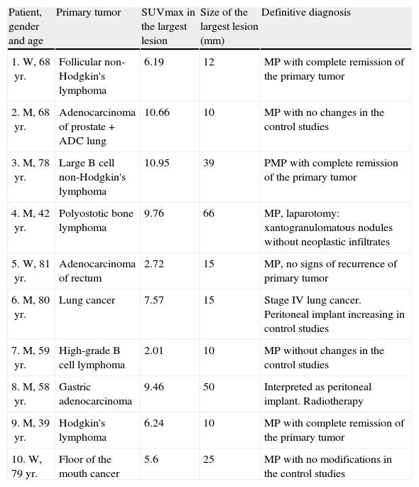
ResultsNo signs of mesenteric tumor involvement were demonstrated during a mean follow-up of 13 months (false positives) in 80% of the Group A patients (mean SUVmax 7.11). Signs of the presence of tumor were only demonstrated in two patients of Group A (SUVmax 7.57 and 9.46) with a positive predictive value of 49.79%. All Group B patients were confirmed to be free of mesenteric involvement.
ConclusionsThe presence of radiological signs suggestive of MP, increase in glycidic metabolism, even intense and focal in these lesions, which may not exclude the possibility of an ongoing tumor process, would have a high likelihood of being indicative of intense inflammatory activity.
Determinar el comportamiento metabólico de la paniculitis mesentérica (PM), los posibles patrones de presentación en la imagen 18F-FDG-PET/TAC y conocer si es una técnica diagnóstica fiable para diferenciar enfermedad tumoral y patología inflamatoria en este contexto.
Material y métodosDos mil seiscientas sesenta y seis exploraciones PET/TAC fueron evaluadas de forma prospectiva desde abril de 2012 hasta agosto de 2013. Se incluyeron en nuestro estudio a 30 pacientes (37 exploraciones) que presentaban signos radiológicos de PM, 8 mujeres y 22 hombres, con edades comprendidas entre 39 y 81 años. Según la captación de 18F-FDG en las lesiones mesentéricas, expresado como SUVmax, los pacientes fueron clasificados en dos grupos: Grupo A: conformado por 10 pacientes con captación aumentada, SUVmax ≥2 o superior a la actividad hallada en el tejido mesentérico sano circundante y Grupo B (conformado por 20 pacientes): SUVmax <2 o indistinguible del tejido sano.
ResultadosEn el 80% de los pacientes del Grupo A (media de SUVmax 7,11) no se demostraron criterios de afectación tumoral mesentérica durante un seguimiento medio de 13 meses (falsos positivos), exceptuando a dos pacientes de este grupo que sí mostraron signos de afectación tumoral (SUVmax 7,57 y 9,46), obteniéndose un valor predictivo positivo de 49,79%. En el 100% de los pacientes del Grupo B se confirmó ausencia de afectación tumoral mesentérica.
ConclusionesAnte la presencia de signos radiológicos de PM, un aumento del metabolismo glucídico, incluso intenso y focal en estas lesiones, aunque no excluye la posibilidad de afectación tumoral, puede corresponder, con una alta probabilidad, a actividad inflamatoria.
Artículo

Revista Española de Medicina Nuclear e Imagen Molecular (English Edition)
Comprando el artículo el PDF del mismo podrá ser descargado
Precio 19,34 €
Comprar ahora


