


El objetivo de este estudio fue investigar la precisión diagnóstica de la PET/TC con 18F-FDG en la estadificación del cáncer de laringe, comparar estos resultados con las técnicas de imagen convencional (CI) y evaluar los parámetros metabólicos para predecir la supervivencia.
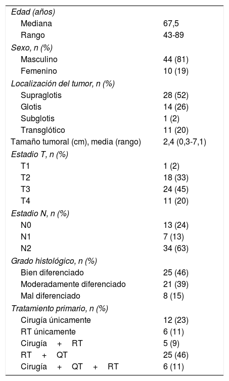
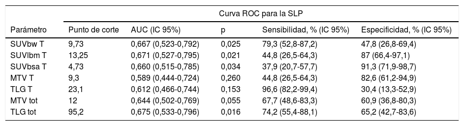
MétodosEstudio retrospectivo que incluye cincuenta y cuatro pacientes con cáncer escamoso de laringe que acudieron para realización de estadificación mediante PET/TC con 18F-FDG. Las imágenes PET se analizaron visualmente y semicuantitativamente midiendo varios parámetros metabólicos. Se tomó como estándar de referencia el seguimiento clínico, por imagen y/o histopatología. Se calculó el tiempo libre de enfermedad y la supervivencia mediante curvas de Kaplan-Meier.
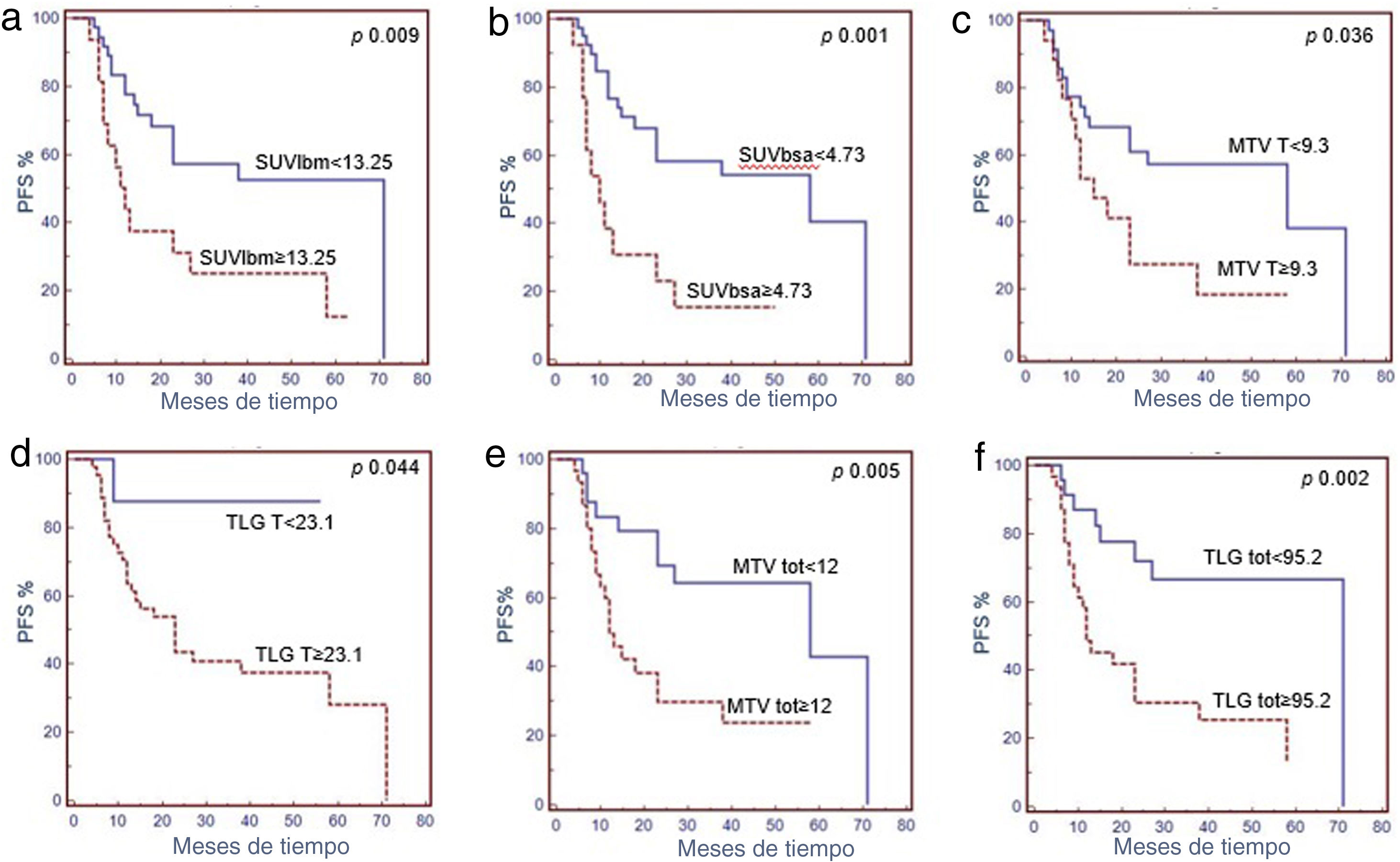
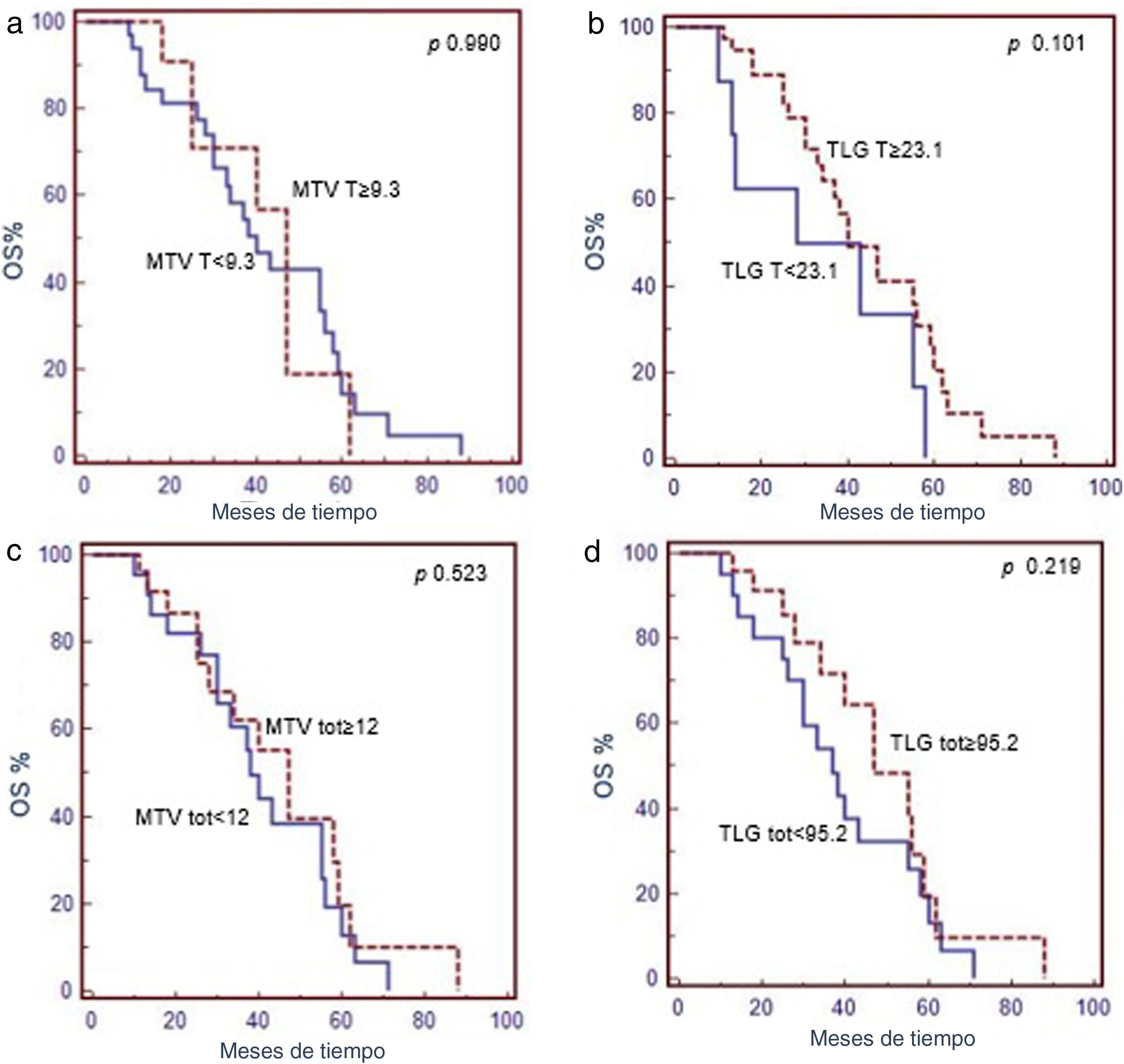
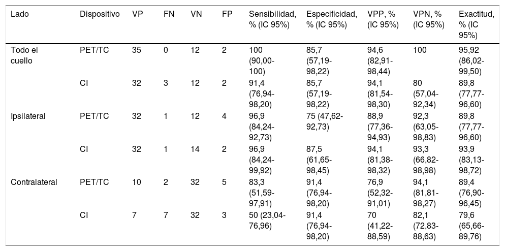
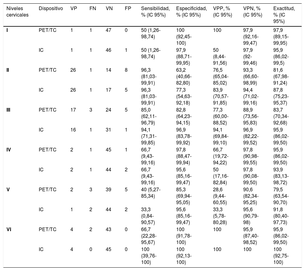
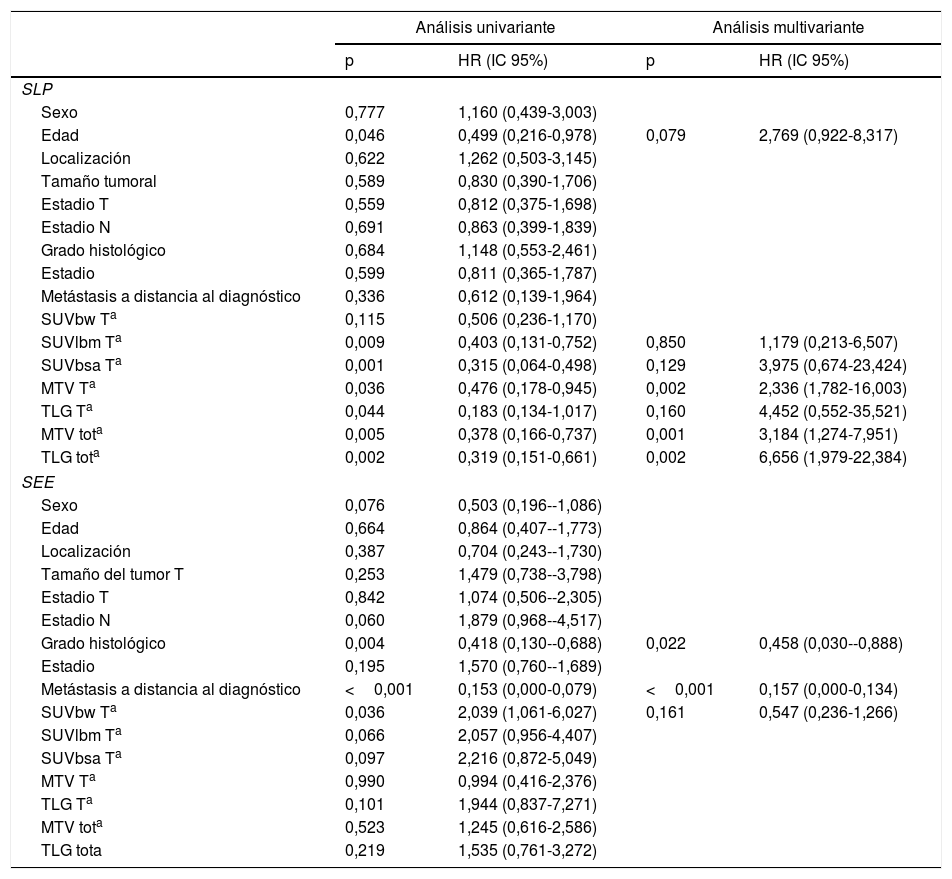
ResultadosTodos los tumores fueron identificados por las CI y 52/54 por la PET/TC con 18F-FDG, con sensibilidad del 96,3%. La sensibilidad, especificidad, valor predictivo positivo, valor predictivo negativo y precisión, basada en el análisis por paciente para la detección de enfermedad ganglionar fue del: 100%, 85,7%, 94,6%, 100% y 95,9% para la PET/TC con 18F-FDG y del 91,4%, 85,7%, 94,1%, 80% y 89,9% para las CI. El rendimiento diagnóstico de la PET/TC y las CI no fue significativamente diferente ni en análisis por paciente, ni en el de la lateralización ni el de los niveles ganglionares. La PET/TC con 18F-FDG detecto metástasis a distancia en 7 pacientes que permitieron una estadificación superior. En una mediana de seguimiento de 27 meses, la recaída/progresión de la enfermedad ocurrió en 31 pacientes y muerte en 32. El volumen metabólico tumoral (VMT) y la glucólisis total (TLG) de la lesión primaria se mostraron como factores pronósticos independientes para el tiempo libre de enfermedad.
ConclusionesTanto las CI como la PET/TC con 18F-FDG mostraron resultados diagósticos en la estadificación del cáncer de laringe. Los parámetros metabólicos basales (MTV y TLG) mostraron valor pronóstico en la evaluación del tiempo libre de enfermedad.
The aim of this study was to investigate the diagnostic accuracy of staging 18F-FDG-PET/CT in laryngeal cancer, compare these results with conventional imaging (CI) and assess the value of 18F-FDG-PET/CT features to predict survival.
MethodsFifty-four patients with laryngeal squamous cell cancer and baseline 18F-FDG-PET/CT were retrospectively enrolled. The PET images were analyzed visually and semi-quantitatively by measuring several metabolic parameters. A combination of clinical follow-up/imaging follow-up and/or histopathology was taken as reference standard. Progression free survival (PFS) and disease specific survival (DSS) were computed using Kaplan-Meier curves.
ResultsAll primary tumors were clearly identified by CI, and 52/54 by 18F-FDG-PET/CT with a sensitivity of 96.3%. Cervical nodal metastases were detected in 40/54 patients at 18F-FDG-PET/CT and in 34/49 patients at CI. Sensitivity, specificity, positive predictive value, negative predictive value, and accuracy on a patient-based analysis for nodal disease were 100%, 85.7%, 94.6%, 100% and 95.9% at 18F-FDG-PET/CT, and 91.4%, 85.7%, 94.1%, 80%, 89.8% at CI. Diagnostic performances of PET/CT and CI were not significantly different on a patient-based, side-by-side and level-by-level analysis. 18F-FDG-PET/CT recognized distant metastases in 7 patients allowing to an upstaging. At a median follow-up of 27 months, relapse/progression of disease occurred in 31 patients and death occurred in 32. Metabolic tumor volume (MTV T), MTV total and total lesion glycolysis (TLG) showed to be independent prognostic factors for PFS.
ConclusionsBoth CI and PET/CT had good diagnostic performances for the staging of laryngeal cancer; baseline metabolic features (MTV and TLG) showed an important prognostic value in assessing the rate of PFS.

