


To evaluate a semi-automated PET-image tumor segmentation algorithm for gross tumor volume (GTV) delineation in patients with locally advanced cervical cancer.
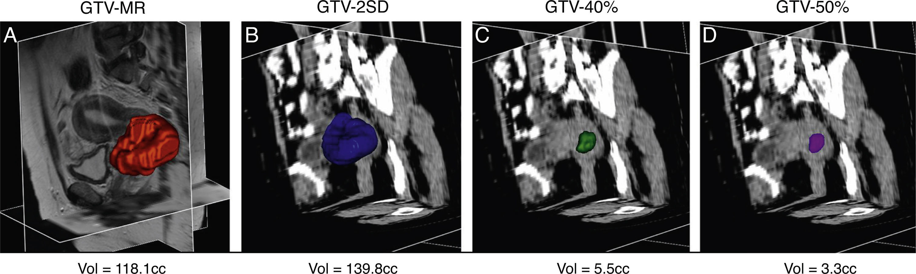
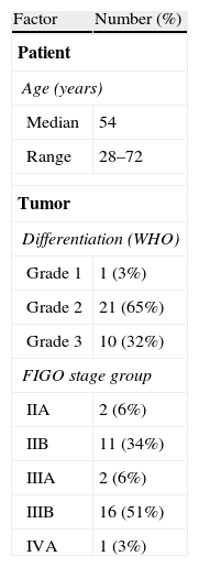
Material and methodsThirty-two patients with locally advanced cervical cancer were retrospectively evaluated. Semi-automated PET-image-based GTV delineation was applied using a previous established algorithm (GTV2SD) and 2 fixed threshold-based methods (GTV40% and GTV50%). GTV2SD was determined as the pixel with the mean value plus 2-standard deviation of the liver intensity, and GTV40% and GTV50% with 40% and 50% of the maximum tumor intensity (Tmax), respectively. The derived volumes were then compared with the GTVs generated manually using MR (GTVMR).
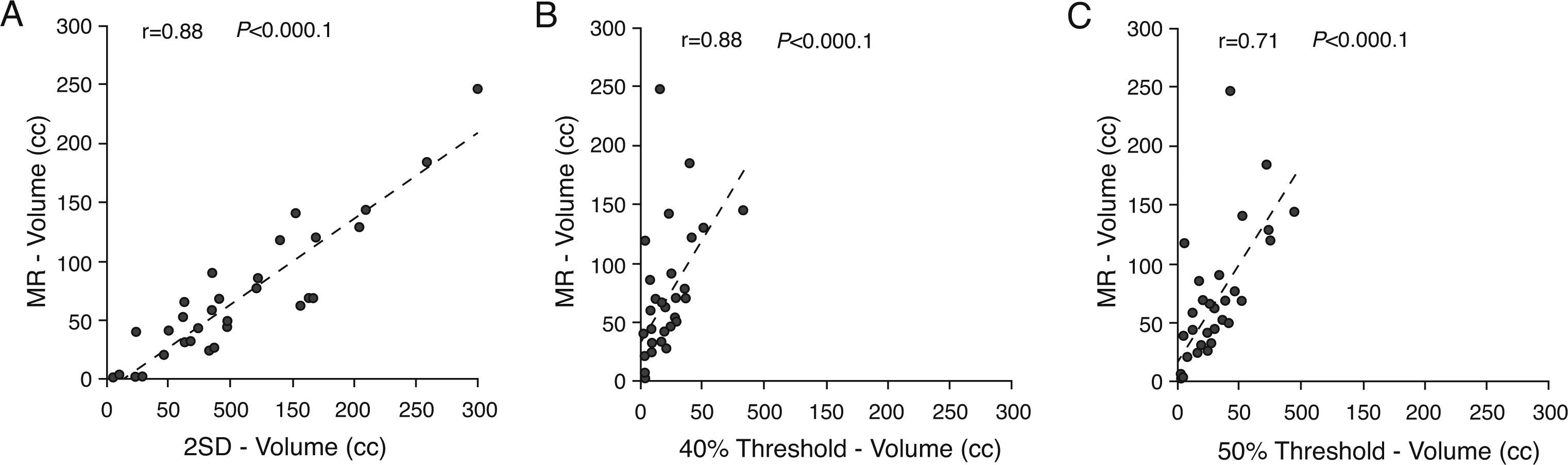
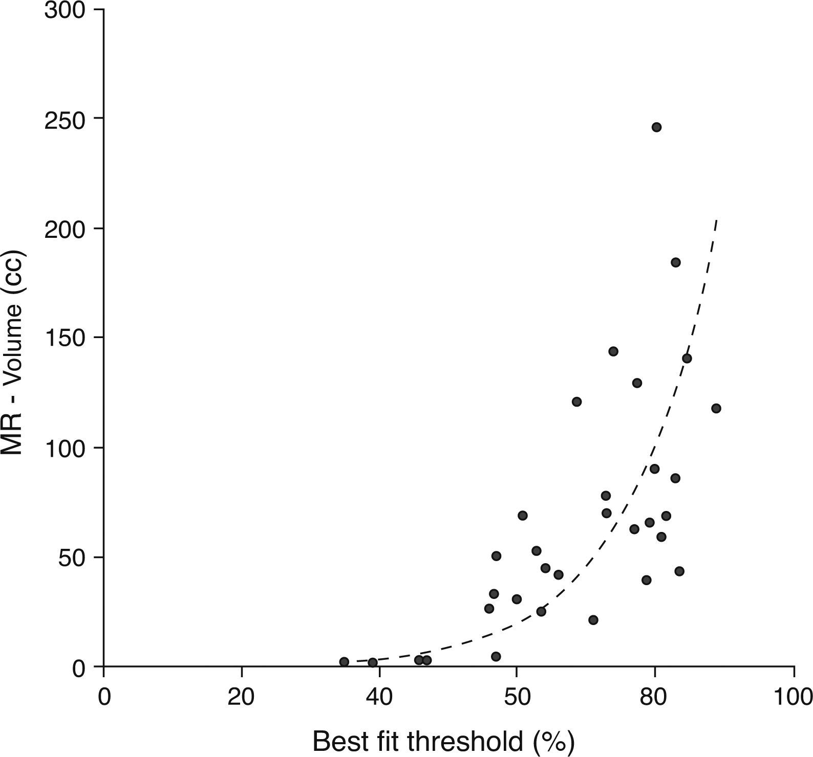
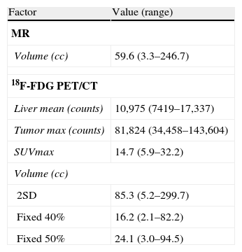
ResultsThe mean value of GTV2SD, GTV40% and GTV50% was 85.3cc, 16.2cc and 24.1cc, respectively. Good agreement was noticed between GTV2SD and GTVMR (ρ=0.88). GTV40% and GTV50% showed weaker correlation with GTVMR (ρ=0.68 and ρ=0.71, respectively).
ConclusionsThis study provides preliminary evidence that metabolic tumor volume delineation is feasible using computer-generated measurements in 18F-FDG PET images. Generation of PET-based tumor volumes is affected by the choice of threshold level used. Metabolic tumor bulk calculated using the pixel with the mean value plus 2-standard deviations of the liver intensity (GTV2SD) correlates better with the MR-derived tumor volumes. The method is a simple and clinically applicable approach to generate PET-derived GTV for radiation therapy planning of cervical cancer.
Evaluar el algoritmo de segmentación tumoral de la imagen PET semiautomatizada para delinear el volumen tumoral grueso (GTV) en pacientes con cáncer cervical localmente avanzado.
Material y métodosSe evaluó retrospectivamente a 32 pacientes con cáncer cervical localmente avanzado. Se utilizó la delineación GTV basada en imagen PET semiautomatizada, utilizando un algoritmo previamente establecido (GTV2SD) y 2 métodos basados en umbrales fijos (GTV40% y GTV50%). Se calculó GTV2SD, como el píxel con el valor medio más 2 desviaciones estándar de la intensidad del hígado, y GTV40% y GTV50% con el 40% y el 50% de intensidad tumoral máxima (Tmáx), respectivamente. A continuación se compararon los volúmenes derivados con los GTV generados manualmente, utilizando RM (GTVMR).
ResultadosEl valor medio de GTV2SD, GTV40% y GTV50% fue de 85,3 cc; 16,2 cc y 24,1 cc, respectivamente. Se halló una buena concordancia entre GTV2SD y GTVMR (ρ=0,88). GTV40% y GTV50% mostraron una menor correlación con GTVMR (ρ=0,68 y ρ=0,71, respectivamente).
ConclusionesEste estudio prueba de modo preliminar que la delimitación del volumen tumoral metabólico es posible utilizando las mediciones generadas informáticamente en las imágenes de 18F-FDG PET. La generación de los volúmenes tumorales basados en PET se ve afectada por la elección del nivel de umbral utilizado. El grueso del tumor metabólico calculado utilizando el píxel con el valor medio más 2 desviaciones de la intensidad del hígado (GTV2SD) guarda una mejor correlación con los volúmenes tumorales derivados de RM. El método constituye un enfoque simple y clínicamente aplicable para generar el GTV derivado del PET, para la planificación de la terapia de radiación del cáncer cervical.
Article

Revista Española de Medicina Nuclear e Imagen Molecular (English Edition)
