metricas
covid
Buscar en
Revista Española de Medicina Nuclear e Imagen Molecular (English Edition)
Toda la web
Inicio Revista Española de Medicina Nuclear e Imagen Molecular (English Edition) Association of PET/CT and VATS findings with histology analysis in the study of ...
Journal Information
Share
Share
Download PDF
More article options
Original Article
Available online 16 September 2024
Association of PET/CT and VATS findings with histology analysis in the study of pleural effusions
Asociación de los hallazgos del PET-TAC y la VATS con la histologia pleural en el estudio de los derrames pleurales
M. Simóa, O. Persivab, L. Sánchezc, J.B. Montorod, I. Sansanoe, A. Vázquezf, F. Ascanioc, C. Alemánf,
Corresponding author
mcarmen.aleman@vallhebron.cat

Corresponding author:
a Departments of Nuclear Imaging, Grupo de Patología Pleural, Universitat Autònoma de Barcelona, University Hospital Vall d’Hebron, Passeig Vall d’Hebron 119-129, Barcelona 08035, Spain
b Departments of Radiology, Grupo de Patología Pleural, Universitat Autònoma de Barcelona, University Hospital Vall d’Hebron, Passeig Vall d’Hebron 119-129, Barcelona 08035, Spain
c Departments of Thoracic Surgery, Grupo de Patología Pleural, Universitat Autònoma de Barcelona, University Hospital Vall d’Hebron, Passeig Vall d’Hebron 119-129, Barcelona 08035, Spain
d Departments of Pharmacy, Grupo de Patología Pleural, Universitat Autònoma de Barcelona, University Hospital Vall d’Hebron, Passeig Vall d’Hebron 119-129, Barcelona 08035, Spain
e Departments of Pathology, Grupo de Patología Pleural, Universitat Autònoma de Barcelona, University Hospital Vall d’Hebron, Passeig Vall d’Hebron 119-129, Barcelona 08035, Spain
f Departments of Internal Medicine, Grupo de Patología Pleural, Universitat Autònoma de Barcelona, University Hospital Vall d’Hebron, Passeig Vall d’Hebron 119-129, Barcelona 08035, Spain
Article information
Abstract
Full Text
Bibliography
Download PDF
Statistics
Figures (2)
Tables (3)
Table 1. Clinical, chest X-ray and pleural analysis findings according to first pleural histology.
Table 2. PET/CT data and VATS findings according to the first histology analysis and the final diagnosis of PE.
Table 3. Explained variation in the final diagnosis of PE according to the PET/CT score, the existence of nodules or pleural masses in the VATS, and the result of the first pleural histology.
Show moreShow less
Abstract
Introduction

Histological analysis of the pleura obtained by video-assisted thoracoscopic surgery (VATS) is the best diagnostic technique in the study of neoplastic pleural effusions. This study evaluates the relationship between Positron Emission Tomography (PET)/Computed Tomography (CT) and VATS findings, the result of the first pleural biopsy, and the final diagnosis of malignancy or non-malignancy.
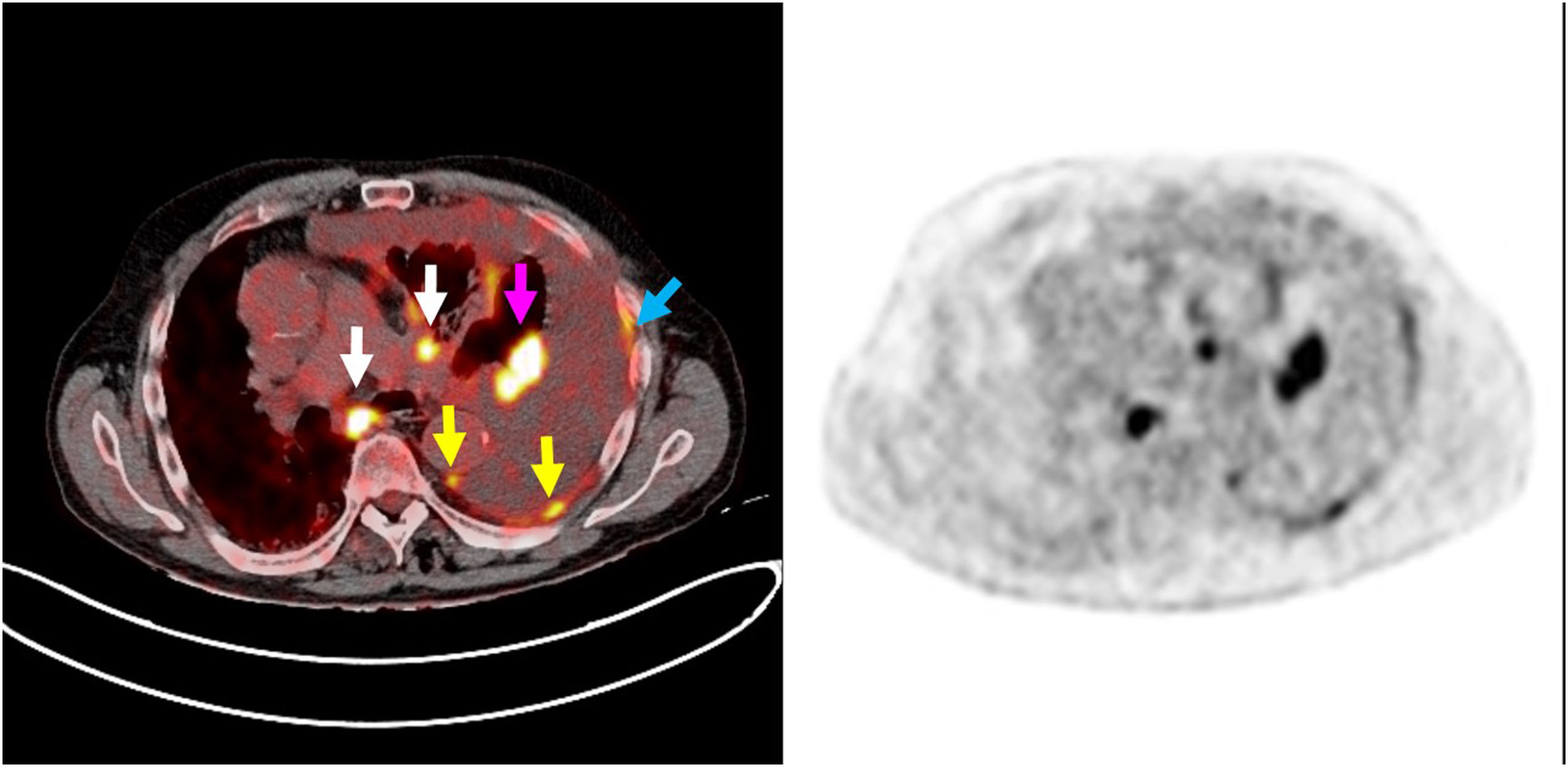
Methods

Prospective study of consecutive patients with pleural effusions undergoing PET/CT and VATS from October 2013 to December 2023. The following variables were recorded: PET/CT score (nodular pleural thickening, pleural nodules with standardized uptake value (SUV) > 7.5, lung mass or extra pleural malignancy, mammary lymph node with SUV > 4.5 and cardiomegaly); VATS data (drained volume, visceral and parietal pleural thickening, nodules or masses, septa, plaques, fluid appearance, trapped lung, and suspected diagnosis of the procedure), as well as the histological study of the first pleural biopsy (benign or malignant) and the final diagnosis of benign or malignant pleural effusion. A logistic regression study of the variables was performed.
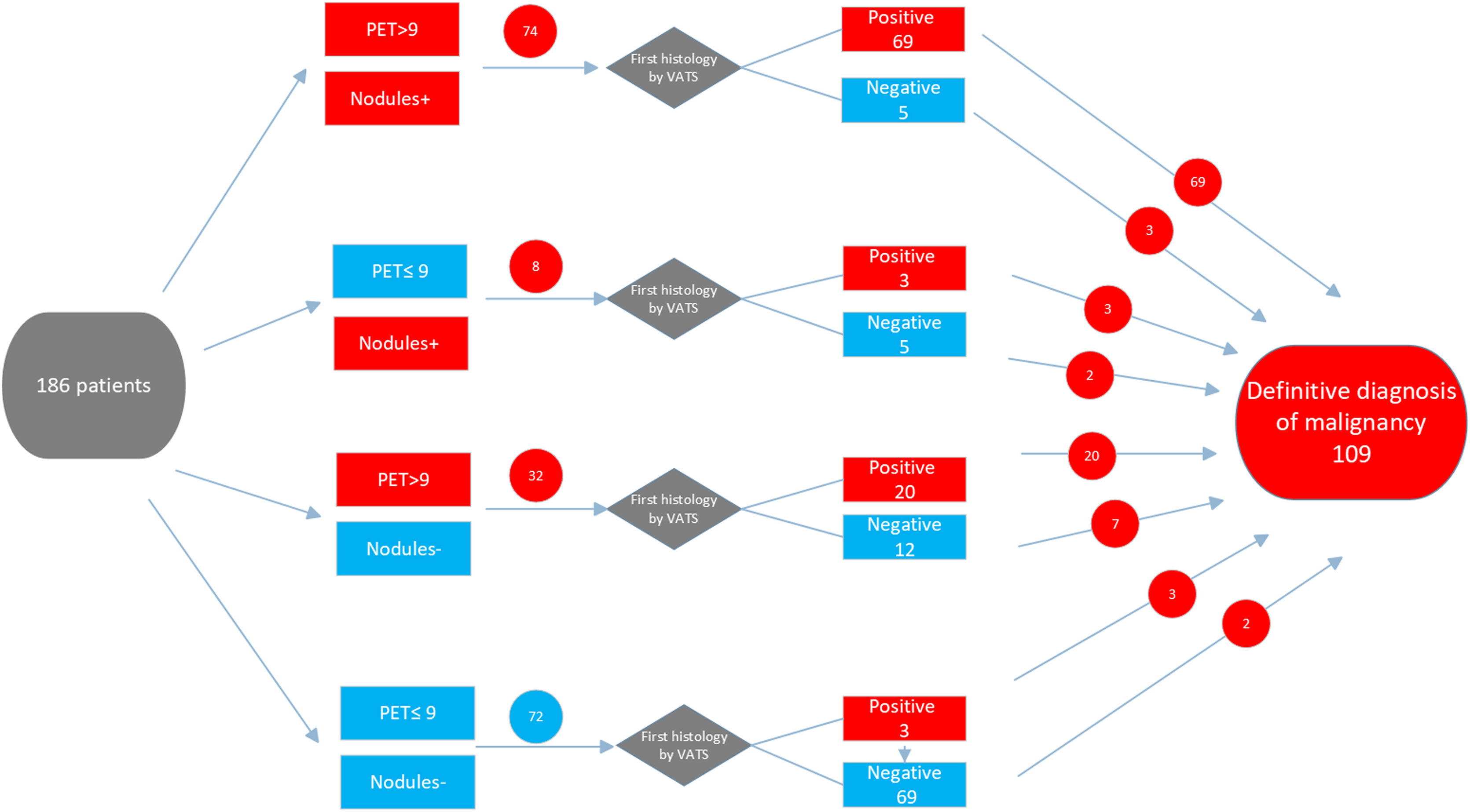
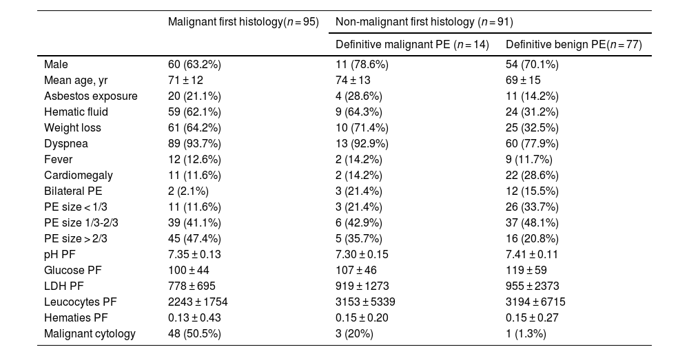
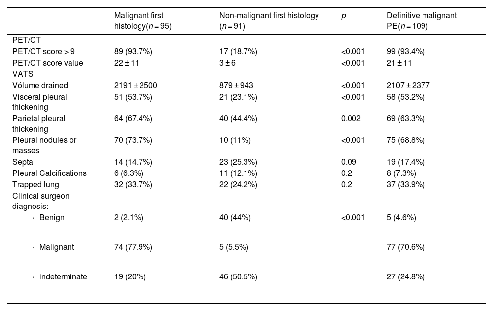
Results

95.8% of the patients with PET/CT and pleuroscopy not suggestive of malignancy had non-malignant histological findings, while 93.2% of the patients with PET/CT and pleuroscopy suggestive of malignancy had malignant histological findings. PET/CT, pleuroscopy, and the result of the first pleural biopsy showed a significant association with the final diagnosis of pleural effusion.

Conclusions

There is a strong association between PET/CT findings, VATS and pleural histology.

Keywords:
PET/CT
VATS
Pleural biopsy
Malignant pleural effusion
Resumen
Introducción

El análisis histológico de la pleura obtenido mediante videotoracoscopia asistida (VATS) es la mejor técnica diagnóstica en el estudio de los derrames pleurales neoplásicos. Este estudio evalúa la relación entre la Tomografía por Emisión de Positrones (PET) - Tomografía Axial Computarizada (TAC) y los hallazgos de VATS, el resultado de la primera biopsia pleural y el diagnóstico final de malignidad o no malignidad.

Métodos

Estudio prospectivo de pacientes consecutivos con derrame pleural sometidos a PET-TAC y VATS desde octubre de 2013 a diciembre de 2023. Se registraron las siguientes variables: puntuación PET-TAC (engrosamiento pleural nodular, nódulos pleurales con valor de SUV > 7,5, masa pulmonar o neoplasia maligna extrapleural, ganglio linfático mamario con SUV > 4,5 y cardiomegalia); datos de la VATS (volumen drenado, engrosamiento pleural visceral y parietal, nódulos o masas, septos, placas, aspecto líquido, pulmón atrapado y diagnóstico de sospecha del procedimiento), así como el resultado histológico de la primera biopsia pleural (benigna o maligna) y el diagnóstico final de derrame pleural benigno o maligno. Se realizó un estudio de regresión logística de las variables.

Resultados

El 95,8% de los pacientes con PET-TAC y pleuroscopia no sugestiva de malignidad tuvieron hallazgos histológicos no malignos, mientras que el 93,2% de los pacientes con PET-TAC y pleuroscopia sugestiva de malignidad tuvieron una histología neoplásica. La PET-TAC, la pleuroscopia y el resultado de la primera biopsia pleural mostraron una asociación significativa con el diagnóstico final de derrame pleural.

Conclusiones

Existe una fuerte asociación entre los hallazgos de PET-TAC, VATS e histología pleural.

Palabras clave:
PET-TAC
VATS
Biopsia pleural
Derrame pleural maligno

Article

These are the options to access the full texts of the publication Revista Española de Medicina Nuclear e Imagen Molecular (English Edition)
Subscriber
Subscriber

If you already have your login data, please click here .

If you have forgotten your password you can you can recover it by clicking here and selecting the option “I have forgotten my password”
Subscribe
Subscribe to

Revista Española de Medicina Nuclear e Imagen Molecular (English Edition)

Purchase
Purchase article

Purchasing article the PDF version will be downloaded

Price 19.34 €

Purchase now
Contact
Phone for subscriptions and reporting of errors
From Monday to Friday from 9 a.m. to 6 p.m. (GMT + 1) except for the months of July and August which will be from 9 a.m. to 3 p.m.
Calls from Spain
932 415 960
Calls from outside Spain
+34 932 415 960
E-mail
Article options
es en pt

¿Es usted profesional sanitario apto para prescribir o dispensar medicamentos?

Are you a health professional able to prescribe or dispense drugs?

Você é um profissional de saúde habilitado a prescrever ou dispensar medicamentos