Paget's disease of bone (PDB) may lead to hearing loss. The present study was conducted with the aim of measuring, characterising and determining the risk factors for hearing loss in a group of subjects with PDB.
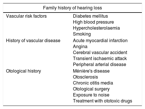
MethodsAn observational, transversal, case–control study was conducted, a cohort of 76 subjects diagnosed with PDB in the case group and a control group of 134 subjects were included. Clinical, demographic and audiometric data were analysed.
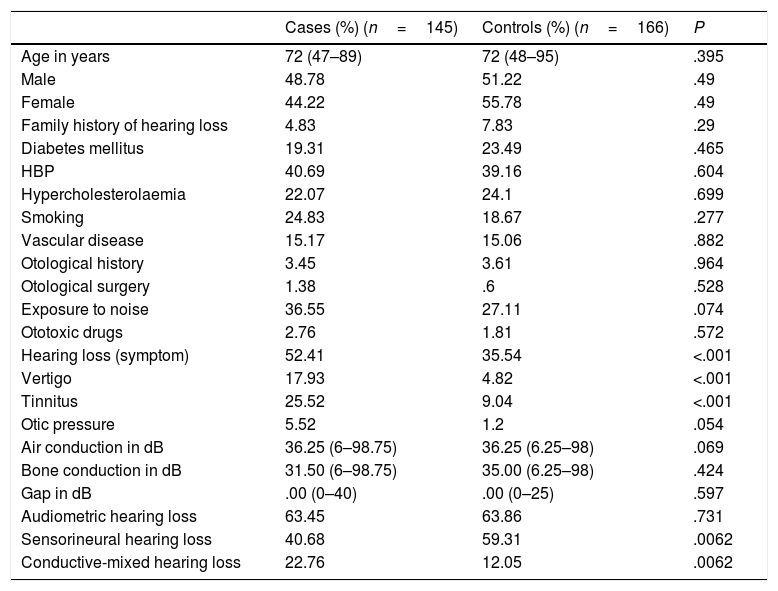
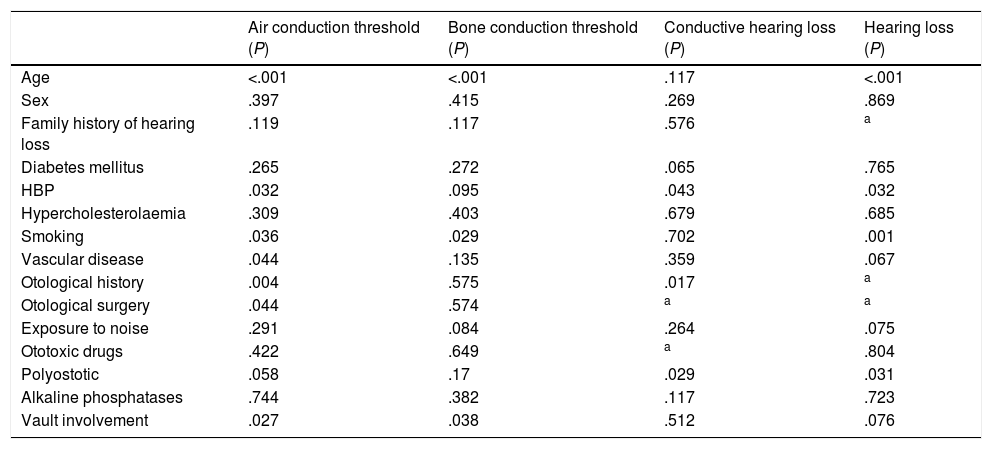
ResultsThe comparative analysis between the subjects in the PDB group and the control group found that the case group showed higher hearing thresholds (39.51dB) compared with the control group (37.28dB) (P=.069) and presented a greater rate of conductive hearing loss (22.76%) than the control group (12.05%) (P=.0062). The study of risk factors for hearing loss found that skull involvement in bone scintigraphy, age and high blood pressure were risk factors for higher impairment in PDB.
ConclusionsThe subjects with PDB showed more profound and a higher proportion of conductive hearing loss than the control group. The patients with PDB and skull involvement presented a more severe hearing loss compared with the subjects without skull involvement. Skull involvement and age were found to be risk factors for hearing loss.
La enfermedad ósea de Paget (EOP) puede cursar con hipoacusia. Con el objetivo de cuantificar, caracterizar y determinar los factores de riesgo de hipoacusia en un grupo de pacientes con EOP se realiza el presente estudio.
MétodosSe realizó un estudio observacional, transversal del tipo casos y controles que incluyó una cohorte de 76 sujetos con diagnóstico de EOP en el grupo caso y un grupo control de 134 sujetos. Se analiza la información clínica, demográfica y audiométrica de los sujetos incluidos.
ResultadosEl análisis comparativo entre el grupo de sujetos con EOP y el grupo control determinó que el grupo caso presentaba un umbral medio auditivo mayor (39,51dB) que el grupo control (37,28dB) (p = 0,069) y que presentaba hipoacusia transmisiva con mayor frecuencia (22,76%) que el grupo control (12,05%) (p = 0,0062). El análisis de los factores de riesgo de hipoacusia determinó que la afectación craneal en la gammagrafía ósea, la edad y la HTA, entre otros, constituían factores de riesgo de mayor pérdida auditiva en la EOP.
ConclusionesLos sujetos con EOP presentaron una pérdida auditiva más severa y con mayor frecuencia de tipo transmisivo que el grupo control. Los sujetos con afectación de la calota craneal por EOP presentaron mayor pérdida auditiva que los sujetos sin afectación craneal. La afectación de la calota craneal por la EOP y la edad constituyeron factores de riesgo de hipoacusia.
Artículo
Comprando el artículo el PDF del mismo podrá ser descargado
Precio 19,34 €
Comprar ahora


