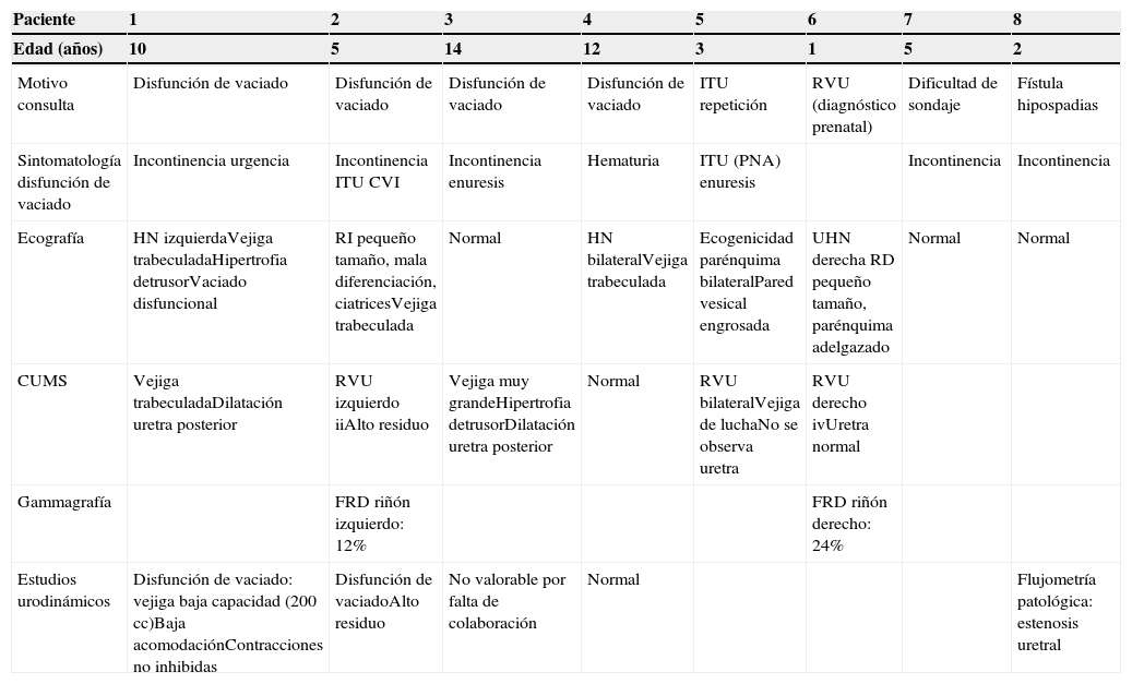
Diagnosticamos 8 pacientes de forma tardía de válvulas de uretra posterior (VUP) entre 1 y 14 años. Cinco pacientes consultaron por sintomatología relacionada con disfunción del vaciamiento vesical. Los otros 3 precisaron una uretrocistoscopia por otro motivo (fístula de hipospadias, dificultad de sondaje y RVU de alto grado), y al rehistoriar a los 2 primeros también presentaban sintomatología de disfunción de vaciado. Todos tenían ecografías preoperatorias: 3 fueron normales y 5 patológicas, con alteraciones a nivel renal o vesical. El diagnóstico se sospechó por cistouretrografía miccional seriada (CUMS) y 4 pacientes tenían estudios urodinámicos. El diagnóstico se confirmó por uretrocistoscopia, realizándose electrofulguración de las válvulas. Realizamos uretrocistoscopia de control a las 3-6 semanas sin observan ninguna estenosis. La sintomatología desapareció en el 100% de los pacientes tras 20 meses de seguimiento. El paciente con RVU se curó. Las ecografías no mostraron progresión de la afectación renal y presentaron mejoría de la afectación vesical. Las flujometrías de control mostraron curvas dentro de la normalidad.
DiscusiónLa mayoría de los niños con VUP se diagnostican ecográficamente en el periodo neonatal. Algunos pacientes manifiestan las VUP a edades más tardías con clínica diversa, lo que dificulta su diagnóstico. Debemos sospecharlas en pacientes varones con síntomas de disfunción de vaciado, tanto si tienen ecografías o cistouretrografía miccional seriada normales o patológicas y recomendamos realizar uretrocistoscopia para descartar obstrucción uretral.
We diagnosed 8 patients with late-stage posterior urethral valves (PUV) between 1 and 14 years of age. Five patients complained of symptoms related to voiding dysfunction. The other 3 patients required urethrocystoscopy for other reasons (hypospadias fistulae, difficulty with catheterisation and high-grade vesicoureteral reflux [VUR]). A second review of the first 2 patients’ medical history showed voiding dysfunction symptoms. All patients underwent preoperative ultrasonography: 3 patients had normal results and 5 had renal or vesical disorders. The diagnosis was reached through voiding cystourethrogram (VCUG), and 4 patients underwent urodynamic studies. The diagnosis was confirmed by urethrocystoscopy, performing valve electrofulguration. We performed urethrocystoscopy during the check-ups at 3-6 weeks and observed no stenosis. The symptoms disappeared for all patients after 20 months of follow-up. The patient with VUR was cured. The ultrasounds showed no progression of the renal involvement and showed improvement in the vesical involvement. The velocimetries during check-ups presented curves within normal ranges.
DiscussionMost children with PUV are diagnosed through ultrasound during the neonatal period. Some patients present PUV at later ages with diverse symptoms, which impedes its diagnosis. We should suspect PUV in male patients with symptoms of voiding dysfunction, either when they have normal or pathological results from ultrasounds or VCUG. We recommend performing urethrocystoscopy to rule out urethral obstruction.
Artículo
Comprando el artículo el PDF del mismo podrá ser descargado
Precio 19,34 €
Comprar ahora


