INTRODUCTION
Cold urticaria is defined as an urticarial and/or angioedematous reaction of the skin to contact with cold objects, water or air. This pathological hyperreactivity is the epiphenomenon for a number of diseases, and it is due to a wide range of causative factors. In many instances, the reason for this cold hypersensitivity is however unknown.
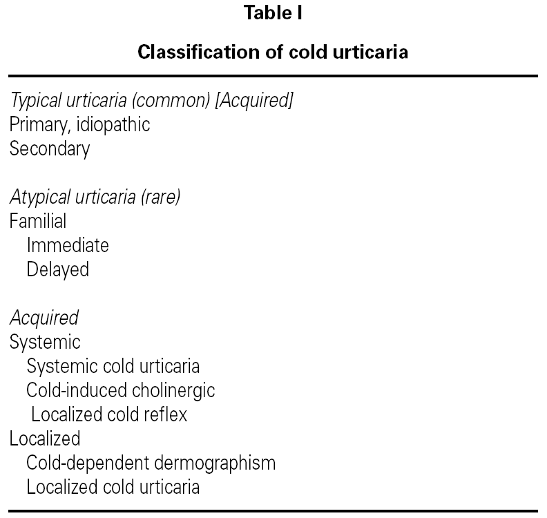
Cold urticaria represents 3-5 % of all physical urticarias. Kontou-Fili et al 1 have proposed a simplified classification (table I) for use in daily practice.
The typical form of cold urticaria presents with pruritus, erythema, and swelling confined to skin areas exposed to cold; the lesions develop 2-5 min or slightly later as the skin starts rewarming. If a large enough portion of the skin is exposed, the patient may develop systemic symptoms analogous to anaphylaxis, and patients are susceptible to shocklike reactions during aquatic activities 2.
The standard clinical test performed to confirm the diagnosis of cold urticaria is the placing of an ice cube on the forearm of the patient for a period of 5-10 minutes, with observation for the development of a weal and flare reaction over the subsequent ten minutes 3.
Secondary acquired cold urticaria includes disorders characterized by cryoglobulinemia, cryofibrinogenia, cold hemolysins, and cold agglutinins of variable duration 1. Types associated with infectious diseases, such as mononucleosis 4, rubeola, varicella, syphilis 2, hepatitis 5,6, and VIH infection 7,8 have been reported.
To our knowledge, no other case of cold urticaria associated with toxoplasmosis has been described. We present the case of an atopic patient who developed a cold urticaria associated with acute serologic toxoplasmosis.
CLINICAL CASE
The patient was a 34 year-old man, electrician, who has been treated in the Allergy Section for the last 3 years due to allergic rhinitis, occasionally associated with asthma symptoms (wheezing and dyspnea). The treatment was based on pollen-immunotherapy, antihistamines, topic nasal steroids and bronchodilators if necessary. He referred oral and pharyngeal pruritus after the intake of nuts and urticaria after touching peach peel.
For two months, he had presented cutaneous pruritus accompanied by several papular lesions in those parts of the skin exposed to cold as well as those in contact with cold water. These lesions disappeared in approximately one hour after the exposure to cold ceased. The patient didn't refer blueberry-muffin lesions, Raynaud's phenomenon or joint affection.
Physical examination was normal and no lymphadenopathies, visceromegaly or other affections were found.
Skin tests were done with a standard battery of aeroallergens (Stallergenes-DHS, Paris, France), that were positive for cat dander, olive tree, grasses, Parietaria and Artemisia pollen. Other tests based on a battery of food showed positive results for hazelnuts, peanuts, almonds, sunflower seeds, pistachios, walnuts and peaches.
An "ice-cube test" was also performed by placing an ice-cube on the anterior surface of the forearm for 10 minutes, obtaining a positive result after the appearance of erythema and edema at warming the zone.
The hematologic laboratory values were: 5.400.000 erythrocytes, 16.5 Hb, 49 Hto. 90 VCM, 30 HCM, 8,300 leukocytes (59 % neutrophils, 30 % lymphocytes, 5 % monocytes, 6 % eosinophils), 241,000 platelets. VSG 7. Blood chemical values: uric acid 8.2, total bilirrubin 1.21 (direct 0.30, indirect 0.91). The rest was normal. Protein and thyroid hormones levels were normal and so were the complement titers. Urine sediment was normal. A test for antinuclear-antibodies was negative as well as antithyroglobuline and antimycrosomal antibodies. Serologic tests performed for Lues, HIV, Epstein-Barr, Mycoplasm and Hidatosis were negative. Hepatitis C antibody was negative and results for Hepatitis B were: HBsAg negative, Anti-HBc negative, Anti-HBsAg > 1000 (the patient had been vaccinated). Serologic tests for Citomegalovirus showed an IgG amount of 15 UA/ml and negative IgM. For Toxoplasma gondii, IgG was 68 UI/ml and IgM was positive. A test for crioglobulines was positive.
Chest radiographs and the ultrasonographic study of the abdomen revealed no abnormalities.
One month later another blood test was performed showing negative crioglobulines. Serologic test for Toxoplasma gondii showed IgG levels of 75 UI/ml and positive IgM. Three months later crioglobulines were also negative, IgG for Toxoplasma gondii was 84 UI/ml and IgM was positive. After six months crioglobulines were still negative, the amount of IgG was 68 UI/ml and IgM was still slightly positive. In the last control, 14 months later, IgG levels were 32 UI/ml and IgM was negative.
The patient has continued to present clinical manifestations of cold urticaria, though he has noted some improvement of his condition, and his tolerance to cold has risen after treatment with cetirizine.
DISCUSSION
This was an atopic patient, who presented allergic asthma and rhinitis due to sensitization to pollen and cat dander plus food allergy. Two months before consulting, he started to show symptoms of cold urticaria. It was a typical urticaria which started a few minutes after the exposure to cold and was positive for the "ice-cube test".
Blood tests were positive for Toxoplasma gondii with an IgG amount of 68 UI/ml and positive IgM. These results indicate an acute infection. IgM levels remained positive for the next three months, being weakly positive after 6 months and finally negative one year later. IgG levels increased to 75 UI/ml during the first month and to 84 UI/ml after three months, descending to those values obtained in the first tests, 6 months later and to 32 UI/ml after a year.
The patient didn't develop any symptoms of toxoplasmosis, as it is usual in patients whose immune system is intact. Despite the negativity of IgM and the decrement of the IgG levels, the patient still presents clinical manifestations of cold urticaria. These findings disagree with those observed in another cases associated with infectious mononucleosis in which cold urticaria is transitory and goes parallel to the course of the infection.
Positive values for cryoglobulines can be found in a 20 % of patients with cold urticaria. In the current case, cryoglobulines were positive on the first blood test tough they became negative one month later, so they were transitory, as it has been reported in another case associated with positive serology for hepatitis B and cryoglobulinemia. Perhaps this is the reason why the patient didn't present symptoms of cryoglobulinemia, such as purpura, Raynaud's phenomenon, polyarthritis or ulcerative lesions. The persistence of cold urticaria symptoms in spite of the absence of cryoglobulines is considered one of the most relevant findings.
Finally, the patient's condition improved and his tolerance to cold increased considerably after initiating treatment with cetirizine (as it has been reported in other studies), a second generation antihistamine which is better tolerated than those usually recommended for this pathology, such as cyproheptadine or difenhidramine.
In summary, this is the first reported case of acquired cold urticaria associated with acute serologic infection by Toxoplasma gondii.


