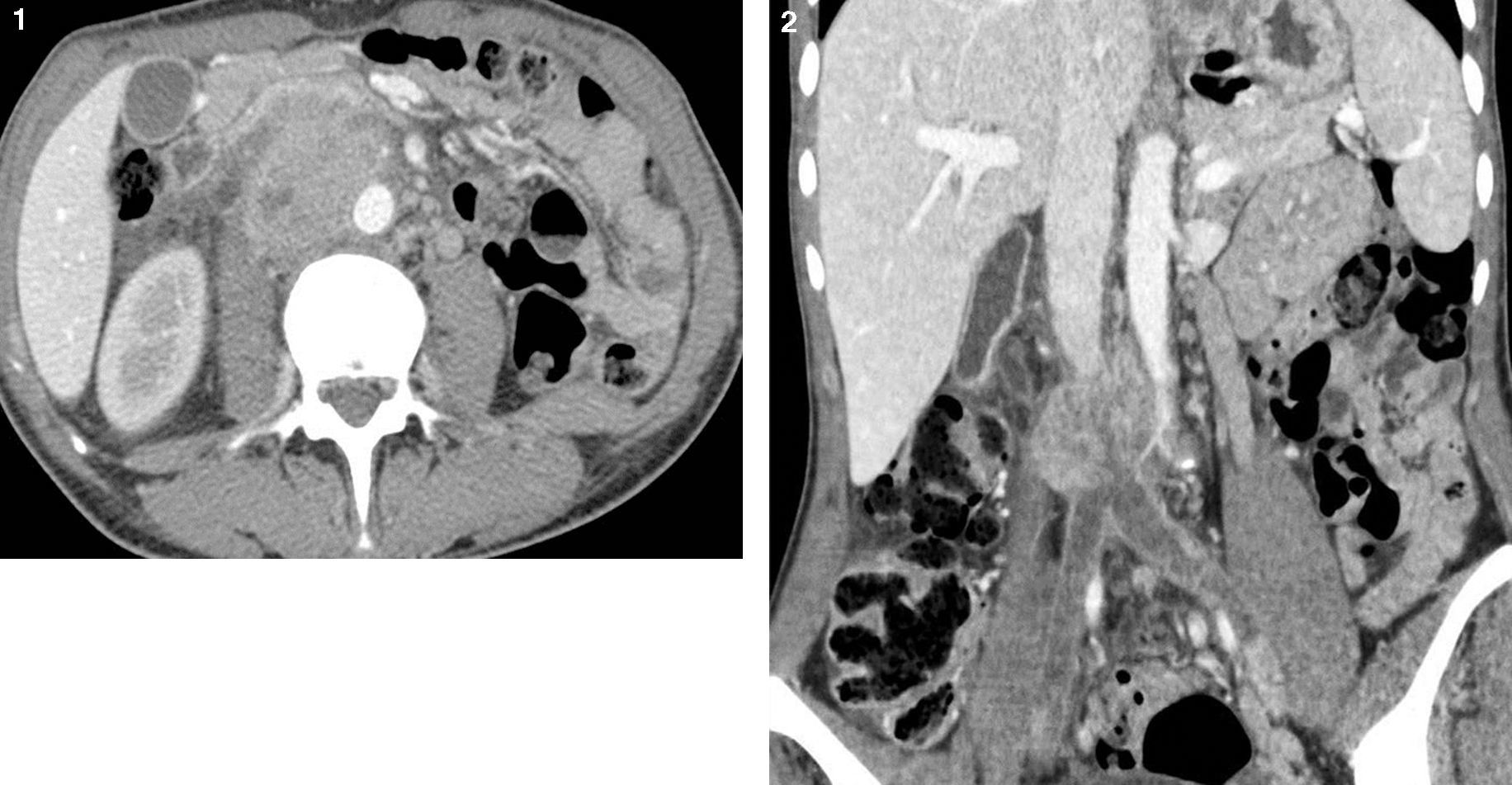
A 43-year-old woman consulted for bilateral lower extremities edema. Questioning revealed abdominal pain, asthenia, anorexia and weight loss of 6 months duration. An abdominal iodine enhanced CT scan was realized and revealed a large solid mass as shown in Figs. 1 and 2.
Question:
What is your diagnosis?
- -
Pheochromocytoma
- -
Leiomyosarcoma of the inferior vena cava
- -
Lymphadenopathy
- -
Gastro-intestinal stromal tumor (GIST)
- -
Abdominal CT scan (axial and frontal). Heterogeneous retroperitoneal solid mass centered in the inferior vena cava (IVC) infrarenal. Iliac vein thrombosis seen. Invasion of the lumbar segment of the ureter and psoas muscle picture, being in contact with the spine. Forward, contact the third duodenal portion. The supra-renal IVC is permeable.


