Abstracts from XVII Mexican Congress of Hepatology
Más datosHepatic hemangiomas (HH) are the most common primary benign tumors of the liver, more frequent in women, attributed to estrogens. Its size can reach up to 30 cm, being considered giant when it measures >4 cm.
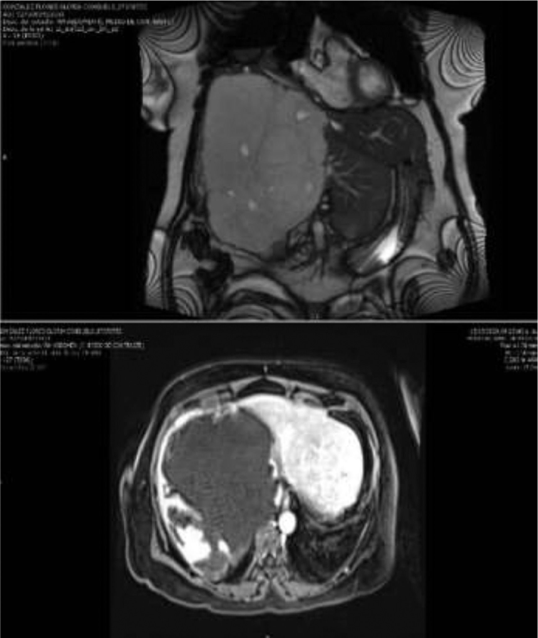
Case ReportA 65-year-old woman with giant cavernous hemangiomas as an incidental finding on liver USG. The abdominal MRI established the diagnosis by spotting two intrahepatic lesions, one of the right lobe occupying all the segments, measuring 18.5 × 16.6 × 15.7 cm, with a volume of 2527.6 cc; another in the left lobe of 7.8 × 7.8 × 6.5 cm, the volume of 206.8 cc; hypointense on T1 sequence, hyperintense on T2, with enhanced contrast medium in the periphery, later it is centripetal, with focal areas without enhancement at 20 minutes.
Physical examination: painful swelling in the left hypochondrium up to the anterior axillary line and epigastrium. Increased alkaline phosphatase and GGT. Surgical management is contraindicated due to the characteristics of the lesion. We decided to send her for a liver transplant.
DiscussionHistologically, they are vascular malformations characterized by caverns covered by a single layer of endothelium. The gold standard is MRI, where we observe peripheral nodular enhancement followed by central enhancement in a well-defined homogeneous mass. Surgical management is indicated in the symptomatic giant HH, with an increase in size or suspicion of malignancy. They require LT if the interventions such as embolization or resection fail to control the disease.
ConclusionsGiant HHs should be treated if they cause symptoms and may require HT when they are unresectable or have complications such as coagulopathy, risk of rupture, or failure of previous management.
FundingThe resources used in this study were from the hospital without any additional financing
Declaration of interestThe authors declare no potential conflicts of interest.


