This study was designed with the goal of adding as much information as possible about the role of pigeons (Columba livia) and chickens (Gallus gallus) in Newcastle disease virus epidemiology. These species were submitted to direct experimental infection with Newcastle disease virus to evaluate interspecies transmission and virus-host relationships. The results obtained in four experimental models were analyzed by hemagglutination inhibition and reverse transcriptase polymerase chain reaction for detection of virus shedding. These techniques revealed that both avian species, when previously immunized with a low pathogenic Newcastle disease virus strain (LaSota), developed high antibody titers that significantly reduced virus shedding after infection with a highly pathogenic Newcastle disease virus strain (São Joao do Meriti) and that, in chickens, prevent clinical signs. Infected pigeons shed the pathogenic strain, which was not detected in sentinel chickens or control birds. When the presence of Newcastle disease virus was analyzed in tissue samples by RT-PCR, in both species, the virus was most frequently found in the spleen. The vaccination regimen can prevent clinical disease in chickens and reduce viral shedding by chickens or pigeons. Biosecurity measures associated with vaccination programs are crucial to maintain a virulent Newcastle disease virus-free status in industrial poultry in Brazil.
Newcastle disease (ND) is an acute, highly contagious viral disease of poultry that can cause high mortality (up to 100%) in chicken, the most important natural host of the disease. The virus can also affect a wide variety of avian species causing severe disease. The disease is regarded as endemic in many countries, and is caused by an avian Paramyxovirus type 1 (APMV 1), a member of the genus Avulavirus, from the Paramyxoviridae family.1 As demonstrated in intensive surveys, nearly 236 free-living species from 27 of the 50 orders of birds have been reported to be susceptible to either natural or experimental infection with ND.2 On several occasions, Newcastle disease virus (NDV) was isolated from wildlife birds,3 and most outbreaks of NDV arise in unvaccinated susceptible animals.4 To keep ND under control, large-scale prophylactic vaccination is used in most member states of the European Union and elsewhere in the world.4,5 Although vaccination in general provides good protection against clinical disease and mortality, it may not provide sufficient protection against virus transmission to prevent ND outbreaks.6,7 The most common strains used worldwide in vaccination are LaSota and B1.5 Conventional vaccine strains may protect against clinical disease caused by virulent NDV, but viral infection can still occur in vaccinated birds,8,9 which may be the route of virus spread in poultry facilities farms.10 However, vaccinated birds may show significantly reduction in virus shedding compared with unvaccinated ones.9,10
The susceptibility of pigeons (Columba livia) and other members of the Columbidae family to NDV has been reported by several authors.11–15 It is now clear that the disease occurs in pigeons as a result of virus dissemination from affected chicken flocks, and it occurs in poultry flocks when the virus is disseminated from domesticated or feral pigeons.16 The source of ND infection to chicken flocks may be food contaminated with feces of feral pigeons infected with NDV.16,17
Many aspects of ND infection in pigeons are unclear, and experimental infection could answer many questions about NDV epidemiology, such as virus pathogenicity, infectivity, and shedding. As previously described by experimental studies, adult pigeons infected via eye drops with a chicken pathogenic APMV-1 strain shed the virus both through the cloaca and the mouth for up to 21 days post-infection (dpi).11 Pigeon Paramyxovirus type 1 (PPMV-1) isolates may be transmitted from infected pigeons to chickens that were in contact with them, with replication in the chickens (as demonstrated by the excretion of the virus by cloacal route), and antibody response against the virus.16 In experimental infections conducted with a PPMV-1 strain, mortality rates greater than 70% were observed in pigeons, but no infected chicken died. In spite of these findings, there are few comparative studies on pigeons and chickens infected with the same PPMV-1 strain, making it difficult to determine the significance of these results.15
Thus, little is known about the course of the infection, the significance of humoral antibody response, viral shedding, clinical signs, and contact transmission of a Brazilian pathogenic NDV strain between pigeons and chickens (experimental infection). To some extent, viral replication complex may play a role in the pigeon-to-chicken transmission, although further studies are needed to investigate the factors that are determinant for interspecies transmission.15
Some studies were carried out to evaluate the prevalence of Newcastle disease in commercial birds in poultry-producing areas in Brazil,18,19 and the results showed that industrial poultry produced in the nine Brazilian states analyzed was free of Newcastle disease.18
The nationwide efficiency of poultry production makes Brazil a competitive nation in international markets, even in the absence of economic subsidies. In order to guarantee better sanitary conditions for Brazilian avian products, the National Program for Poultry Health (PNSA) was implemented for the control of Newcastle disease in the country.19
Considering the potential risk of contamination of poultry species by pigeons carrying NDV, it is important to study the pathogenesis of the disease both in pigeons and in chickens. This study was designed to evaluate humoral immune response, viral shedding, and contact transmission after experimental infection of pigeons and chickens with a pathogenic NDV isolate of chicken origin under experimental and controlled conditions.
Materials and methodsBirdsIn an attempt to reproduce natural conditions of NDV transmission, a total of fifty-two free-living adult domestic pigeons (C. livia) were used in this study. Animals were clinically healthy, had non-specific levels of hemagglutination inhibition (HI) antibodies (HI Titers≤2), and were negative in reverse transcriptase polymerase chain reaction (RT-PCR) for the presence of NDV in cloacal swab samples. After capture, pigeons were housed in adequate facilities (away from chicken facilities) for 90 days to be adapted to captivity. Similarly, twenty-nine 90-day-old, SPF (specific pathogen free) chickens were used in the study. The two species were kept in separate facilities until the beginning of the experimental study.
On the day of experimental infection, pigeons and chickens divided in four experimental groups were moved to Negative Pressure Isolators (Alesco®, Brazil), under controlled laboratory and biosafety conditions.
All animal procedures were performed according to the Ethical Principles in Animal Research adopted by the Brazilian College of Animal Experimentation, and to the 2000 Report of the AVMA Panel on Euthanasia.20
VirusesThe experimental infection was performed using the Sao Joao do Meriti (SJM) strain (Gene Bank Number: EF534701), a highly pathogenic NDV strain (APMV-1) for chickens (mean death time in chicken embryos=48h; ICPI in day-old chicken=1.78). The virus was propagated twice in the allantoic cavity of 9 to 10-day-old embryonated SPF eggs by inoculation of 0.1mL of infectious allantoic fluid. A virus stock was produced by harvesting allantoic fluid from chicken embryos, freezing it at −70°C, and storing it. This virus stock titer was 109.0 median embryo lethal dose/mL (ELD50), determined on day three before experimental infection. All virus dilutions were carried out with sterile phosphate buffered saline (PBS), pH 7.2.
Live vaccines containing LaSota (LS) NDV strain (New Vac-LS – Fort Dodge Saúde Animal Ltda®, Brazil) were used in the vaccination procedures. This strain is commercially used in the immunization of chickens in Brazil.
Experimental infection and samplingPigeons and chickens were randomly divided into four groups, as described below. After vaccination/experimental infection, birds were monitored daily for presence of any clinical signs, such as diarrhea, torticollis, incoordination, apathy, tremors, ocular and nasal discharge, abnormal posture, and flying difficulties. For the evaluation of viral shedding, cloacal swabs were collected daily from all groups (pigeons and chickens), until the last day of analysis, in each experimental model. Swabs were placed in tubes containing 1.0mL sterile PBS, pH 7.2, and stored at −70°C until the moment of analysis.
Experiment 01 – Infectivity of the challenge NDV strain in chickens and pigeons: This group was used to validate the Experimental Infection. Ten pigeons and five chickens were inoculated by ocular and oral routes with 0.1mL/route of a 10−1 virus dilution from a 109.0 ELD50/0.1mL stock suspension of Sao Joao do Meriti strain. The control group consisted of five pigeons and two SPF chickens that were inoculated with sterile phosphate buffered saline (PBS), pH 7.2, and kept in separate facilities.
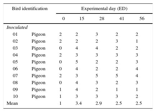
Blood samples were collected from the brachial vein of each bird on day 0 (zero), 15, 28, 41, and 56 ED (Experimental day). Samples were allowed to clot. Serum samples were stored at −20°C and used in the evaluation of the immune response, as shown in Table 1.
Antibody titers (Log2X) of inoculated (SJM strain) pigeons during the experimental period in Experimental Model 1.
| Bird identification | Experimental day (ED) | |||||
|---|---|---|---|---|---|---|
| 0 | 15 | 28 | 41 | 56 | ||
| Inoculated | ||||||
| 01 | Pigeon | 2 | 2 | 3 | 2 | 2 |
| 02 | Pigeon | 2 | 2 | 2 | 3 | 1 |
| 03 | Pigeon | 0 | 4 | 4 | 2 | 2 |
| 04 | Pigeon | 2 | 3 | 3 | 3 | 3 |
| 05 | Pigeon | 0 | 5 | 2 | 2 | 3 |
| 06 | Pigeon | 0 | 4 | 2 | 2 | 4 |
| 07 | Pigeon | 2 | 3 | 5 | 5 | 4 |
| 08 | Pigeon | 0 | 4 | 3 | 2 | 3 |
| 09 | Pigeon | 1 | 4 | 2 | 1 | 1 |
| 10 | Pigeon | 1 | 3 | 3 | 3 | 2 |
| Mean | 1 | 3.4 | 2.9 | 2.5 | 2.5 | |
Experiment 02 – Transmission of NDV by vaccinated pigeons following challenge infection: This group consisted of nine pigeons and four chickens. Pigeons were vaccinated with LaSota Strain of NDV, with one drop of the vaccine by ocular route, on day 0 (zero) and 35. On ED 61, pigeons were inoculated by ocular and oral routes with 0.1mL/route of a 10−1 virus dilution from a 109.0 ELD50/0.1mL stock suspension of Sao Joao do Meriti strain. The four remaining non-inoculated SPF chickens were used as sentinels, and kept in separate facilities until ED 66, when pigeons and sentinel chickens were placed in contact to detect lateral spread of the virus (infectivity). The control group consisted of five pigeons vaccinated with LaSota strain on day 0 (zero) and 35, and two SPF chickens that were inoculated with sterile phosphate buffered saline (PBS), pH 7.2, and kept in separate facilities.
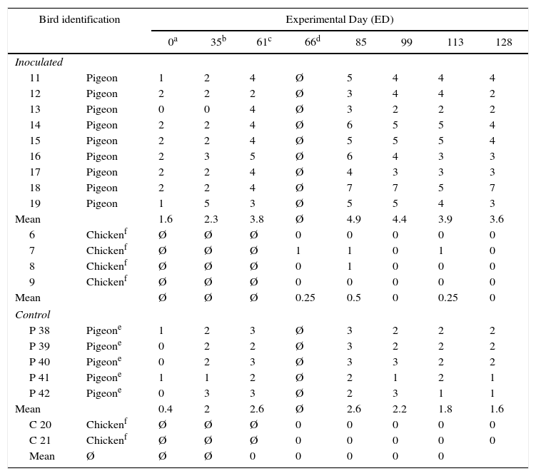
Blood samples were collected from the brachial vein of each bird on day 0 (zero), 35, 61, 85, 99, 113, and 128 ED. Serum samples were stored at −20°C and used in the evaluation of immune response, as shown in Table 2.
Antibody titers (Log2X) of vaccinated (LS strain) and inoculated (SJM strain) birds during the experimental period, in Experimental Model 2.
| Bird identification | Experimental Day (ED) | ||||||||
|---|---|---|---|---|---|---|---|---|---|
| 0a | 35b | 61c | 66d | 85 | 99 | 113 | 128 | ||
| Inoculated | |||||||||
| 11 | Pigeon | 1 | 2 | 4 | Ø | 5 | 4 | 4 | 4 |
| 12 | Pigeon | 2 | 2 | 2 | Ø | 3 | 4 | 4 | 2 |
| 13 | Pigeon | 0 | 0 | 4 | Ø | 3 | 2 | 2 | 2 |
| 14 | Pigeon | 2 | 2 | 4 | Ø | 6 | 5 | 5 | 4 |
| 15 | Pigeon | 2 | 2 | 4 | Ø | 5 | 5 | 5 | 4 |
| 16 | Pigeon | 2 | 3 | 5 | Ø | 6 | 4 | 3 | 3 |
| 17 | Pigeon | 2 | 2 | 4 | Ø | 4 | 3 | 3 | 3 |
| 18 | Pigeon | 2 | 2 | 4 | Ø | 7 | 7 | 5 | 7 |
| 19 | Pigeon | 1 | 5 | 3 | Ø | 5 | 5 | 4 | 3 |
| Mean | 1.6 | 2.3 | 3.8 | Ø | 4.9 | 4.4 | 3.9 | 3.6 | |
| 6 | Chickenf | Ø | Ø | Ø | 0 | 0 | 0 | 0 | 0 |
| 7 | Chickenf | Ø | Ø | Ø | 1 | 1 | 0 | 1 | 0 |
| 8 | Chickenf | Ø | Ø | Ø | 0 | 1 | 0 | 0 | 0 |
| 9 | Chickenf | Ø | Ø | Ø | 0 | 0 | 0 | 0 | 0 |
| Mean | Ø | Ø | Ø | 0.25 | 0.5 | 0 | 0.25 | 0 | |
| Control | |||||||||
| P 38 | Pigeone | 1 | 2 | 3 | Ø | 3 | 2 | 2 | 2 |
| P 39 | Pigeone | 0 | 2 | 2 | Ø | 3 | 2 | 2 | 2 |
| P 40 | Pigeone | 0 | 2 | 3 | Ø | 3 | 3 | 2 | 2 |
| P 41 | Pigeone | 1 | 1 | 2 | Ø | 2 | 1 | 2 | 1 |
| P 42 | Pigeone | 0 | 3 | 3 | Ø | 2 | 3 | 1 | 1 |
| Mean | 0.4 | 2 | 2.6 | Ø | 2.6 | 2.2 | 1.8 | 1.6 | |
| C 20 | Chickenf | Ø | Ø | Ø | 0 | 0 | 0 | 0 | 0 |
| C 21 | Chickenf | Ø | Ø | Ø | 0 | 0 | 0 | 0 | 0 |
| Mean | Ø | Ø | Ø | 0 | 0 | 0 | 0 | 0 | |
Ø =Not tested.
Experiment 03 – Transmission of NDV by unvaccinated pigeons following experimental infection: This group consisted of ten pigeons and five chickens. Three SPF chickens were vaccinated with LaSota Strain of NDV using one drop of the vaccine by ocular route, on ED 0 (zero) and 21. On ED 40, pigeons were inoculated by ocular and oral routes with 0.1mL/route of a 10−1 virus dilution from a 109.0 ELD50/0.1mL stock suspension of Sao Joao do Meriti strain, and inoculated pigeons, three vaccinated and two unvaccinated SPF chickens were placed in contact to detect lateral spread of the virus (infectivity) and vaccine protection. The control group consisted of two chickens vaccinated with LaSota strain on day 0 (zero) and 21, and two SPF chickens and five pigeons that were inoculated with sterile phosphate buffered saline (PBS), pH 7.2, and kept in separate facilities.
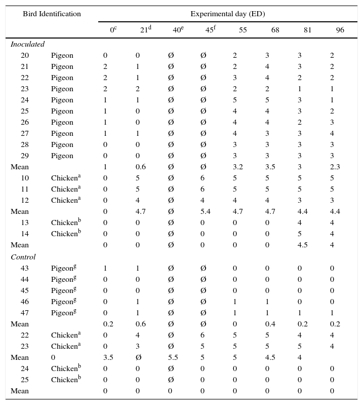
Blood samples were collected from the brachial vein of each bird on day 0 (zero), 21, 40, 45, 55, 68, 81, and 96 ED. Serum samples were stored at −20°C and used in the evaluation of immune response, as shown in Table 3.
Antibody titers (Log2X) of vaccinated (LS strain) and inoculated (SJM strain) birds during the experimental period, in Experimental Model 3.
| Bird Identification | Experimental day (ED) | ||||||||
|---|---|---|---|---|---|---|---|---|---|
| 0c | 21d | 40e | 45f | 55 | 68 | 81 | 96 | ||
| Inoculated | |||||||||
| 20 | Pigeon | 0 | 0 | Ø | Ø | 2 | 3 | 3 | 2 |
| 21 | Pigeon | 2 | 1 | Ø | Ø | 2 | 4 | 3 | 2 |
| 22 | Pigeon | 2 | 1 | Ø | Ø | 3 | 4 | 2 | 2 |
| 23 | Pigeon | 2 | 2 | Ø | Ø | 2 | 2 | 1 | 1 |
| 24 | Pigeon | 1 | 1 | Ø | Ø | 5 | 5 | 3 | 1 |
| 25 | Pigeon | 1 | 0 | Ø | Ø | 4 | 4 | 3 | 2 |
| 26 | Pigeon | 1 | 0 | Ø | Ø | 4 | 4 | 2 | 3 |
| 27 | Pigeon | 1 | 1 | Ø | Ø | 4 | 3 | 3 | 4 |
| 28 | Pigeon | 0 | 0 | Ø | Ø | 3 | 3 | 3 | 3 |
| 29 | Pigeon | 0 | 0 | Ø | Ø | 3 | 3 | 3 | 3 |
| Mean | 1 | 0.6 | Ø | Ø | 3.2 | 3.5 | 3 | 2.3 | |
| 10 | Chickena | 0 | 5 | Ø | 6 | 5 | 5 | 5 | 5 |
| 11 | Chickena | 0 | 5 | Ø | 6 | 5 | 5 | 5 | 5 |
| 12 | Chickena | 0 | 4 | Ø | 4 | 4 | 4 | 3 | 3 |
| Mean | 0 | 4.7 | Ø | 5.4 | 4.7 | 4.7 | 4.4 | 4.4 | |
| 13 | Chickenb | 0 | 0 | Ø | 0 | 0 | 0 | 4 | 4 |
| 14 | Chickenb | 0 | 0 | Ø | 0 | 0 | 0 | 5 | 4 |
| Mean | 0 | 0 | Ø | 0 | 0 | 0 | 4.5 | 4 | |
| Control | |||||||||
| 43 | Pigeong | 1 | 1 | Ø | Ø | 0 | 0 | 0 | 0 |
| 44 | Pigeong | 0 | 0 | Ø | Ø | 0 | 0 | 0 | 0 |
| 45 | Pigeong | 0 | 0 | Ø | Ø | 0 | 0 | 0 | 0 |
| 46 | Pigeong | 0 | 1 | Ø | Ø | 1 | 1 | 0 | 0 |
| 47 | Pigeong | 0 | 1 | Ø | Ø | 1 | 1 | 1 | 1 |
| Mean | 0.2 | 0.6 | Ø | Ø | 0 | 0.4 | 0.2 | 0.2 | |
| 22 | Chickena | 0 | 4 | Ø | 6 | 5 | 5 | 4 | 4 |
| 23 | Chickena | 0 | 3 | Ø | 5 | 5 | 5 | 5 | 4 |
| Mean | 0 | 3.5 | Ø | 5.5 | 5 | 5 | 4.5 | 4 | |
| 24 | Chickenb | 0 | 0 | Ø | 0 | 0 | 0 | 0 | 0 |
| 25 | Chickenb | 0 | 0 | Ø | 0 | 0 | 0 | 0 | 0 |
| Mean | 0 | 0 | 0 | 0 | 0 | 0 | 0 | 0 | |
Ø=Not tested.
Experiment 04 – Transmission of NDV by vaccinated chickens following challenge infection: This group consisted of eight pigeons and five chickens. Three SPF chickens were vaccinated with LaSota NDV Strain, with one drop of the vaccine by ocular route, on ED 0 (zero) and 21. On ED 40, vaccinated chickens were inoculated by ocular and oral routes with 0.1mL/route of a 10−1 virus dilution from a 109.0 ELD50/0.1mL stock suspension of Sao Joao do Meriti strain, and non-inoculated pigeons and two unvaccinated SPF chickens were placed in contact to detect lateral spread of the virus (infectivity) and vaccine protection. The control group consisted of two chickens vaccinated with LaSota strain on days 0 (zero) and 21, and two SPF chickens and five pigeons that were inoculated with sterile phosphate buffered saline (PBS), pH 7.2, and kept in separate facilities.
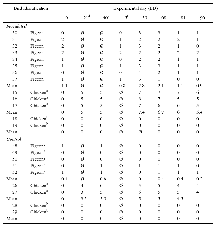
Blood samples were collected from the brachial vein of each bird on day 0 (zero), 21, 40, 45, 55, 68, 81, and 96 ED. Serum samples were stored at −20°C and used in the evaluation of immune response, as shown in Table 4.
Antibody titers (Log2X) of vaccinated (LS strain) and inoculated (SJM strain) birds during the experimental period, in Experimental Model 4.
| Bird identification | Experimental day (ED) | ||||||||
|---|---|---|---|---|---|---|---|---|---|
| 0c | 21d | 40e | 45f | 55 | 68 | 81 | 96 | ||
| Inoculated | |||||||||
| 30 | Pigeon | 0 | Ø | Ø | 0 | 3 | 3 | 1 | 1 |
| 31 | Pigeon | 2 | Ø | Ø | 1 | 2 | 2 | 2 | 1 |
| 32 | Pigeon | 2 | Ø | Ø | 1 | 3 | 2 | 1 | 0 |
| 33 | Pigeon | 2 | Ø | Ø | 2 | 2 | 2 | 2 | 2 |
| 34 | Pigeon | 1 | Ø | Ø | 0 | 2 | 2 | 1 | 1 |
| 35 | Pigeon | 1 | Ø | Ø | 1 | 3 | 3 | 1 | 1 |
| 36 | Pigeon | 0 | Ø | Ø | 0 | 4 | 2 | 1 | 1 |
| 37 | Pigeon | 1 | Ø | Ø | 1 | 3 | 1 | 0 | 0 |
| Mean | 1.1 | Ø | Ø | 0.8 | 2.8 | 2.1 | 1.1 | 0.9 | |
| 15 | Chickena | 0 | 5 | 5 | Ø | 7 | 7 | 7 | 6 |
| 16 | Chickena | 0 | 5 | 5 | Ø | 8 | 7 | 5 | 5 |
| 17 | Chickena | 0 | 5 | 5 | Ø | 7 | 6 | 6 | 5 |
| Mean | 0 | 5 | 5 | Ø | 7.4 | 6.7 | 6 | 5.4 | |
| 18 | Chickenb | 0 | 0 | 0 | Ø | 0 | 0 | 0 | 0 |
| 19 | Chickenb | 0 | 0 | 0 | Ø | 0 | 0 | 0 | 0 |
| Mean | 0 | 0 | 0 | Ø | Ø | 0 | 0 | 0 | |
| Control | |||||||||
| 48 | Pigeong | 1 | Ø | 1 | Ø | 0 | 0 | 0 | 0 |
| 49 | Pigeong | 0 | Ø | 0 | Ø | 0 | 0 | 0 | 0 |
| 50 | Pigeong | 0 | Ø | 0 | Ø | 0 | 0 | 0 | 0 |
| 51 | Pigeong | 0 | Ø | 1 | Ø | 1 | 1 | 1 | 0 |
| 52 | Pigeong | 1 | Ø | 1 | Ø | 0 | 1 | 1 | 1 |
| Mean | 0.4 | Ø | 0.6 | Ø | 0 | 0.4 | 0.4 | 0.2 | |
| 26 | Chickena | 0 | 4 | 6 | Ø | 5 | 5 | 4 | 4 |
| 27 | Chickena | 0 | 3 | 5 | Ø | 5 | 5 | 5 | 4 |
| Mean | 0 | 3.5 | 5.5 | Ø | 5 | 5 | 4.5 | 4 | |
| 28 | Chickenb | 0 | 0 | 0 | Ø | 0 | 0 | 0 | 0 |
| 29 | Chickenb | 0 | 0 | 0 | Ø | 0 | 0 | 0 | 0 |
| Mean | 0 | 0 | 0 | Ø | 0 | 0 | 0 | 0 | |
Ø=Not tested.
Microtiter HI test was performed using 4 UHA of the LaSota NDV vaccine strain propagated in the allantoic cavity of 9 to 10-day-old embryonated SPF eggs. Results were recorded as log2X of the highest reciprocal of the dilution that showed hemagglutination inhibition. HI titers equal to or greater than 4 log2 were considered positive for NDV.21
Tissue samplesAt the end of the study, after the last blood and swab collection, all birds in all four experimental models were killed in a CO2 chamber, which causes immediate death. Tissue samples from birds that died due to the pathogenic action of NDV were also collected. Immediately after death, post-mortem examination was carried out, and samples of spleen, liver, lung, and trachea were collected. These samples were placed in labeled flasks and stored at −70°C.
Statistical analysisStatistical significance was determined at 1% probability level. Antibody titers (log2X) of each group were evaluated using the Statistical Analysis Systems − SAS® (Statistical Analysis Systems Institute Inc., Cary, NC, USA). To evaluate the interaction between time and treatment, Tukey–Kramer test was used.
Viral sheddingThis assay was based on the fusion (F) gene of NDV. RNA extraction from cloacal swab samples was performed using the QIamp Viral RNA Mini Kit (Qiagen, USA), according to the manufacturer's protocol. For tissue samples, the Total RNA Extraction Kit (Mini), Real Genomics®, was used according to the manufacturer's protocol. The LaSota NDV vaccine strain (New Vac-LS® – Fort Dodge Saúde Animal Ltda) and ultra-pure water were used as positive and negative controls, respectively. Controls were submitted to the same procedures carried out with experimental chickens and pigeons. RT-PCR was performed using primers targeting a conserved region of the NDV genome (Fusion – F gene), as previously described.22 These primers are targeted to a conserved genome region that is able to amplify any NDV strain, no matter the pathogenicity, yielding a 362-bp fragment,22 and were validated in other studies.14,23–25 The primer sequence was as follows: P1F (sense) 5′-TTG ATG GCA GGC CTC TTG C-3′ and P2R (anti-sense) 5′-GGA GGA TGT TGG CAG CAT T-3′. cDNA synthesis and PCR were performed according to Jestin and Jestin.23 Samples were analyzed by electrophoresis on 1.5% agarose (w/v) gels (Invitrogen®, USA) stained with ethidium bromide 0.5μg/mL (Invitrogen®). The electrophoresis run was carried out at 100volts/50min. Positive samples showed a 362-bp DNA fragment under UV light.
For the validation of our results, the genes that encode protein F were sequenced from the material shed in the feces of the animals. These sequences were compared with standard samples: the pathogenic NDV strain São Joao do Meriti (EF534701), and the LaSota vaccine strain (EF534702). Positive samples in RT-PCR were sequenced using the BigDye Terminator Kit (Applied Biosystems, Foster City, CA, USA).
Results obtained were analyzed with the aid of the BioEdit® software. The sequences obtained were analyzed in an online database,26 confirming nucleotide homology with São Joao do Meriti and LaSota strains.
ResultsSerologyIn Experimental Model 1, non-immunized pigeons and chickens were experimentally infected. Although pigeons remained clinically healthy during the whole trial, antibody production was fast, reaching high HI titers from ED 15 to ED 56, as presented in Table 1. However, all five chickens died on the 7th day after virus inoculation (ED 7), and blood samples could not be collected.
In pigeons of Experimental Model 2, vaccination led to high antibody titers even before experimental infection. After experimental infection (ED 61), higher titers of HI antibodies were detected on ED 85, which remained practically stable until the end of the study, on ED 128. In sentinel chickens of Experimental Model 2, which were placed in contact with infected pigeons on ED 66, no seroconversion (Table 2) and no clinical signs compatible with ND were observed.
In Experimental Model 3, at the moment the chickens were placed in contact with infected birds (ED 45), vaccinated chickens already presented high levels of antibodies, as shown in Table 3. In unvaccinated chickens (sentinels), seroconversion was only observed on ED 81, whereas in experimentally infected pigeons, high titers of antibodies were detected from ED 55 on, that is, since the 15th day after the inoculation with strain SJM.
In Experimental Model 4, it was observed that vaccinated chickens presented high titers of antibodies, and the highest level was observed on ED 55 (Table 4) due to the experimental infection carried out after vaccination. Sentinel pigeons seroconverted, although they did not present high antibody titers. Similar to what was observed for vaccinated chickens, higher antibody titers in pigeons were observed on ED 55. However, no antibodies were detected in sentinel chickens throughout the experimental model.
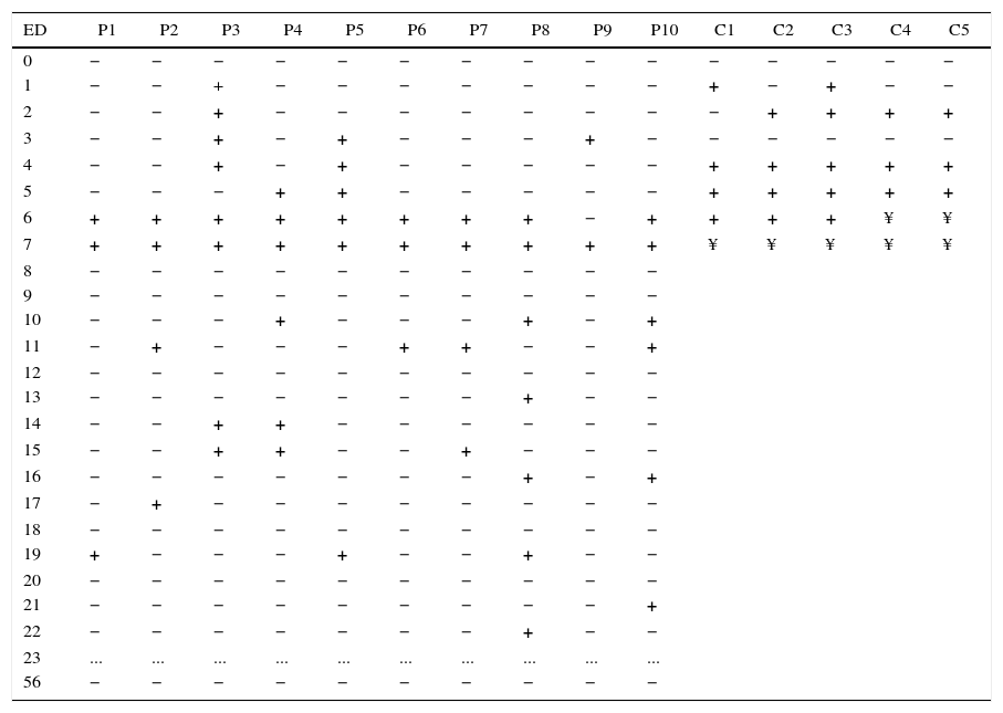
Evaluation of viral sheddingIn birds belonging to Experimental Model 1, NDV was detected in cloacal swabs collected after the first 24h of experimental infection. Thus, NDV was detected in all chickens on ED 4 and 5, and in all pigeons on ED 7. Besides, all chickens died on ED 7. In pigeons, sporadic and intermittent shedding of the NDV was observed from ED 5 to ED 22 (Table 5).
Cloacal shedding of NDV as determined by RT-PCR in samples collected from pigeons and chickens experimentally infected with SJM strain, in Experimental Model 1.
| ED | P1 | P2 | P3 | P4 | P5 | P6 | P7 | P8 | P9 | P10 | C1 | C2 | C3 | C4 | C5 |
|---|---|---|---|---|---|---|---|---|---|---|---|---|---|---|---|
| 0 | − | − | − | − | − | − | − | − | − | − | − | − | − | − | − |
| 1 | − | − | + | − | − | − | − | − | − | − | + | − | + | − | − |
| 2 | − | − | + | − | − | − | − | − | − | − | − | + | + | + | + |
| 3 | − | − | + | − | + | − | − | − | + | − | − | − | − | − | − |
| 4 | − | − | + | − | + | − | − | − | − | − | + | + | + | + | + |
| 5 | − | − | − | + | + | − | − | − | − | − | + | + | + | + | + |
| 6 | + | + | + | + | + | + | + | + | − | + | + | + | + | ¥ | ¥ |
| 7 | + | + | + | + | + | + | + | + | + | + | ¥ | ¥ | ¥ | ¥ | ¥ |
| 8 | − | − | − | − | − | − | − | − | − | − | |||||
| 9 | − | − | − | − | − | − | − | − | − | − | |||||
| 10 | − | − | − | + | − | − | − | + | − | + | |||||
| 11 | − | + | − | − | − | + | + | − | − | + | |||||
| 12 | − | − | − | − | − | − | − | − | − | − | |||||
| 13 | − | − | − | − | − | − | − | + | − | − | |||||
| 14 | − | − | + | + | − | − | − | − | − | − | |||||
| 15 | − | − | + | + | − | − | + | − | − | − | |||||
| 16 | − | − | − | − | − | − | − | + | − | + | |||||
| 17 | − | + | − | − | − | − | − | − | − | − | |||||
| 18 | − | − | − | − | − | − | − | − | − | − | |||||
| 19 | + | − | − | − | + | − | − | + | − | − | |||||
| 20 | − | − | − | − | − | − | − | − | − | − | |||||
| 21 | − | − | − | − | − | − | − | − | − | + | |||||
| 22 | − | − | − | − | − | − | − | + | − | − | |||||
| 23 | ... | ... | ... | ... | ... | ... | ... | ... | ... | ... | |||||
| 56 | − | − | − | − | − | − | − | − | − | − |
ED, experimental day; ¥, dead; −, negative; +, positive; P, pigeon; C, chicken.
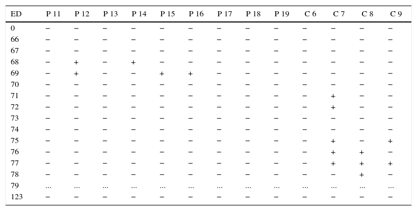
In birds of Experimental Model 2, low frequency of detection of NDV in pigeons was observed on ED 68, persisting for only two days. On the other hand, shedding of the virus was observed in 75% (3/4) of the unvaccinated chickens (sentinels). It was also observed that virus shedding in the chickens began after experimentally infected birds stopped shedding it, that is, on ED 71 (Table 6).
Cloacal shedding of NDV as determined by RT-PCR in samples collected from sentinel birds and from pigeons and chickens vaccinated (LS strain) and experimentally infected with SJM strain, in Experimental Model 2.
| ED | P 11 | P 12 | P 13 | P 14 | P 15 | P 16 | P 17 | P 18 | P 19 | C 6 | C 7 | C 8 | C 9 |
|---|---|---|---|---|---|---|---|---|---|---|---|---|---|
| 0 | − | − | − | − | − | − | − | − | − | − | − | − | − |
| 66 | − | − | − | − | − | − | − | − | − | − | − | − | − |
| 67 | − | − | − | − | − | − | − | − | − | − | − | − | − |
| 68 | − | + | − | + | − | − | − | − | − | − | − | − | − |
| 69 | − | + | − | − | + | + | − | − | − | − | − | − | − |
| 70 | − | − | − | − | − | − | − | − | − | − | − | − | − |
| 71 | − | − | − | − | − | − | − | − | − | − | + | − | − |
| 72 | − | − | − | − | − | − | − | − | − | − | + | − | − |
| 73 | − | − | − | − | − | − | − | − | − | − | − | − | − |
| 74 | − | − | − | − | − | − | − | − | − | − | − | − | − |
| 75 | − | − | − | − | − | − | − | − | − | − | + | − | + |
| 76 | − | − | − | − | − | − | − | − | − | − | + | + | − |
| 77 | − | − | − | − | − | − | − | − | − | − | + | + | + |
| 78 | − | − | − | − | − | − | − | − | − | − | − | + | − |
| 79 | ... | ... | ... | ... | ... | ... | ... | ... | ... | ... | ... | ... | ... |
| 123 | − | − | − | − | − | − | − | − | − | − | − | − | − |
ED, experimental day; −, negative; +, positive; P, pigeon; C, chicken.
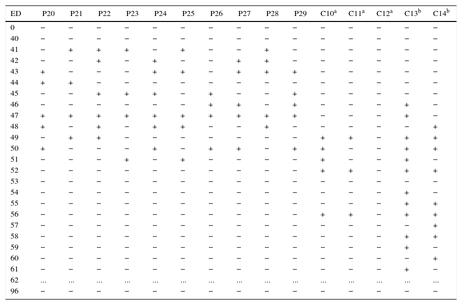
In Experimental Model 3, it was observed that challenged pigeons shed NDV 24h after the challenge, similar to that observed in Experimental Model 1. In spite of the short duration, the greatest number of pigeons shedding the virus occurred on ED 47, that is, seven days after initial virus infection, persisting until ED 51 in only two birds (Table 7). The two unvaccinated chickens presented the greatest frequency of virus shedding when compared with the vaccinated ones. Only one of them did not show the virus in cloacal swabs.
NDV as determined by RT-PCR in cloacal samples collected from sentinel birds and from chickens vaccinated (LS strain) and pigeons and chickens experimentally infected with SJM strain, in Experimental Model.
| ED | P20 | P21 | P22 | P23 | P24 | P25 | P26 | P27 | P28 | P29 | C10a | C11a | C12a | C13b | C14b |
|---|---|---|---|---|---|---|---|---|---|---|---|---|---|---|---|
| 0 | − | − | − | − | − | − | − | − | − | − | − | − | − | − | − |
| 40 | − | − | − | − | − | − | − | − | − | − | − | − | − | − | − |
| 41 | − | + | + | + | − | + | − | − | + | − | − | − | − | − | − |
| 42 | − | − | + | − | + | − | − | + | + | − | − | − | − | − | − |
| 43 | + | − | − | − | + | + | − | + | + | + | − | − | − | − | − |
| 44 | + | + | − | − | − | − | − | − | − | − | − | − | − | − | − |
| 45 | − | − | + | + | + | − | + | − | − | + | − | − | − | − | − |
| 46 | − | − | − | − | − | − | + | + | − | + | − | − | − | + | − |
| 47 | + | + | + | + | + | + | + | + | + | + | − | − | − | + | − |
| 48 | + | − | + | − | + | + | − | − | + | − | − | − | − | − | + |
| 49 | − | + | + | − | − | − | − | − | − | − | + | + | − | + | + |
| 50 | + | − | − | − | + | − | + | + | − | + | + | − | − | + | + |
| 51 | − | − | − | + | − | + | − | − | − | − | + | − | − | + | − |
| 52 | − | − | − | − | − | − | − | − | − | − | + | + | − | + | + |
| 53 | − | − | − | − | − | − | − | − | − | − | − | − | − | − | − |
| 54 | − | − | − | − | − | − | − | − | − | − | − | − | − | + | − |
| 55 | − | − | − | − | − | − | − | − | − | − | − | − | − | + | + |
| 56 | − | − | − | − | − | − | − | − | − | − | + | + | − | + | + |
| 57 | − | − | − | − | − | − | − | − | − | − | − | − | − | − | + |
| 58 | − | − | − | − | − | − | − | − | − | − | − | − | − | + | + |
| 59 | − | − | − | − | − | − | − | − | − | − | − | − | − | + | − |
| 60 | − | − | − | − | − | − | − | − | − | − | − | − | − | − | + |
| 61 | − | − | − | − | − | − | − | − | − | − | − | − | − | + | − |
| 62 | ... | ... | ... | ... | ... | ... | ... | ... | ... | ... | ... | ... | ... | ... | ... |
| 96 | − | − | − | − | − | − | − | − | − | − | − | − | − | − | − |
ED, experimental day; −, negative; +, positive; P, pigeon; C, chicken.
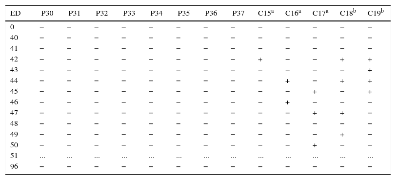
In Experimental Model 4 (Table 8), NDV was detected both in vaccinated and unvaccinated chickens, but not in sentinel pigeons. In vaccinated chicken, shedding of the virus began on ED 42, that is, 48h after the experimental infection of the three vaccinated and challenged chicken. Viral shedding was fast and did not last longer than three alternate days. However, the two sentinel chickens that shed the virus during four days did not die.
NDV as determined by RT-PCR in cloacal samples collected from chickens vaccinated (LS strain) and experimentally infected with SJM strain in contact with sentinel pigeons and chickens, in Experimental Model 4.
| ED | P30 | P31 | P32 | P33 | P34 | P35 | P36 | P37 | C15a | C16a | C17a | C18b | C19b |
|---|---|---|---|---|---|---|---|---|---|---|---|---|---|
| 0 | − | − | − | − | − | − | − | − | − | − | − | − | − |
| 40 | − | − | − | − | − | − | − | − | − | − | − | − | − |
| 41 | − | − | − | − | − | − | − | − | − | − | − | − | − |
| 42 | − | − | − | − | − | − | − | − | + | − | − | + | + |
| 43 | − | − | − | − | − | − | − | − | − | − | − | − | + |
| 44 | − | − | − | − | − | − | − | − | − | + | − | + | + |
| 45 | − | − | − | − | − | − | − | − | − | − | + | − | + |
| 46 | − | − | − | − | − | − | − | − | − | + | − | − | − |
| 47 | − | − | − | − | − | − | − | − | − | − | + | + | − |
| 48 | − | − | − | − | − | − | − | − | − | − | − | − | − |
| 49 | − | − | − | − | − | − | − | − | − | − | − | + | − |
| 50 | − | − | − | − | − | − | − | − | − | − | + | − | − |
| 51 | ... | ... | ... | ... | ... | ... | ... | ... | ... | ... | ... | ... | ... |
| 96 | − | − | − | − | − | − | − | − | − | − | − | − | − |
ED, experimental day; −, negative; +, positive; P, pigeon; C, chicken.
In relation to the evaluation of the presence of the virus in tissue samples from pigeons, detection was not uniform in the four experimental models. From the samples of organs collected from 37 pigeons (not considering the animals in the control group), NDV was detected in 29.73% (11/37) of spleen samples, in 16.22% (6/37) of liver samples, in 10.81% (4/37) of lung samples, and in 2.7% (1/37) of trachea samples. Viruses were observed in all tissue samples of only one individual in Experimental Model 2.
In chickens, the distribution of positive tissue samples in each experimental model was also uneven. The frequency of positive results in the spleen, liver, lungs, and trachea was, respectively, 42.1% (8/19), 36.85% (7/19), 31.5% (6/19), and 5.26% (1/19). None of the tissue samples that came from control birds was positive for NDV.
All genetic material shed in the samples (swabs and tissues) that were positive in RT-PCR was sequenced and showed to be genetically homologous with São Joao de Meriti (EF534701) strain.
DiscussionThis study was designed to evaluate, under experimental and controlled conditions, humoral antibody response, virus excretion, and contact transmission of pathogenic Newcastle disease Virus of chicken origin used in the experimental infection of pigeons (C. livia) and chickens (Gallus gallus). A pathogenic strain that was isolated from a Brazilian outbreak in the 1970s was used in the experimental infection.
In general, it is accepted that the dissemination of NDV among pigeons and chickens takes place in natural conditions by indirect contact, or when these species get in contact with each other.27 Experimental models of infection, under controlled conditions, enable better evaluation of the infectivity and transmissibility of the virus, besides the evaluation of vaccine effects (particularly in relation to mortality), onset of clinical signs of the disease, and prevention and reduction of virus shedding. The oculonasal route was used to reproduce the natural route of infection. This type of transmission by direct contact has already been described among ducks and chickens, as well as environmental contamination by viruses shed in the feces.28 It is important to determine whether pigeons would be able to assemble adequate antibody response against LaSota vaccine strain and get protected against the challenge with APMV-1, considering the absence of commercial vaccines for PPMV and APMV in pigeons.
Therefore, given the results observed in Experimental Model 1 and the already expected death of all chickens on ED 6, it was confirmed that São Joao do Meriti (SJM) strain is highly pathogenic to this species, as it had already been demonstrated.14 Besides, these results validate those obtained in the other Experimental Models (models 2, 3, and 4). Chickens that died showed respiratory signs, and secondarily, nervous signs, both of which are compatible with ND.
In relation to the analysis of the genome of the virus, it was demonstrated that pigeons shed the virus intermittently throughout the experimental period since ED 1, persisting until ED 22. Peak virus shedding was observed on ED 7, when NDV was detected in cloacal swabs collected from all pigeons. In chickens, virus detection took place from ED 1 to ED 6. On ED 5 and 6, virus was detected in all birds until they died. The results obtained for the virus shedding period after experimental infection in pigeons in this study were similar to those reported by other authors.12,29,30 However, the use of an exotic Newcastle disease virus strain that was responsible for the most recent outbreak in California (USA) in an experimental infection of adult pigeons yielded a morbidity rate of 20% and two pigeons were euthanized because they displayed severe clinical signs of ND.13 These results are important to compare the different strains of NDV responsible for outbreaks in commercial poultry from different countries, and to evaluate their virulence to different avian species. The fact that NDV was excreted for up to 20 days (24 dpi) without any clinical signs of the disease is particularly significant for the organization of control programs and knowledge on NDV epidemiology.14 These data reinforce the importance of studying virus-host relationships focusing on each individual virus strain, as each strain shows specific pathobiological patterns.
The different behavior of NDV strain when inoculated in different hosts was also observed in the present study, once 100% of the chickens died in a short period of time, whereas pigeons that were in contact with these chickens (or were experimentally infected) remained healthy, without any clinical signs compatible with ND.
Chickens infected by ocular and oral route with strain APMV-1, which is pathogenic to pigeons, did not present clinical signs of ND, either.30 Still, these findings confirm the results that showed that chicken infected with a PPMV-1 strain of high pathogenicity to pigeons remained clinically healthy after the challenge.15,31 It was reported that virus strains isolated from wild birds need some passages in chicken to be adapted to this species and, consequently, to show increased pathogenicity.32 Therefore, it may be inferred that, in natural field conditions, ND outbreaks may occur if a pathogenic strain is introduced in a new area that, until then, had not been challenged.33
The reason for such different viral behavior in different bird species has been investigated. The analysis of the molecular basis of Paramyxovirus pathogenesis shows that virulence is a result of a complex relationship between the virus and its host, so that the ability of a virus strain to induce the disease is intimately related with the presence of proteases that are able to lyse some polypeptide structures of the virus.34,35 Nevertheless, host specificity is not a typical characteristic of APMV-1, since a great number of hosts are susceptible to different NDV strains.34
From a serological viewpoint, the levels of antibodies detected by HI in Experimental Model 1 presented a common pattern observed in this disease, with the peak HI around ED 15 (Table 1), followed by a gradual decrease. On the other hand, the other models showed that when SJM strain was used in vaccinated animals (chickens and pigeons), the presence of antibodies led to a significant reduction in virus shedding. Besides, it was observed that LaSota strain was able to protect the chickens during ND clinical phase, even when they were in contact with infected birds. These results had already been reported by Jeon et al.9
Persistent virus shedding, even in immunized animals, may suggest that vaccinated birds may remain as reservoirs or sources of infection, especially their feces and/or contaminated material.9,36 On the other hand, these authors reported that, in some ND outbreaks, reduction in virus shedding is of crucial importance in the control of virus dissemination. That is, although vaccination protects the bird against the disease, it does not prevent infection and consequent virus shedding.8,10,36
In fact, there is a correspondence between high titers of hemagglutination inhibition antibodies and resistance of the bird to the challenge of a virulent NDV strain, once high HI titers may lead to protection lasting for up to six months.37 In this context, HI antibody titers over 64 were able to protect the birds from the challenges, as demonstrated in Models 2, 3, and 4 (Tables 2–4). Kapczynski and King8 agree with this finding and show that high titers of HI antibodies (≥28) rule out the use of larger doses of the vaccines, because they provide similar protection against the challenge, including in terms of virus shedding. In broilers, a large part of the flock (≥85%) has to have high antibody titers (log2 titer≥3) after vaccination to prevent any epidemic spread in vaccinated populations.6
In relation to prophylactic measures used against ND, vaccination is indispensable and routinely applied in the poultry production sector. Therefore, in the design of the present study, LaSota virus strain was chosen because it is the most common strain used in the immunization of poultry flocks, given its ability to induce adequate protection level, no matter the pathogenic strain involved.4,9 However, there are reports on failures in immunization programs in outbreaks involving highly virulent strains.30,36 This fact, though, was not observed in the present study, in which LaSota strain was able to prevent the occurrence of clinical signs in chickens infected with highly pathogenic strains (SJM). Mild infections caused by NDV vaccine can stimulate local immunity and prevent the occurrence of clinical signs. It should be emphasized that these results are related to a vaccination program carried out in controlled conditions, in which the correct vaccine dose was used in all animals. According to Dortmans et al.,4 a live vaccine adapted to an outbreak strains does not increase protection nor reduce viral shedding compared with a classic live vaccine. Therefore, in a challenge in field conditions, the immune response of a flock against a vaccine strain may not be uniform. Vaccination of a large number of chickens against ND is usually carried out using non-virulent live virus administrated by spray or in drinking water. These administration techniques usually produce considerable variation in individual antibody responses.6
Although vaccination protected the animals, NDV was detected in the feces of 4 from 10 vaccinated pigeons (Model 2) only on ED 3 and 4, that is, when shedding was significantly lower (p<0.01), compared with unvaccinated pigeons of Experimental Model 1. Besides, it should be emphasized that 3 from the 4 chickens in contact with these pigeons have shed NDV without seroconversion or clinical signs of the disease. Conventional vaccine strains may protect against clinical disease caused by virulent NDV, but virus infection can still occur in vaccinated birds.6,8,9 However, virus shedding may be significantly reduced when compared with unvaccinated chickens.9
The fact of some birds shed NDV without seroconversion or clinical signs of the disease may be due to a change in the infectivity of the pathogenic strain, with the consequent shedding of defective virus particles or particles modified by vaccination. In these conditions, a modified virus strain would be unable to stimulate the immune system of the bird, as proven by the lack of antibodies or clinical signs of the disease (Tables 2 and 4). Another hypothesis is related to the low fidelity of RNA polymerase, as well as to the possibility of production of new virus particles that are intimately related, but different from each other, called quasispecies.38 This finding was not observed in the present study, once positive samples in RT-PCR were genetically homologous with the São Joao do Meriti strain.
In this context, it is important to emphasize that RT-PCR enables the detection of virus genome fragments, but not the detection of the virion. Therefore, sentinel chickens would surely get in contact with fragments of the NDV genome that are unable to cause the disease and, consequently, to produce an immune response in these birds. In these conditions, fragments of the NDV genome have to be detected by a technique of high analytical sensitivity, such as RT-PCR. This characteristic of molecular techniques is one of the differentials of these techniques compared with virus isolation, as samples that lost their infectivity may still be detected by RT-PCR, as reported by Kho et al.39 Care should be taken when drawing conclusions based on RT-PCR or Real Time PCR, especially when considering testing of virus infection and elucidation of virus epidemiology. Although these methods are sensitive, results obtained with them sometimes contradict clinical studies on virus isolation in NDV/PPMV-1 outbreaks.15,40
In the evaluation of the presence of the virus in tissue samples, the different organs showed different rates of detection. In general, frequency was greater in samples of spleen, followed by liver, lung, and trachea, both in pigeons and chickens. In general, non-uniform distribution of positive tissue samples may be attributed to the lack of infection of the tissues analyzed at the moment of collection, or the action of the immune system in the different organs. In an experimental infection with a PPMV-1 strain, not all tissue samples from dead pigeons had detectable virus RNA, which was only detected in six tissue samples, including the kidneys, lungs, brain, and trachea.15 The greatest frequency of positive spleen samples found in the present study corroborated the results obtained by Wakamatsu et al.,41 indicating the spleen as the main target organ affected by the virus. Accordingly, pigeons experimentally infected with PPMV-1 strains and analyzed by RT-PCR presented greater detection rates of NDV in the spleen, lungs, cecum, tonsils, and kidneys.31 Therefore, these authors recommend that these organs are preferentially considered in NDV detection. Kho et al.39 and Barbezange and Jestin42 observed that RT-PCR was able to detect NDV in brain, lung, trachea, and spleen samples, at the same rates in these different organs. Differently, Creelan et al.43 observed that detection of NDV in tissue samples should not be the main focus in an attempted diagnosis, due to the fact that there is no tissue tropism pattern in different virus strains.
Several differences have been observed in virus strains isolated from different species of birds, mainly free-living birds in different locations throughout the planet. It is very important to devise more accurate and precise methods to evaluate the virulence of NDV isolates, especially in hosts other than chicken, and further studies are needed to investigate the determinant factors of interspecies transmission.15 These strains circulate in bird populations, generally without causing the disease, in a parasite vs. host balance. When these free-living birds get in contact with commercial birds, outbreaks may occur, with considerable losses to countries that raise and export poultry and poultry products. Biosecurity measures associated with vaccination programs as postulated by the International Animal Health Code are crucial for the preservation of the virulent NDV-free status for industrial poultry in Brazil.19
Conflicts of interestThe authors declare no conflicts of interest.
The authors wish to thank Bosque Municipal Dr. Fábio Barreto, Ribeirão Preto, São Paulo, Brazil. This research was carried out with financial support from the Fundação de Amparo à Pesquisa do Estado de São Paulo and from the Conselho Nacional de Desenvolvimento Científico e Tecnológico-CNPq, Brazil.


